Share
Start here
Soft-tissue sarcoma at a glance
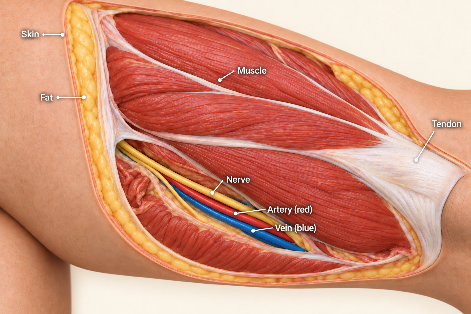
Soft-tissue sarcoma is a group of uncommon cancers that start in the tissues that support, connect or move the body. These include muscle, fat, blood vessels, the coverings around nerves and fibrous tissue, such as tendons that connect muscle to bone. Sarcoma can develop in an arm or leg, the body wall, the head and neck, or deep inside the abdomen (belly).
The exact kind of sarcoma is called its subtype. Its grade describes how abnormal the tumour looks under a microscope and helps estimate how quickly it may grow or spread. The subtype, grade and location all affect treatment. A medicine that helps one kind of sarcoma may not help another.
This guide mainly covers soft-tissue sarcoma in adults. Cancers that begin in bone have a separate bone cancer guide. Gastrointestinal stromal tumour (GIST), which starts in the wall of the digestive tract, has its own GIST guide. Some childhood sarcomas and related soft-tissue conditions also need different care; these are explained below.
Specialist assessment before removal
A lump that is growing, lies deep beneath the skin or seems unusual should be assessed before an operation to remove it. Sarcomas can be small or painless. Most lumps are not cancer, but the absence of pain does not tell you whether a lump is harmless.
Scans show the lump and the tissues around it. A biopsy removes a small sample so that a specialist can examine it. Planning the scans and biopsy with a sarcoma team helps them choose the right operation and avoid a cut or needle route that could complicate later surgery.
If sarcoma is discovered only after a lump has been removed, the team will review the tissue report, scans and operation notes. Another operation may be needed to remove tissue around the earlier surgery. This is called re-excision. An unexpected diagnosis after surgery does not mean that effective treatment is no longer possible.
Symptoms and warning signs
A lump that slowly or quickly gets bigger is a common symptom. Other symptoms depend on where the tumour is:
- A tumour deep in the abdomen may cause fullness, pain or a change in bowel habits. It may become quite large before it is noticed.
- Pressure on a nerve may cause pain, numbness or weakness.
- A tumour near a joint may make movement difficult.
A persistent or changing lump should be checked, even if it does not hurt. These symptoms can have causes other than sarcoma.
When to seek urgent help
Seek urgent medical help for severe pain with sudden swelling, heavy bleeding, repeated vomiting with a swollen abdomen, or new weakness or loss of bladder or bowel control. A newly painful, swollen arm or leg can also be caused by a blood clot or infection and needs prompt assessment. Do not wait for your next routine appointment.
Where to begin
You do not need to learn every treatment name at once. Start with three things: the full name of the diagnosis, where the cancer is, and what the team recommends next. A relative or friend can join an appointment if you would find that helpful.
Keep copies of your reports and a list of medicines, allergies and other health conditions. Include supplements, herbal remedies and medicines bought without a prescription. Tell the team about earlier cancer treatment, pregnancy or breastfeeding, and anything that could make attending treatment difficult. Do not stop prescribed medicines without advice.
This guide describes possible treatments. Your test results, general health and priorities determine which ones fit your situation. If advice differs from something you have read, ask the team to explain how it applies to your particular sarcoma.
Understand
What the different sarcoma names mean
Sarcoma is a family of cancers, rather than one disease. The laboratory report identifies the subtype by looking at the cells and, when needed, testing their genes. Genes are instructions inside cells. You do not need to memorise the names, but it helps to know the exact name on your report.
- Liposarcoma is a cancer with features of fat cells. It has several forms. Well-differentiated cells still look quite like fat cells; dedifferentiated areas have changed into a more aggressive-looking form. Myxoid and pleomorphic liposarcomas are other distinct forms. “Myxoid” describes a jelly-like material around the cells; “pleomorphic” means the cells vary greatly in size and shape.
- Leiomyosarcoma has features of smooth muscle, the muscle that works without conscious control in places such as the womb, bowel and blood vessels. It can start in the womb (uterus) or elsewhere.
- Undifferentiated pleomorphic sarcoma has very varied-looking cancer cells that do not clearly match a particular normal tissue type.
- Synovial sarcoma is a distinct subtype that often occurs near joints, but can also develop elsewhere. Its name does not mean that it must start inside a joint.
- Angiosarcoma has features of cells that line blood vessels or lymph vessels, which help drain fluid from tissues.
- Malignant peripheral nerve sheath tumour is a cancer involving the tissues around a nerve.
The name may describe how the tumour cells look rather than the part of the body where the lump was noticed. These differences matter when choosing surgery, medicines, follow-up tests or a clinical trial, which is a research study involving patients.
Grade, stage and spread: different pieces of information
Grade helps estimate how a tumour may behave. In a commonly used sarcoma grading system, the pathologist, a doctor who examines tissue, considers three features:
- How closely the tumour resembles normal tissue, called differentiation.
- How many cells are dividing to make new cells, called the mitotic count.
- How much of the tumour contains dead cells, called necrosis.
These features are combined to give grade 1 (low), grade 2 (intermediate) or grade 3 (high). A higher grade generally means a greater chance of growth or spread. This grading system does not apply to every subtype. Even a low-grade tumour may need treatment because it can grow into nearby tissues.
Stage brings together the tumour's size, grade and whether it has spread. It is different from grade. A grade 3 result does not automatically mean stage 3. The staging diagram under Planning your treatment explains the numbered stages and the exceptions for different body sites.
Cancer that has stayed where it began is called localised. Cancer that has spread to a distant part of the body is called metastatic; the new cancer deposits are called metastases. A sarcoma deposit in the lung is still sarcoma, rather than a new lung cancer. The lungs are a common place for many sarcomas to spread, but some subtypes have other patterns.
Lymph nodes are small structures that help the body fight infection. Most soft-tissue sarcomas do not commonly spread to nearby lymph nodes, so removing nodes is not part of every operation. The team checks the nodes when the subtype or test findings make this necessary.
Why sarcoma happens
Most people with sarcoma have no clear cause or recognised risk factor. An ordinary injury is not generally the cause, although it may draw attention to a lump that is already there.
A small number of sarcomas are associated with earlier radiotherapy, which uses radiation to treat cancer; long-lasting swelling from poor lymph drainage, called lymphoedema; or an inherited condition. Examples of inherited conditions include neurofibromatosis type 1, which can affect nerves, and Li–Fraumeni syndrome, which increases the risk of several cancers.
If your personal or family history suggests an inherited risk, genetic counselling can explain whether testing might help you or your relatives. Most gene changes found in a sarcoma developed within the tumour itself. They are not necessarily present in the rest of the body and do not automatically mean that your children are at risk.
Diagnoses that need a different approach
Some conditions are treated by sarcoma teams but follow their own treatment plans:
- GIST often has changes in genes called KIT or PDGFRA. These genes help control cell growth, and their exact changes can guide medicines. The separate GIST guide explains this in detail.
- Rhabdomyosarcoma has features of developing skeletal muscle, the muscle used to move the body. Children and many teenagers need treatment planned with childhood cancer specialists.
- Ewing sarcoma can start in bone or soft tissue. When it starts outside bone, it may be called extraskeletal Ewing sarcoma. It uses Ewing-specific treatment, rather than the usual adult soft-tissue sarcoma plan.
- Desmoid tumour can grow into nearby tissues but does not spread to distant organs. Some tumours can be monitored before treatment is needed.
- Tenosynovial giant cell tumour grows in or around joints and tendon coverings. It can damage a joint and affect movement, but is usually not cancer. Its treatment is different from treatment for malignant sarcoma.
Tests and diagnosis
What the scans show
Scans help the team see the tumour's size, its position and whether it has spread. They are usually planned before the biopsy so that the sample can be taken safely from the most useful area.
- Magnetic resonance imaging (MRI) uses a strong magnet to produce detailed pictures. It is often used for an arm, leg or body-wall tumour to show its relationship to muscles, nerves and blood vessels.
- Computed tomography (CT) combines X-rays into detailed pictures. It is often used for tumours in the abdomen or the retroperitoneum, the space behind the abdominal organs. A chest CT commonly checks the lungs for spread.
- Positron emission tomography combined with CT (PET-CT) uses a small amount of radioactive tracer to show areas of activity alongside detailed pictures. It can help in selected situations, but is not needed for every sarcoma.
Some subtypes need scans of other areas because of where they tend to spread. Ask what each scan is intended to show. A second abnormal area on a scan is not automatically another cancer deposit; it may need further assessment.
How the biopsy is done
A core needle biopsy removes small cylinders of tissue through a hollow needle. It is often the first tissue test for a suspected sarcoma. The team plans the route carefully, avoiding unnecessary tissues and allowing the needle track to be removed during the later operation if needed.
Occasionally, a different biopsy method is more suitable, for example for a very small lump just under the skin or when a needle sample cannot give an answer. The choice should be planned with the specialist team. If a sample is too small or does not represent the tumour well, another sample may be needed.
Understanding your pathology report
The pathology report gives the result of examining the biopsy. Sarcoma types can look similar, so review by a pathologist experienced in sarcoma is particularly useful. The report usually identifies the subtype and, where appropriate, the grade.
The laboratory may use special stains to identify proteins in the cells. This is called immunohistochemistry. It may also use gene tests to look for a fusion, where parts of two genes have joined, or an amplification, where extra copies of a gene are present. These findings can help confirm the diagnosis. They do not, by themselves, mean that the condition was inherited.
Your report may mention differentiation, mitotic count and necrosis, the features used in grading described above. If the wording is unclear, ask the team to explain which findings affect your treatment and which are simply describing the sample.
Gene and protein tests that may guide medicines
Molecular testing looks for particular changes in the tumour's genes or proteins. Some changes help identify the subtype. Others may reveal a target for a medicine. A targeted treatment acts on a particular growth signal or weakness in the cancer.
For example, a fusion involving an NTRK gene, or a suitable change involving the ALK gene, may allow a targeted medicine in certain circumstances. NTRK and ALK are gene names used on laboratory reports; both are involved in cell-growth signals. Finding any change in one of these genes is not enough: the exact change and diagnosis matter.
For advanced cancer, meaning cancer that cannot readily be removed or has spread, a test that checks several genes together may help identify a medicine or clinical trial. Not every result leads to a useful treatment. Ask whether there is enough biopsy tissue for the tests and how a result could change the plan.
Some cell treatments require separate blood and tumour tests. One example is afamitresgene autoleucel, often shortened to afami-cel, for selected people with advanced synovial sarcoma after chemotherapy. Chemotherapy uses medicines to kill cancer cells or stop them multiplying.
A blood test checks the human leukocyte antigen (HLA) tissue type, which helps the immune system recognise cells. A tumour test checks for melanoma-associated antigen A4 (MAGE-A4), a protein this treatment targets. The protein's name does not mean that the sarcoma is melanoma. Both results must be suitable. The treatment and its other requirements are explained under Treatment options.
Checking your health before treatment
Blood tests and other checks show how well your heart, kidneys and other organs are working. The team also considers your movement, nutrition, symptoms and everyday activities. Some chemotherapy medicines, including doxorubicin, need particular attention to heart health.
If treatment may affect a limb, organ or nerve, a rehabilitation assessment can help plan exercises, equipment or support for recovery. If having children in the future matters to you, discuss fertility before treatment starts. Some options to protect fertility need to be arranged beforehand.
Preparing for your appointment
For a consultation or second opinion, bring a compact disc (CD) containing your scan images in Digital Imaging and Communications in Medicine (DICOM) format and the software needed to view them, along with the written scan reports. DICOM is the standard format used to store medical scan images. The actual images help the team compare findings and plan treatment.
Bring the biopsy report too. Ask whether the specialist needs the pathology slides or tissue blocks, the glass slides and stored tissue used for laboratory examination. Keep originals safe and agree how borrowed material will be returned. Bring your medicine list and the questions that matter most to you.
Before a scan or biopsy, mention kidney problems, earlier reactions to contrast dye, implanted devices, blood-thinning medicines, diabetes medicines and any possibility of pregnancy. Contrast is a substance used in some scans to make structures easier to see. Follow the centre's instructions about eating, drinking and medicines; do not make changes without advice.
Ask when and how results will be explained. An uncertain result does not automatically mean that the cancer is worse. Sometimes a repeat test or discussion between specialists is needed to reach a clear answer.
Planning your treatment
Understanding your stage
Site-specific staging for soft tissue sarcoma
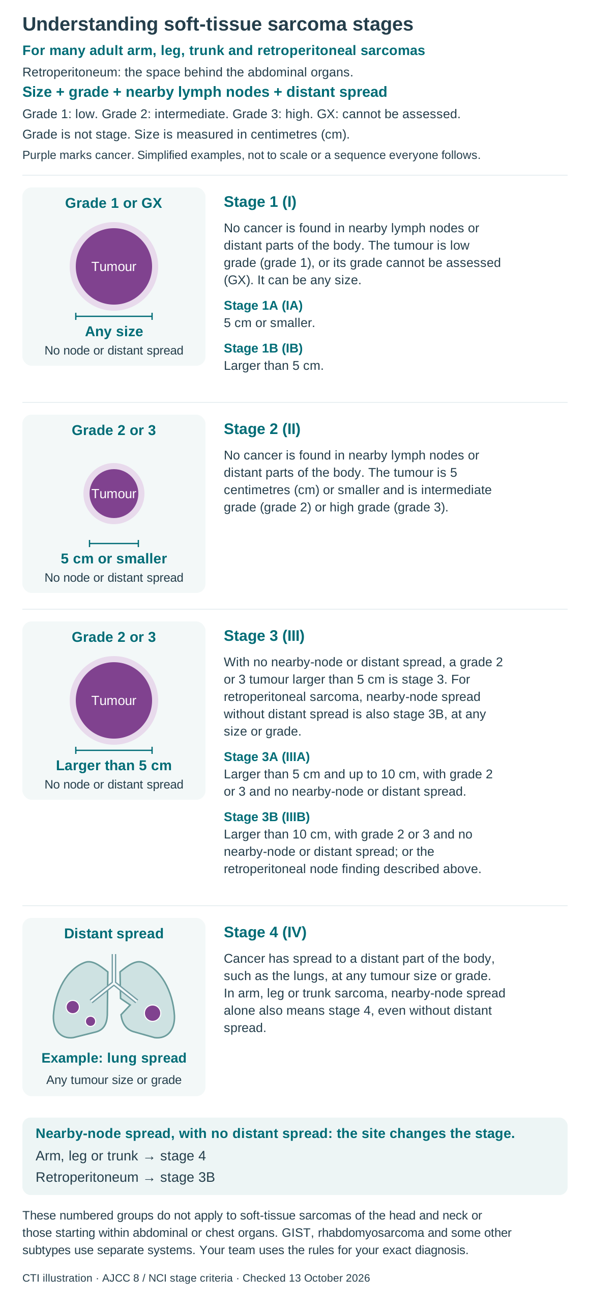
Stage brings together the tumour’s size, its grade and whether it has spread. The diagram below applies to many adult sarcomas in an arm or leg, the trunk (body wall), or the retroperitoneum (the space behind the abdominal organs). Different rules apply to some other sites and subtypes.
Lymph nodes help the body fight infection. Regional nodes are the nearby nodes that collect fluid from the area where the tumour began. Distant spread means cancer has reached other parts of the body, such as the lungs. Stages describe what has been found; they are not steps that everyone will pass through.
Grade describes how abnormal the tumour looks under a microscope and helps estimate how it may behave. In this system, grade 1 is low, grade 2 intermediate and grade 3 high. Grade is one part of stage: a grade 3 tumour is not automatically stage 3. GX means the grade cannot be assessed. Tumour size is measured in centimetres (cm).
Purple marks cancer. These are simplified examples, not to scale or steps that everyone passes through.
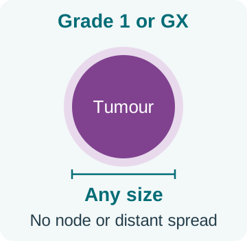
Stage 1 (I)
No cancer is found in nearby lymph nodes or distant parts of the body. The tumour is low grade (grade 1), or its grade cannot be assessed (GX). It can be any size.
- Stage 1A (IA): 5 cm or smaller.
- Stage 1B (IB): Larger than 5 cm.
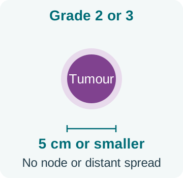
Stage 2 (II)
No cancer is found in nearby lymph nodes or distant parts of the body. The tumour is 5 centimetres (cm) or smaller and is intermediate grade (grade 2) or high grade (grade 3).
Stage 3 (III)
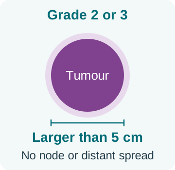
With no nearby-node or distant spread, a grade 2 or 3 tumour larger than 5 cm is stage 3. For retroperitoneal sarcoma, nearby-node spread without distant spread is also stage 3B, at any size or grade.
- Stage 3A (IIIA): Larger than 5 cm and up to 10 cm, with grade 2 or 3 and no nearby-node or distant spread.
- Stage 3B (IIIB): Larger than 10 cm, with grade 2 or 3 and no nearby-node or distant spread; or the retroperitoneal node finding described above.
Stage 4 (IV)
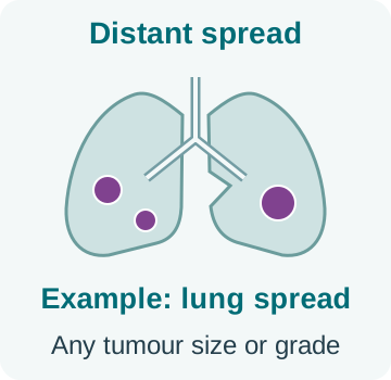
Cancer has spread to a distant part of the body, such as the lungs, at any tumour size or grade. In arm, leg or trunk sarcoma, nearby-node spread alone also means stage 4, even without distant spread.
These numbered groups do not apply to soft-tissue sarcomas of the head and neck or those starting within abdominal or chest organs. GIST, rhabdomyosarcoma and some other subtypes use separate systems. Your team uses the rules for your exact diagnosis.
The stage estimated from examination, biopsy and scans is called the clinical stage. Tissue removed during surgery may give more detail; this is called the pathological stage. The letters yp before a stage mean it was assessed from tissue after treatment such as chemotherapy or radiotherapy. A change in the assessment can reflect better information, rather than the cancer growing while you waited.
How your treatment plan is chosen
A sarcoma multidisciplinary team is a group of specialists who review your results together. It usually includes surgeons, doctors who give cancer medicines and radiotherapy, scan specialists and pathologists. Rehabilitation and supportive-care professionals help plan recovery and day-to-day needs.
The team considers the subtype, grade, stage, tumour location, your health and your priorities. They also assess whether surgery can remove the cancer and how treatment may affect movement or organ function. The first operation is planned as part of the full course of treatment.
For many localised adult sarcomas, surgery is the main treatment. Radiotherapy may reduce the chance of the cancer returning near the operation site. Chemotherapy is helpful in selected situations rather than being needed by everyone. A deep abdominal sarcoma may need a very different plan from a small tumour near the skin of a leg.
Balancing cancer control with everyday life
Whenever possible, treatment aims to control the cancer while preserving useful movement and function. Surgery may include rebuilding tissues, called reconstruction, or removing a nearby organ that is involved with the tumour. Ask what this would mean for your recovery and longer-term life.
Most arm and leg sarcomas can be treated without removing the limb. Amputation, removal of part or all of a limb, is occasionally the safest option when other surgery cannot adequately control the cancer or leave a useful limb. If it is being considered, the team should explain the alternatives and the rehabilitation and artificial-limb support available.
For advanced cancer, the immediate aim may be to shrink a tumour that is threatening an organ or causing symptoms. In other situations, a gentler treatment that controls slower-growing cancer may fit better. More medicines or a stronger treatment do not automatically mean a better outcome.
Questions to help you decide
- What is the full subtype and grade, and has a sarcoma pathologist reviewed the tissue?
- Is the aim to cure the cancer, reduce its chance of returning, control it or relieve symptoms?
- Are any important test results still pending?
- Why do you recommend this treatment, and what are the alternatives?
- If radiotherapy is needed, why give it before or after surgery?
- What benefit might chemotherapy add in my situation, and what side effects should I expect?
- How could treatment affect movement, work, fertility, sexual health or independence?
- If the cancer has spread to only a few places, could treatment to those areas help?
- How will we know whether treatment is working, and who should I contact between visits?
Clinical trials and supportive care
A clinical trial may be an option at diagnosis or when treatment needs to change. Some studies compare established treatments; others investigate a new approach. Ask whether the study includes your subtype, what is known so far, and what extra visits, tests or side effects it involves. In some studies, treatment groups are assigned by chance; this is called randomisation. Taking part is voluntary, and declining does not remove your right to usual care.
Supportive and palliative care help with pain, other symptoms, sleep, mood and family concerns. They can be offered alongside treatment intended to cure or control cancer. A referral does not mean that treatment is stopping.
Tell the team what you most hope treatment will allow you to do. Choices can be revisited if your health or priorities change. When a cure is unlikely, ask which options may help you live longer or more comfortably and how their likely benefits compare with the time and side effects involved.
Treatment options
Surgery for cancer that has not spread
The surgeon aims to remove the tumour with an appropriate border of surrounding healthy tissue, called a margin. The amount of tissue needed depends on the location and subtype; there is no single distance that suits every sarcoma.
After surgery, the pathologist examines the cut edges. A clear or negative margin means no cancer cells are seen at the edge. A positive or involved margin means cancer cells reach the edge, so further surgery or other treatment may be considered. A close margin means cancer is near the edge but does not reach it. The team weighs the chance of cancer returning against the effects of further treatment.
A close margin beside an important nerve or organ can sometimes be part of a planned approach that also uses radiotherapy. After an unexpected sarcoma diagnosis following removal of a lump, review of the scans, tissue and operation area helps decide whether re-excision is needed. The visible lump being gone does not, by itself, prove that all cancer cells have been removed.
Radiotherapy before or after surgery
Radiotherapy uses carefully planned radiation to treat a particular area. It can reduce local recurrence, meaning cancer returning near where it began. Whether you need it depends on the tumour and operation; some small, low-risk sarcomas do not need it.
Radiotherapy before surgery often treats a smaller area, but can increase wound-healing problems after the operation. Radiotherapy after surgery usually treats a larger area and may use a higher dose, with a different risk of later stiffness or swelling. The team considers the location, any tissue reconstruction and your health when choosing the timing.
For a tumour deep behind the abdominal organs, nearby bowel, kidneys and other tissues can limit what can safely be treated. Radiotherapy is not automatically part of treatment for every sarcoma in this area. It may also be used when surgery is not possible or to relieve symptoms such as pain.
Chemotherapy before or after surgery
Treatment before surgery is called neoadjuvant treatment. Treatment after surgery to reduce the chance of cancer returning is called adjuvant treatment. Chemotherapy at either time may be discussed for selected sarcomas with a higher risk of spread and a subtype likely to respond to it.
It is not needed for every localised adult sarcoma. The possible benefit depends on the subtype, size, grade and location, as well as your ability to tolerate treatment. Ask how much benefit the team expects and what remains uncertain.
Chemotherapy before surgery may shrink some tumours, but it cannot guarantee that an operation will become possible. If a trial is offered, ask how its approach differs from usual care. The decision should be based on your particular sarcoma, rather than an assumption that chemotherapy always prevents recurrence.
Sarcoma deep inside the abdomen
Retroperitoneal sarcoma develops in the space behind the abdominal organs. It may grow around important structures before it causes symptoms. Surgery can be complex and is best planned by a team experienced in this type of sarcoma. Sometimes nearby organs need to be removed with the tumour; this depends on the anatomy and subtype.
Cancer may return in the same area, in distant parts of the body or both. The pattern differs between subtypes, including liposarcoma and leiomyosarcoma. If another operation is considered, its purpose should be clear: for example, lasting cancer control or relief of a particular problem. Growth rate, symptoms and the likely effect on daily life all matter.
A large tumour does not automatically benefit from radiotherapy or chemotherapy. The specialist team will explain whether either is useful for your diagnosis and treatment aim.
Medicines for advanced sarcoma
Medicines that travel through the body to treat cancer are called systemic treatments. These include chemotherapy, targeted medicines and immunotherapy, which helps the immune system act against cancer. Treatment may aim to shrink cancer, slow its growth, relieve symptoms or help you live longer.
Doxorubicin is a common first medicine for several advanced adult sarcomas. Sometimes it is combined with another medicine, such as ifosfamide, when a higher chance of shrinkage is particularly important. The combination also causes more side effects and is not the best choice for everyone.
Later options may include gemcitabine-based chemotherapy, trabectedin, pazopanib, eribulin or dacarbazine. Their usefulness depends on the subtype, earlier treatments and your health. For example, some medicines used for liposarcoma are not useful for other sarcomas, and vice versa. You do not need to compare every drug yourself; ask why the recommended one fits your diagnosis.
The team checks symptoms, scans and side effects to decide whether to continue, adjust or change treatment. A medicine may be helping even if a tumour is not shrinking, if it is keeping previously growing cancer under control. The expected benefit must still be weighed against how treatment affects you.
How the subtype can change the medicine choice
Some sarcomas have options that differ from the general approach:
- Leiomyosarcoma: for some people with advanced disease, doxorubicin together with trabectedin, followed by trabectedin alone, is an option. It requires careful selection and monitoring because the combination can cause substantial side effects.
- Angiosarcoma: medicines called taxanes, such as paclitaxel, can be useful in appropriate situations.
- Alveolar soft part sarcoma: this rare subtype may be treated with immunotherapy such as atezolizumab when it cannot be removed by surgery or has spread. It has different treatment options from many other sarcomas.
- Inflammatory myofibroblastic tumour: a rare tumour made of cells with features of connective-tissue repair cells, with immune cells mixed in. Some have a suitable ALK gene change and can be treated with a medicine that blocks that growth signal when the tumour cannot be removed, has returned or has not responded to treatment.
- Malignant perivascular epithelioid cell tumour (PEComa): a rare cancer with cells that have characteristic features around blood vessels. It has selected targeted-treatment options that differ from routine sarcoma chemotherapy.
Epithelioid sarcoma and other rare subtypes also need an individual specialist plan. A drug mentioned in older information may no longer be recommended, so treatment choices should be checked against current evidence.
Immunotherapy is helpful for some specific subtypes or test results, but is not routinely useful for every sarcoma. A promising result in a different cancer does not prove that the same medicine will help yours.
Synovial sarcoma and treatment using your own immune cells
Synovial sarcoma has chemotherapy and local-treatment options. Afamitresgene autoleucel (afami-cel) is another option for selected patients. It uses a person's own T cells, a type of immune cell, changed in a laboratory to recognise the MAGE-A4 protein in cancer cells.
It may be considered from age 12 onwards for synovial sarcoma that cannot be removed by surgery (unresectable) or has spread to distant sites (metastatic), after previous chemotherapy. The blood test must show a compatible HLA-A*02 tissue type and the tumour must contain MAGE-A4. The specialist checks the exact result: not every HLA-A*02 type is suitable. This treatment is not suitable for all sarcomas.
This is not a chimeric antigen receptor T-cell (CAR-T) therapy. It is an engineered T-cell receptor (TCR) therapy. Both approaches change immune cells, but use different ways to recognise their target. Afami-cel recognises a piece of MAGE-A4 displayed on the cancer cell by a suitable HLA protein.
Treatment involves collecting T cells from the blood, changing and growing them in a laboratory, giving preparatory chemotherapy, and returning the cells through a vein. Manufacturing takes time, so how quickly the cancer is changing matters. The team also considers travel, access and the support you will need nearby.
Close monitoring is essential. Serious side effects include cytokine release syndrome, an inflammatory reaction that may cause fever, low blood pressure or breathing difficulty. Effects on the nervous system, infections and blood counts that remain low can also occur. Follow the specialist centre's instructions about observation, caregiver support and urgent help.
When cancer has spread to only a few places
For some people with a small number of cancer deposits in the lungs or elsewhere, treatment directly to those areas may help. Options include surgery, focused radiotherapy or ablation, which destroys a tumour using heat, cold or another local technique.
The team considers the subtype, how long the cancer was controlled before it spread, whether all important areas can be treated, and your general health. This approach can give useful control for selected patients, but it does not guarantee a cure.
Medicines may be given before, after or instead of these local treatments. The whole plan should be reviewed together. Treatment to one area can also relieve pain or prevent a particular complication even when cancer is more widespread.
GIST, desmoid and other separate treatment plans
For GIST, changes in KIT, PDGFRA or other genes help guide treatment. Imatinib can work well for some changes but not others. Gene testing may therefore be needed before choosing a medicine. Later medicines depend on what has already been tried and why it stopped working. GIST is generally treated differently from the adult sarcomas described above. See the GIST guide for its full treatment pathway.
For desmoid tumour, regular checks without immediate treatment, called active surveillance, are often appropriate. Some tumours remain stable or shrink on their own. A tumour that is growing or causing problems may need medicines, ablation, surgery or another selected treatment.
Nirogacestat is one medicine for adults with progressing desmoid tumour who need systemic treatment. It can affect ovarian function and fertility, as well as cause other side effects. Discuss fertility and pregnancy plans before treatment.
For tenosynovial giant cell tumour, surgery may help. Selected patients can receive medicines that block the colony-stimulating factor 1 receptor (CSF1R), a signal involved in the condition. The choice depends on symptoms and whether surgery would seriously damage movement or function. This is a separate pathway from treatment for malignant sarcoma.
Rhabdomyosarcoma and Ewing sarcoma also need their own treatment plans, often with childhood or young-adult cancer specialists. An ordinary adult sarcoma drug sequence is not a substitute for those plans.
Managing side effects
Recovery, swelling and movement
After surgery or radiotherapy, pain, stiffness, weakness and swelling may take time to improve. Physiotherapy helps with movement and strength. Occupational therapy helps you manage everyday tasks and choose useful equipment. A lymphoedema specialist can help with swelling caused by poor lymph drainage.
Follow the wound-care and activity instructions, especially after reconstruction. Increasing redness, fluid or pus from the wound, fever or suddenly worsening pain needs prompt review. A suddenly swollen limb should not be assumed to be an ordinary treatment effect; a clot or infection may need treatment.
Radiotherapy can cause skin changes and fatigue, meaning tiredness that may not improve fully with rest. Later it can cause fibrosis, a stiffening or scarring of tissues, which may restrict movement. Rehabilitation can help, but exercises should respect any surgical restrictions. Ask for practical advice about lifting, sport, driving and work. Recovery may continue over months.
Side effects of medicines
The effects depend on the medicine, dose and your health. The team will explain the risks and monitoring for your own treatment. Examples include:
- Doxorubicin can lower blood counts and affect heart function. Blood tests and heart checks help the team monitor this.
- Ifosfamide can affect the kidneys, bladder or nervous system. Protective medicines, fluids and monitoring are part of its treatment plan.
- Other medicines may cause liver problems, high blood pressure, sore or peeling skin on the hands and feet, or lung inflammation. Tablets still need blood tests and checks for interactions with other medicines.
- Cell therapy needs the centre's specific monitoring plan. Fever after afami-cel may be due to infection or cytokine release syndrome and needs urgent assessment.
Report side effects early. Do not change cancer-medicine doses, restart a stopped medicine or take leftover antibiotics without instructions. A planned delay or dose reduction may allow treatment to continue more safely.
Symptoms during treatment that need urgent help
Keep the team's contact details and out-of-hours plan where you and your caregiver can find them. Do not wait for a routine appointment if you develop a serious new symptom.
During treatment that can lower white blood cells, which help fight infection, a temperature of 38°C (100.4°F) or higher needs urgent advice. Follow any lower temperature limit or additional instructions your team gives you. Chills, confusion, sudden weakness or feeling very unwell also need urgent assessment, even without fever. Contact the team before taking medicine just to bring the temperature down.
Seek emergency help for severe breathlessness, chest pain, a new seizure, sudden weakness, heavy bleeding or collapse. New severe back pain with leg weakness, numbness around the bottom or loss of bladder or bowel control also needs immediate assessment.
Contact the team promptly for much less urine than usual, severe diarrhoea, repeated vomiting, inability to keep down fluids or important medicines, or unusual bleeding. A website form or routine message is not an emergency service.
Eating, drinking and energy
Small, frequent meals may be easier when appetite is poor. Include foods containing protein and ask for dietitian support if you are losing weight, struggling to eat or avoiding many foods. There is no single diet that treats sarcoma. Severe food restriction can make it harder to recover and tolerate treatment.
Take anti-sickness medicine as advised. Mouth sores, pain or infection that interfere with eating deserve treatment. Check supplements with the team because some interact with cancer medicines. The amount you should drink may need adjustment if you have heart or kidney problems.
Gentle movement, short walks and planned rest can help with fatigue, within the limits of your recovery plan. Tiredness can also be caused by low red blood cells, called anaemia, as well as infection, pain, poor sleep or low mood. Tell the team about a marked or lasting change so they can assess the cause.
Pain, body image and longer-term effects
Scars, changes in movement, organ removal or amputation can affect confidence, relationships and intimacy. Rehabilitation, advice about an artificial limb where needed, and psychological support are part of care. Persistent pain may come from nerves, muscles or other structures and should be assessed rather than simply endured.
Some treatment effects develop months or years later. Follow-up may include heart health, fertility, movement and other checks, particularly for people treated when young. Keep a record of your chemotherapy and radiotherapy for future healthcare. Ask about the safe timing of pregnancy or a return to strenuous activity.
Live well
Your follow-up plan
Follow-up checks look for cancer returning and help you manage recovery and treatment effects. The schedule depends on the subtype, grade, location and earlier treatment. It may include examinations, scans of the treated area and chest scans, with other tests for particular sarcomas.
Low-grade and high-grade tumours do not always need the same schedule. Some sarcomas can return many years later, so follow-up may continue long term. Ask which tests you need, how often, and who will explain the results.
Report a new lump near the operation site, increasing pain, a persistent cough, breathlessness or unexplained abdominal symptoms between appointments. A new symptom does not always mean recurrence, but should be checked. Follow-up should also address movement, swelling, organ function and emotional wellbeing.
Finding information that applies to you
Because sarcoma is uncommon, information online may describe a different subtype. Use the full name on your pathology report when discussing evidence, support groups or clinical trials. Ask whether a study included people with your type and stage of sarcoma. Your team can help explain why advice for another patient may differ from yours.
Emotional support and help for caregivers
Feeling frightened, angry, sad or uncertain is understandable. You do not have to appear positive all the time. Ask about counselling, a cancer support group or a social worker if worry is affecting sleep, relationships or everyday life. Persistent depression or panic deserves professional help. Thoughts of self-harm need urgent professional help.
Caregivers also need rest, information and support. It can help to share tasks such as transport, meals and appointments instead of leaving everything to one person. With the patient's agreement, a caregiver can take notes and help keep track of medicines and contacts. Let the patient decide how much information to share with relatives and friends.
If concentration is difficult, use reminders or a calendar and bring someone you trust to important visits. Children usually benefit from simple, honest explanations suited to their age and reassurance about who will care for them.
Relationships, fertility and everyday health
Cancer and treatment can affect sexual desire, comfort, body image and the ability to have children. These are valid medical concerns. Tell the team about pain, dryness, erection difficulties or other changes in intimacy; treatment, rehabilitation or counselling may help. You can choose how much to involve a partner.
Discuss fertility before treatment when possible. Do not assume that absent periods or reduced fertility means pregnancy is impossible. Ask about contraception and the safe timing of pregnancy for your medicines. Check before starting hormone treatments or supplements.
Continue care for other health conditions. Ask about vaccinations, because their timing and suitability can change during cancer treatment. Avoid tobacco and ask for support to stop if needed. Eating, activity and sleep plans should fit your health and surgical restrictions. Accepting help can leave more energy for the things that matter to you.
Planning ahead
If treatment continues for a long time, make space to discuss what is helping, what is difficult and what you want next. You can talk about future care preferences and who should speak for you if you become too unwell to make decisions. These discussions can be revisited and can happen while treatment and everyday life continue.
Sources and further reading
For more information, explore these resources from the National Cancer Institute (NCI) and other medical organisations. The linked organisations have not reviewed or endorsed this CTI guide. Your care team can explain how the information applies to you.
- NCI: Soft Tissue Sarcoma Treatment (PDQ®)–Patient Version
- ASCO guideline collection and update status
- ESMO–EURACAN–GENTURIS soft-tissue sarcoma guideline
- EMA: nirogacestat for progressing desmoid tumours
- FDA: afamitresgene autoleucel (Tecelra), current prescribing information
- D’Angelo et al. SPEARHEAD-1: afamitresgene autoleucel in advanced sarcoma (Lancet, 2024)
- Pautier et al.: doxorubicin and trabectedin in leiomyosarcoma (NEJM, 2024)
- FDA: nirogacestat for progressing desmoid tumours
- FDA: vimseltinib for symptomatic tenosynovial giant cell tumour
- FDA: atezolizumab for alveolar soft part sarcoma
- FDA: crizotinib for ALK-positive inflammatory myofibroblastic tumour
- NLM DailyMed: albumin-bound sirolimus for malignant PEComa
- FDA: tazemetostat safety alert and withdrawal
Staging and risk-group references
AJCC 8th edition.
Medicine approval and prescribing references
These official records describe licensed uses and prescribing information for the medicines named in this guide. Approval depends on the cancer type, biomarkers, previous treatment, combination and formulation. Some uses discussed in the guide are off-label: outside the medicine’s licence, with evidence from clinical guidelines or studies. FDA or EMA approval does not establish approval or availability in India.
- Afamitresgene autoleucel — FDA approval record
- Atezolizumab — FDA prescribing information · Approval history
- Dacarbazine — FDA approval record
- Doxorubicin — FDA prescribing information · Approval history
- Eribulin — FDA prescribing information · Approval history
- Gemcitabine — FDA prescribing information · Approval history
- Ifosfamide — FDA prescribing information · Approval history
- Imatinib — FDA prescribing information · Approval history
- Nirogacestat — FDA prescribing information · Approval history
- Pazopanib — FDA prescribing information · Approval history
- Trabectedin — FDA prescribing information · Approval history
Additional medicine approval notices
These records explain the specific medicine uses and eligibility requirements discussed in this guide.
Related care
Meet our doctors


Where to get care