
Share
Start here
Ovarian germ cell tumours at a glance
Ovarian germ cell tumours start in the cells that normally develop into eggs. They are uncommon and often affect teenagers and younger adults, although they can occur at other ages. Some germ cell tumours are non-cancerous. This guide focuses on malignant, or cancerous, ovarian germ cell tumours.
These cancers are different from the more common epithelial ovarian cancers, which start in another cell type. Many ovarian germ cell cancers respond very well to treatment, including some that have spread. Protecting fertility, the ability to have a pregnancy, and long-term health is part of care whenever this can be done safely.
- The exact tumour type matters. Dysgerminoma, yolk sac tumour and immature teratoma can need different plans.
- Surgery often removes the affected ovary and fallopian tube while preserving the womb and the other ovary when appropriate.
- Some early tumours can be followed closely after surgery without immediate chemotherapy. Others need chemotherapy, medicines that treat cancer, even when the cancer appears limited to an ovary.
- Many ovarian germ cell cancers respond well to chemotherapy containing a platinum medicine. Pure immature teratoma, which has no other malignant germ cell type mixed in, may need a different approach.
- Blood tests called tumour markers may help with diagnosis and follow-up, but not every tumour raises them.
- Ask about fertility before treatment begins, and about a written follow-up plan after it ends.
Symptoms and when to seek help
Possible symptoms include abdominal pain or swelling, a lump or feeling of pressure, feeling full quickly, changes in bowel or bladder habits, and menstrual changes. Symptoms can develop over a relatively short time. Most people with these symptoms do not have an ovarian germ cell cancer, but persistent or worsening symptoms need assessment.
Sudden severe lower abdominal pain, especially with nausea (feeling sick) or vomiting, needs urgent assessment. An enlarged ovary can twist and interrupt its blood supply, called ovarian torsion. Other causes, including bleeding, also need prompt attention. Seek emergency care for collapse, severe pain or heavy bleeding.
What to bring to a specialist appointment
Bring your written scan reports and a compact disc (CD) containing the scan images. Ask the scan centre for images in Digital Imaging and Communications in Medicine (DICOM) format, with the software needed to view them.
Also bring blood-test results with their dates and your tissue test (pathology) report. If you have already had surgery, bring the operation note too. The team may need your tissue samples, stored on slides or in blocks, for specialist review.
A pathologist is a doctor who examines tissue to diagnose disease. Ask whether one with experience in ovarian germ cell tumours should review your samples. Write down your medicines, menstrual history, whether pregnancy is possible and any concerns about future fertility.
For teenagers and young adults, care may involve specialists in cancers of the reproductive organs, cancer specialists for children or young people, and fertility services. Ask who is coordinating your care and whom to call between appointments.
Understand
The ovaries and germ cells
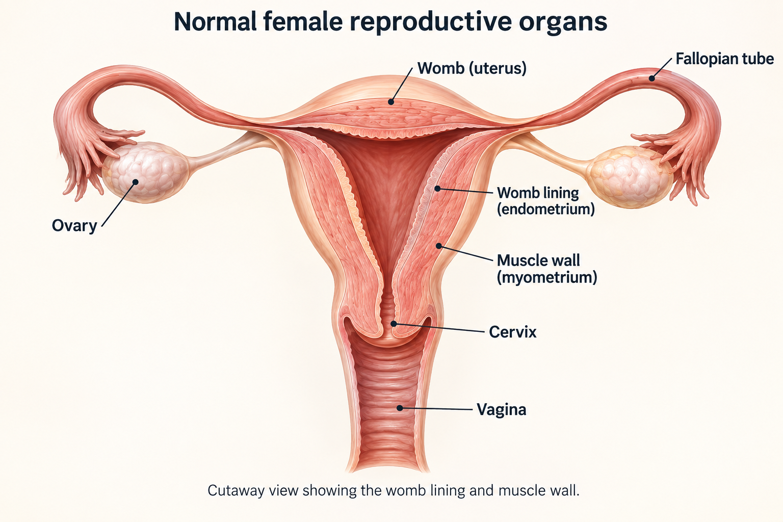
The ovaries are two small organs on either side of the womb, also called the uterus. They contain eggs and make hormones. The fallopian tubes run from near the ovaries towards the womb. The cervix is the lower part of the womb and connects to the vagina.
Germ cells are the cells from which eggs develop. A tumour arising from these cells is named according to the tissue it contains. The word germ in this name does not mean infection.
The main tumour types
| Type | What it means for the discussion |
|---|---|
| Dysgerminoma | A malignant germ cell tumour that is usually very sensitive to chemotherapy. Selected early cases can be monitored after surgery. |
| Yolk sac tumour | Often grows quickly and commonly raises a blood marker called alpha-fetoprotein. Chemotherapy is usually an important part of treatment. |
| Immature teratoma | Contains tissue that has not fully developed. Its grade, stage, your age and whether other cancerous cell types are present all affect treatment. Pure immature teratoma needs expert review. |
| Mixed malignant germ cell tumour | Contains more than one type of cancerous germ cell. All the types present can affect treatment, even if one makes up only a small part of the tumour. |
| Other rare types | Examples include embryonal carcinoma and choriocarcinoma unrelated to pregnancy. These need specialist diagnosis and planning. |
A mature cystic teratoma, often called a dermoid cyst, is usually benign, meaning non-cancerous. It is different from an immature teratoma. Rare complications or unusual tissue findings need their own assessment.
Grade is different from stage
Stage describes where the cancer is and whether it has spread. For immature teratoma, grade describes how much of a particular kind of immature tissue is seen under the microscope. A higher grade is not the same as a higher stage.
Understanding tumour markers
Tumour markers are substances measured in the blood that some tumours produce or cause to rise. They can help with diagnosis, treatment checks and follow-up. Not every tumour raises them.
| Marker | What the result may tell the team |
|---|---|
| Alpha-fetoprotein, or AFP | Often raised in yolk sac tumours and some mixed tumours. Pure dysgerminoma should not produce AFP. A raised result needs checking for another cause or another tumour type mixed in. |
| Beta-human chorionic gonadotrophin, or beta-hCG | Raised in choriocarcinoma and some embryonal carcinomas; it can also rise in some dysgerminomas. Pregnancy also raises it. |
| Lactate dehydrogenase, or LDH | Can be raised in dysgerminoma but is less specific; many other conditions can affect it. |
These markers are ideally checked before treatment and followed afterwards when useful. How quickly a raised marker falls can help show whether treatment is working.
One abnormal result does not prove the cancer has returned, and normal results do not rule out a tumour. Pure immature teratoma may have normal markers. A modest rise in AFP needs specialist interpretation.
Other blood tests check your blood cells, kidneys, liver and body salts. Cancer antigen 125 (CA-125), a marker used in common ovarian cancer, is sometimes measured too. It does not replace the germ cell marker tests above.
Why these tumours happen
Usually, there is no clear cause and nothing you did to bring this on. These tumours are not caused by sexual activity and are not contagious. They do not usually have the inherited risk pattern seen with some epithelial ovarian cancers.
Rarely, a germ cell tumour occurs with differences in how the ovaries or other reproductive structures developed. Advice from a genetics or hormone specialist may then be appropriate. Testing should be guided by the diagnosis and personal circumstances; relatives do not automatically need cancer screening.
Tests and diagnosis
Scans and examination
An ultrasound is often the first scan. It uses sound waves to look at the ovaries and any mass. It may be performed over the abdomen or, when appropriate and acceptable to you, with a vaginal probe. The team should explain the examination, obtain consent and discuss alternatives if you are uncomfortable. You can ask for a chaperone, a trained staff member who stays during the examination to support you.
Computed tomography (CT) uses X-rays to make detailed pictures of the abdomen, pelvis and usually the chest to check for spread.
In selected cases, your team may also recommend positron emission tomography combined with CT (PET–CT). This uses a small amount of a radioactive substance, called a tracer, to show areas of increased activity. It is not needed routinely for every ovarian germ cell tumour.
Magnetic resonance imaging (MRI) uses magnets to make detailed pictures. It may help the team understand a lump or look more closely at the pelvis. The choice of scan depends on your age, whether pregnancy is possible and what the team needs to find out.
Tell the team if you could be pregnant before imaging or treatment. A blood or urine pregnancy test may be part of the assessment. A positive hormone marker needs interpretation because both pregnancy and certain tumours can raise it.
How tissue establishes the diagnosis
The diagnosis often comes from tissue removed during surgery. If a lump in the ovary looks removable, the team usually plans an operation rather than routinely passing a biopsy needle through it. A biopsy takes a small sample of tissue for examination.
Avoiding an unnecessary tear in the tumour’s covering matters because rupture can affect the stage. When cancer is more widespread or the diagnosis is uncertain, a biopsy before treatment may be appropriate. This needs specialist planning.
The pathologist checks each type of tissue in the tumour and, for immature teratoma, its grade. A quick check during the operation may not give the full answer. The final examination takes longer. Ask whether a specialist experienced in ovarian germ cell tumours has reviewed the diagnosis.
Assessments before chemotherapy
Before chemotherapy, you may need hearing and lung tests and checks of kidney function. These show how things are before medicines that can affect these organs. Tell the team about lung disease, smoking, hearing problems and all medicines or supplements you take.
Discuss fertility and whether pregnancy is possible before treatment starts. The team may arrange a pregnancy test.
Planning your treatment
Understanding the stage
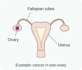
Ovarian germ cell cancers usually use the staging system of the International Federation of Gynaecology and Obstetrics (FIGO). The team uses findings from surgery, tissue tests and scans to work out the stage. The pictures below show the main groups; your full stage may include more detail.
The FIGO stage describes where the cancer is and whether it has spread. Blood tumour-marker levels do not change this stage, but they still help the team assess risk and plan treatment.
Abdominal washings are fluid used to rinse the inside of the abdomen during surgery, then collected and checked for cancer cells. Retroperitoneal lymph nodes are small structures behind the abdominal lining that help the body defend against infection.
Purple shows cancer. Each picture is an example, not every substage.
Stage 1 (I)
Cancer is confined to one or both ovaries. Surface involvement, rupture and cells in abdominal fluid or washings help define the substage.
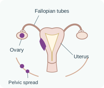
Stage 2 (II)
Cancer has spread to other tissues within the pelvis, such as the womb or pelvic lining.
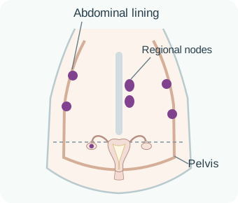
Stage 3 (III)
Cancer involves the abdominal lining beyond the pelvis and/or lymph nodes behind that lining. Deposits on the liver or spleen surface can be stage III.
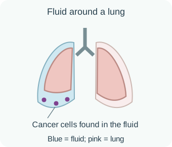
Stage 4A (IVA)
Cancer cells are found in fluid around a lung. Fluid alone does not establish stage IVA: it must contain cancer cells.
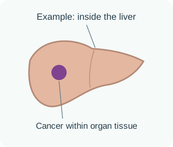
Stage 4B (IVB)
Examples include spread within liver or lung tissue, or to other distant sites or lymph nodes. A deposit inside liver tissue differs from a deposit on its surface.
A large ovarian tumour is not automatically an advanced-stage cancer. Even when a germ cell cancer has spread, treatment may still aim for a cure. The tumour type and how it responds to treatment both matter.
How stage and tumour markers shape your plan
Stage is one part of the assessment. Your team also considers the tumour type, its grade where relevant, any cancer left after surgery, and changes in blood-marker levels. Together, these help assess how treatment may work and the risk of the cancer returning.
If AFP or beta-hCG was raised, the team checks whether it falls as expected after surgery or chemotherapy. Levels that stay raised or rise again can suggest active cancer and affect the treatment plan, even if scans look clear. Other causes of an abnormal result must also be checked.
For patients who need chemotherapy, specialists may use a risk model developed for ovarian germ cell tumours. It groups patients by their expected outlook with treatment. Dysgerminoma has good- and intermediate-risk groups. Other malignant germ cell tumours have good-, intermediate- and poor-risk groups, using marker levels and where the cancer has spread.
These risk groups are separate from the FIGO stage. This model has less evidence than the similar model for testicular cancer, so your specialist needs to interpret it for you.
Children and adolescents may follow a different staging and treatment system. Your age, general health and fertility priorities also matter. Ask your team which approach applies to you.
Fertility-preserving surgery
When appropriate, the surgeon removes the affected ovary and its fallopian tube while keeping the womb and the other ovary. This is called a unilateral salpingo-oophorectomy.
With one functioning ovary, many people still make hormones, have periods and can become pregnant. Fertility cannot be guaranteed, so ask about your own situation. Keeping a functioning ovary can also protect hormone production, whether or not you plan a pregnancy.
Keeping the womb and an unaffected ovary may sometimes be possible even if the cancer has spread, particularly when it responds well to chemotherapy. The extensive surgery used for some epithelial ovarian cancers is not automatically needed.
The surgeon checks inside the abdomen, takes appropriate samples and examines any suspicious lymph nodes. A biopsy of the other ovary is usually avoided if it looks normal.
Discuss fertility before treatment starts
Ask early about fertility preservation, such as freezing eggs or embryos for possible future use. An embryo is an egg that has been fertilised. The options depend on your age, the time available, the tumour and your ovarian reserve, meaning your remaining egg supply.
You can ask about these options even if you are unsure about future parenthood. Other approaches may suit some patients. Freezing ovarian tissue and putting it back later needs particular caution when the cancer involves an ovary.
Medicines that temporarily reduce ovarian activity do not replace established ways of preserving fertility. Any delay to cancer treatment must be agreed with your oncology team.
Questions worth asking your doctor
- Which tumour types were found, and has a specialist reviewed the tissue?
- What are my stage and, if relevant, grade?
- Can the womb and an unaffected ovary be preserved safely?
- Can I have close monitoring after surgery instead of immediate chemotherapy, and what would follow-up involve?
- If chemotherapy is advised, why this combination of medicines, and how many courses of treatment will I need?
- What are the expected chances of cure, short-term risks and long-term effects in my situation?
Treatment options
Surgery
Surgery can both establish the diagnosis and remove the tumour. The surgeon plans how much to remove based on the tumour and your fertility priorities. If you had surgery elsewhere, bring the operation note and tissue-test report. Ask whether the team also needs the tissue slides or blocks.
Surgery may also be needed for a lump left after treatment, complications or cancer that returns. A lump remaining after chemotherapy does not always contain active cancer. It may be scar tissue or mature teratoma. The tissue type, marker results and scans help the team decide what to do next.
Surveillance after surgery
Surveillance means close, planned monitoring without immediate further treatment. It can avoid chemotherapy when surgery may already have cured the disease, while aiming to detect any return of the cancer promptly.
Common situations in which surveillance is considered include completely removed stage IA dysgerminoma and stage IA grade 1 immature teratoma. Stage IA means disease is confined to one ovary with an intact covering, no surface tumour and no cancer cells in abdominal fluid or washings.
For other stage I pure immature teratomas, recommendations differ between adult and children’s cancer services and between expert groups.
The Malignant Germ Cell International Consortium, a group of specialists in these tumours, published guidance in 2024 supporting surveillance more broadly after complete removal of stage I pure immature teratoma, including higher grades, in suitable circumstances. Some adult guidelines recommend chemotherapy more often.
This difference needs a specialist discussion, review of the tissue and a reliable follow-up plan. Advice for pure immature teratoma cannot automatically be applied to a mixed malignant germ cell tumour.
Surveillance is active care. You must be able to attend the agreed visits, have relevant marker tests and scans, and report symptoms between appointments. If follow-up is difficult, discuss this before selecting the plan.
Chemotherapy
Many ovarian germ cell cancers are treated with bleomycin, etoposide and cisplatin (BEP). Cisplatin is a platinum medicine. This combination works very well for many germ cell cancers, but its benefit differs by tumour type.
BEP is usually given through a vein in repeated cycles. A cycle includes treatment days and time to recover.
In a common adult schedule, cisplatin and etoposide are given on days 1 to 5, and bleomycin on days 1, 8 and 15. The next cycle usually begins about three weeks after the previous one started. Other schedules are used too.
Three or four cycles are often used, depending on the tumour, surgery findings and treatment plan. Blood tests, fluids to protect the kidneys and anti-sickness medicines are part of care.
Ask whether you can go home after treatment each day or will need to stay in hospital, and request a written calendar. Another person’s schedule may not be right for you.
Bleomycin may be unsuitable for some patients because it can affect the lungs. A specialist may recommend a different combination or schedule. Do not leave out a medicine without an agreed plan. Children and adolescents may receive treatment adapted to their age and circumstances.
When chemotherapy comes before further surgery
If the cancer is widespread or major surgery would be unsafe, the team may take a tissue sample to confirm the diagnosis and give chemotherapy first. They then check how the cancer has responded and whether surgery is needed. Specialists experienced in germ cell cancers should plan the order of treatment.
Immature teratoma and growing teratoma syndrome
An experienced team should guide treatment for pure immature teratoma. If it also contains another cancerous type, such as yolk sac tumour, this can change the plan. Even a tiny area needs to be considered alongside the rest of the tumour.
Occasionally, lumps grow during or after chemotherapy even though previously raised markers have returned to normal. One possible reason is growing teratoma syndrome: the lumps contain mature teratoma rather than the original cancer that responded to chemotherapy.
Other causes must also be checked. Surgery is often important for growing teratoma syndrome; more of the same chemotherapy may not help.
If the cancer returns
Recurrence means the tumour has come back after treatment. Some recurrent germ cell cancers remain curable. The options depend on the tissue type, earlier medicines, response, marker pattern and time since treatment. Specialist review is particularly important.
Options may include surgery or further chemotherapy containing a platinum medicine. In selected patients, high-dose chemotherapy may be followed by returning their own previously collected blood-forming stem cells. These cells help restore blood-cell production after intensive treatment.
One combination used in some situations is paclitaxel, ifosfamide and cisplatin. These approaches can carry substantial risks. Some evidence comes from germ cell cancers in other parts of the body. Ask about the team’s experience, clinical trials (research studies of treatments) and why a particular plan is recommended.
Radiotherapy and targeted treatments
Radiotherapy uses radiation to treat cancer. Although dysgerminoma can respond to it, radiotherapy is rarely used routinely because chemotherapy is effective and radiation can damage fertility and nearby tissues.
Medicines approved for epithelial ovarian cancer are not automatically suitable for ovarian germ cell tumours. They may be intended for a different cell type or a particular gene or protein finding.
Managing side effects
What to expect with BEP
| Possible effect | What can help |
|---|---|
| Nausea, vomiting and appetite changes | Take prescribed anti-sickness medicines and contact the team if you cannot keep fluids down. A dietitian can help with weight loss. |
| Lower blood counts | Low white blood cells can increase infection risk. Low red blood cells (anaemia) can cause tiredness or breathlessness, while low platelets can cause bruising or bleeding. Blood tests guide treatment. Keep the emergency contact number available. |
| Hair loss, tiredness and mouth soreness | Ask about hair support, mouth care, rest and activity suited to your energy. Many short-term effects improve after treatment. |
| Kidney or body-salt changes | Cisplatin requires fluids and blood-test monitoring. Tell the team about reduced urine or inability to drink. |
| Hearing changes or nerve symptoms | Report ringing in the ears, hearing loss, tingling or numbness promptly. Some effects can persist. |
| Lung inflammation | New cough, breathlessness or fever during or after bleomycin needs urgent assessment. |
If you have received bleomycin, tell future surgeons and anaesthetists, even years later. Anaesthetists are doctors who give medicines to keep you comfortable or asleep during surgery. They can plan oxygen and fluid care appropriately. Necessary oxygen must not be withheld in an emergency.
When to call urgently
Contact your cancer team immediately for a temperature of 38°C (100.4°F) or higher during chemotherapy, or any lower threshold they have given you. Also call for shaking chills, a new cough or breathlessness, repeated vomiting, severe diarrhoea, unusual bleeding or passing much less urine.
Feeling very unwell can signal a serious infection even without a high temperature. Do not wait for your next appointment.
A new painful or swollen arm or leg needs urgent assessment because it may indicate a blood clot. Sudden chest pain or breathlessness can be signs of a clot in the lung and need emergency care. Collapse, severe breathing difficulty or sudden severe abdominal pain also need emergency assessment. Keep a treatment summary and the hospital contact route with you.
Periods, contraception and menopause symptoms
Periods may stop during treatment and return later. Some people develop a lasting reduction in ovarian function or an earlier menopause, when hormone production falls and periods stop permanently.
Having periods does not guarantee fertility. Missing periods does not always mean pregnancy is impossible. Discuss suitable contraception (birth control) during treatment and for the period your team recommends afterwards.
Report hot flushes, vaginal dryness, pain with sex or changes in mood and sleep. The team can advise on symptom management and, when appropriate, hormone treatment. Do not assume that these concerns are too minor to raise.
Live well
Follow-up after treatment
Follow-up visits check symptoms and may include an examination, relevant tumour-marker tests and scans. Visits are usually closer together early on, when the chance of the cancer returning is higher, then become less frequent.
Your schedule depends on the tumour type, treatment and whether you are on surveillance. Normal markers do not replace scans when the original tumour did not raise them. Surveillance without chemotherapy often means particularly close follow-up during the first two years.
Ask for a written schedule and what to do if you miss a visit or move to another city. Keep a treatment summary with your diagnosis, surgery details, treatment dates, medicine names and total doses.
Long-term care also checks hearing, kidneys, nerves, lungs, hormones and fertility. Some treatments carry a small risk of later cancers, including blood cancers. Your team can explain this alongside the benefits of treatment aimed at a cure.
Fertility and future pregnancy
Many people continue to have periods and may be able to become pregnant after treatment that preserves fertility. Ask when it is appropriate to try for pregnancy; timing depends on the medicines used, recovery and the follow-up plan. There is no single waiting period that suits everyone. A fertility specialist can help if becoming pregnant is difficult or if you want to understand ovarian reserve.
If both ovaries or the womb have been removed, ask for advice about the options for future parenthood and hormone treatment that may be relevant to you. Discuss these issues privately if you prefer, even if relatives attend other appointments.
Returning to everyday life
Build activity gradually, eat regularly and seek help for lasting tiredness or weight change. School, university, work, relationships and body image may all need time and support. You do not need to feel positive all the time to recover well. Counselling and support suited to your age can help with fear of the cancer returning or feeling different from other people your age.
Caregivers can help with appointments, meals and transport. Ask the person with cancer what they want shared and with whom. Young people should have opportunities for private conversations with their clinical team, with support suited to their age. Our caregiver toolkit offers practical guidance and charts for use at home.
Sources and further reading
These patient resources, clinical guidance and medicine information support this guide. The organisations have not reviewed or endorsed the CTI text. For this rare cancer, some recommendations draw on studies in children or germ cell cancers elsewhere in the body. Your care team can explain how they apply to you.
Information for patients and caregivers
- Macmillan Cancer Support — Germ cell ovarian cancer.
- Cancer Research UK — Germ cell ovarian tumours.
- eviQ, Cancer Institute NSW — Patient information for ovarian germ cell BEP.
- Macmillan Cancer Support — BEP treatment, practical information and side effects.
- Cancer Council Australia — Ovarian cancer overview. Used for general anatomy, symptom and support information; its common epithelial cancer treatment pathway is not used as a germ cell treatment guideline.
Clinical guidance and research
- National Cancer Institute — Ovarian germ cell tumours treatment: professional information.
- European Society for Medical Oncology — Non-epithelial ovarian cancer guideline.
- Sessa and colleagues — European guidance for teenagers and young adults with non-epithelial ovarian cancers, 2020.
- Malignant Germ Cell International Consortium — Consensus on ovarian immature teratoma, 2024.
- Liu and colleagues — Risk assessment in female germ cell tumours using a modified prognostic classification, 2023.
- Su and colleagues — American Society of Clinical Oncology fertility preservation guideline update, 2025, with its published correction.
- National Cancer Institute — Childhood germ cell tumours outside the brain: professional information.
Medicine information
- eviQ, Cancer Institute NSW — Ovarian germ cell BEP protocol.
- United States Food and Drug Administration — Approved medicine information.
- European Medicines Agency — Medicines and product information.
Medically reviewed and approved by Dr (Prof) Vijay Agarwal
Professor and Lead Consultant – Medical Oncology
Related care
Meet our doctors



Where to get care