
Share
Start here
About this guide
A uterine or womb cancer diagnosis can feel overwhelming. This guide explains endometrial cancer, which starts in the womb lining, and a related type called carcinosarcoma. Cancers that start in the womb’s muscle or supporting tissue, called uterine sarcomas, need a different specialist treatment plan.
Start with your biopsy result: a biopsy takes a tissue sample to check for cancer. Scans, examination of the tissue (pathology), and tests of the cancer’s genes and proteins (molecular tests) guide the next decision about treatment.
This guide takes you and those supporting you through the results and choices step by step.
Bring your reports, medicines and questions to your appointment. Tell the team about your priorities, including having children, menopause, sexual health and practical support.
A family member or caregiver can help take notes if you wish. Ask who is coordinating your care and whom to contact between appointments.
Uterine cancer at a glance
Most endometrial cancers cause abnormal bleeding and are found while treatment can still aim to cure the cancer.
- Any bleeding after menopause needs assessment. Bleeding between periods, unusually heavy bleeding or a persistent watery or blood-stained discharge also needs review.
- An ultrasound can show a thickened womb lining, but a tissue sample is needed to diagnose cancer. This may be taken during a biopsy or a camera examination of the womb (hysteroscopy).
- The plan depends on the cancer type, how abnormal its cells look, how far it has grown or spread, and its molecular test results. Stage is only one part of the decision.
- Surgery usually removes the womb and cervix (total hysterectomy), both fallopian tubes and both ovaries, often through small cuts. The team may also check nearby lymph nodes, small structures that are part of the immune system.
- Many low-risk cancers need no further treatment after surgery. Others may need radiation, cancer-killing medicines (chemotherapy), medicines that help the immune system attack cancer (immunotherapy), or a combination.
- For advanced cancer or cancer that has returned, mismatch repair (MMR) testing checks a system that repairs errors in the cell’s genetic material. Chemotherapy with a type of immunotherapy called a checkpoint inhibitor is an important initial treatment option.
- For selected small, low-grade endometrioid cancers limited to the womb lining, hormone treatment may temporarily delay surgery to allow a pregnancy. This needs specialist assessment and close monitoring.
Aim of treatment
When cancer is limited to the uterus, treatment generally aims to cure it. The outlook varies even within the same stage. Ask how your results affect the treatment goal and the risk of cancer returning.
Diagnosis
A tissue sample confirms the cancer type and how abnormal its cells look. Ultrasound and other scans cannot replace a biopsy.
Treatment readiness
Fitness, other illnesses, fertility and menopause priorities, nutrition and support can all shape the safest plan.
Molecular testing
Molecular testing is a cornerstone of understanding endometrial cancer and planning treatment. Alongside the biopsy findings, it helps identify the cancer group, estimate the risk of it returning and guide treatment choices.
Bring your scan images
For your consultation or a second opinion, bring a compact disc (CD) containing your scan images in Digital Imaging and Communications in Medicine (DICOM) format and the software needed to view them, along with the written scan reports. DICOM is the standard format for medical scans.
Understand
Topics in this section
The uterus and endometrium
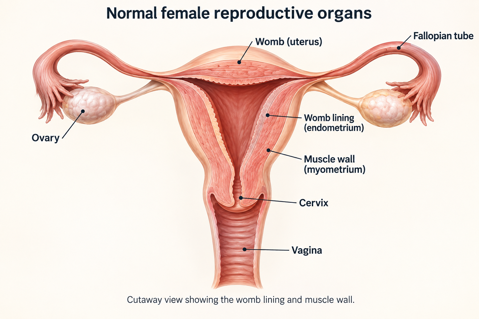
The uterus is a hollow muscular organ in the pelvis. Endometrial cancer starts in its inner lining.
Endometrium
The lining changes during the menstrual cycle. Most uterine cancers begin in its gland-forming cells.
Myometrium
The muscle wall surrounds the lining. How deeply cancer enters it is an important staging and risk feature.
Cervix and vagina
The cervix is the lower part of the uterus. Cervical cancer begins there and is treated differently.
Tubes, ovaries and nodes
Cancer may spread to the fallopian tubes or ovaries, nearby pelvic tissues, or lymph nodes in the pelvis and higher in the abdomen beside the main artery, the aorta.
The exact tumour type matters
Cancers in the same organ can behave differently. Histology means the type of cancer seen under a microscope. Grade describes how abnormal the cells look. Molecular tests help identify a group with features that may affect treatment.
Pathology label | What it means for care |
|---|---|
Endometrioid carcinoma | The most common type. Often low grade, with proteins called hormone receptors that can respond to hormones. Its behaviour ranges from lower risk to high risk. |
Serous carcinoma | Usually high risk, often with abnormal p53, a protein involved in controlling cell growth. Spread to the abdominal lining can occur even with a small womb tumour. Testing for human epidermal growth factor receptor 2 (HER2), a growth-related protein, may guide treatment. |
Clear cell carcinoma | An uncommon type, generally treated as high risk. The stage and molecular test results help shape treatment. |
Carcinosarcoma | A high-risk cancer with a mixture of cell appearances, including areas that resemble sarcoma. It is generally treated as endometrial cancer. |
Undifferentiated or dedifferentiated | Rare cancers that can grow quickly. A specialist should review the tissue and molecular test results. |
Uterine sarcoma | Starts in muscle or supporting tissue, such as leiomyosarcoma or stromal sarcoma. These cancers have different staging and medicine treatment from those described here. |
Four molecular groups explain more
Tests of the cancer’s genes and proteins add information to its appearance under a microscope. They can help estimate the outlook and how much treatment is needed.
POLE gene changes
The POLE gene helps make polymerase epsilon, which copies and proofreads genetic material, called deoxyribonucleic acid (DNA).
Certain cancer-related changes in its proofreading region can signal a very favourable outlook in early cancer. Expert review may support less treatment; not every POLE change has this meaning.
MMR-deficient (dMMR)
MMR-deficient (dMMR) means the repair system is not working properly, allowing many DNA errors to build up. It may suggest an inherited condition called Lynch syndrome. Advanced cancers in this group are particularly likely to benefit from checkpoint immunotherapy.
No specific molecular profile (NSMP)
These cancers do not fit the other three groups. They vary in behaviour, so grade, stage, hormone-receptor results and other features remain important.
p53-abnormal
Abnormal p53 usually indicates a cancer that is more likely to grow quickly or return. Serous cancers often belong to this group, but other cancer types can too.
How the group is assigned
- MMR immunohistochemistry (IHC), a tissue-staining test, checks four repair proteins: MLH1, PMS2, MSH2 and MSH6. Microsatellite instability (MSI) testing looks for related changes in the tumour DNA and can help in some situations.
- Staining for p53 protein helps identify cancers with changes related to the TP53 gene.
- POLE testing reads the gene’s proofreading region, called the exonuclease domain. A specialist checks whether any change found is one known to affect cancer risk and treatment.
- Some cancers have more than one of these findings. A pathologist, the doctor who examines tissue, interprets them together using established rules.
For your outlook
Early cancers that look similar on scans may have very different risks of returning because of their POLE or p53 results.
For treatment
MMR status helps guide immunotherapy. Oestrogen receptor (ER) and progesterone receptor (PR) tests look for proteins that respond to hormones. These and HER2 results help select treatment in some high-risk cancers or cancers that have returned.
Symptoms and warning signs
Abnormal bleeding is the key warning sign. It should be investigated rather than repeatedly treated without a diagnosis.
After menopause
Any vaginal bleeding or blood-stained discharge after menopause is abnormal, even if it happens once or is only spotting.
Before menopause
Bleeding between periods, a major change in cycle pattern, very heavy bleeding or bleeding that persists despite treatment needs review.
Other pelvic symptoms
Watery discharge, pelvic pressure or pain, pain during sex, increasing abdominal size or a pelvic mass can occur.
Later symptoms
Unexplained weight loss, poor appetite, cough, breathlessness, bone pain or leg swelling may occur if disease has spread, but have many other causes.
Write down the pattern
Note when bleeding started, whether it is spotting or heavy, clots, discharge, pain and any dizziness.
Tell your doctor
Mention menopause status, pregnancy possibility, hormones, tamoxifen, blood thinners and any previous abnormal biopsy.
Risk factors
Usually there is no single cause. Risk factors affect the chance of developing cancer; they do not explain every individual diagnosis.
Factor | What is known | Practical response |
|---|---|---|
Age and menopause | Risk rises with age; most cases are diagnosed after menopause. | Report any postmenopausal bleeding promptly. |
Excess body fat | Fat tissue increases exposure to oestrogen. Higher body fat is an important risk factor that can sometimes be changed. | Avoid blame. Safe activity and weight support help overall health at any stage. |
Unopposed oestrogen | Oestrogen without adequate progestogen can stimulate the endometrium. | Use menopausal hormones only with appropriate medical advice. |
Irregular ovulation | Polycystic ovary syndrome (PCOS), long gaps between periods, an early first period, late menopause or never having been pregnant can increase exposure to oestrogen over a lifetime. | Persistent abnormal bleeding still needs assessment. |
Diabetes and metabolic health | Diabetes and related metabolic factors are associated with higher risk. | Manage blood sugar, blood pressure and heart health with your care team. |
Tamoxifen | It lowers breast-cancer recurrence but slightly raises endometrial cancer risk. | Do not stop it yourself. Report bleeding; routine ultrasound without symptoms is not usually useful. |
Lynch syndrome | An inherited change in a mismatch-repair gene can greatly increase womb and bowel cancer risk. | Tumour MMR testing and genetic counselling can identify families at risk. |
Lynch syndrome and family risk
Most endometrial cancers are not inherited. MMR testing of the tumour helps identify people who may need further tests for an inherited risk.
What happens after an abnormal MMR result
- The tumour is checked for loss of MLH1, PMS2, MSH2 or MSH6, or for microsatellite instability.
- If MLH1 and PMS2 proteins are missing, the tumour is usually checked for a chemical change that switches off MLH1, called promoter methylation. This often means the change arose in the tumour rather than being inherited.
- Unexplained loss of repair proteins, a diagnosis at a young age, several Lynch-related cancers or a strong family pattern prompts genetic counselling and blood or saliva testing for inherited changes, called germline testing.
- If Lynch syndrome is confirmed, relatives can choose targeted testing. Screening and risk-reduction plans are then individualised.
Cancers linked with Lynch
Colon and rectum, endometrium, ovary, stomach, small bowel, urinary tract and several other sites can be involved.
Family clues
Cancer at a young age, several related cancers in one person, or repeated bowel and uterine cancers across generations.
Tumour and inherited-risk tests answer different questions
A tumour can be MMR-deficient without an inherited gene change. Blood or saliva testing helps establish whether the change can run in the family.
No family history does not rule it out
Lynch syndrome can be missed in small families or when relatives’ medical histories are incomplete. This is why tumour MMR testing is recommended even without a known family history.
Your own prevention plan
A confirmed inherited gene change may affect advice about bowel, ovarian and other cancer screening, or surgery to reduce risk.
Relatives choose testing
After genetic counselling, adult relatives can choose testing for the specific gene change found in the family.
Children are not automatically affected
A child may inherit the gene change, but this is not certain. The right age for testing depends on the gene and the recommended prevention plan.
Keep the laboratory report
The exact gene, variant wording and classification are needed if another clinic or relative arranges testing.
Tests and diagnosis
Topics in this section
How cancer is diagnosed
You may not need every possible test. Each step should answer a question that changes treatment.
- Discuss bleeding or discharge, periods, menopause, possible pregnancy, medicines including tamoxifen or hormones, and family history. Mention tiredness, dizziness or breathlessness that could indicate anaemia, a low red blood cell level.
- Examine the abdomen and pelvis when appropriate. The doctor checks the cervix, vagina and womb and feels for any lump. A normal examination cannot rule out cancer.
- Use transvaginal ultrasound, with a probe placed in the vagina, to look at the womb lining and ovaries. It helps decide which further tests are needed but cannot confirm cancer.
- Take a tissue sample, either with a biopsy in the clinic or during hysteroscopy. Additional tissue may be collected by gently scraping the lining, called curettage, when needed.
- Confirm the cancer type and grade. Arrange MMR and p53 testing, and POLE testing when available or needed to complete the risk assessment.
- Use magnetic resonance imaging (MRI) of the pelvis and/or computed tomography (CT) of the chest, abdomen and pelvis according to risk and symptoms. MRI uses magnets and CT uses X-rays to make detailed pictures. Specialists review the results together to plan care.
Keep copies
Keep the biopsy and molecular test reports, scan reports, operation note and final tissue report from surgery together.
Ask which results may change
Scans help estimate the stage before treatment. Examining the tissue after surgery can change the final stage and risk assessment.
Core specialists
Your team may include a gynaecological oncologist (a surgeon specialising in these cancers), a pathologist (tissue tests), a radiologist (scans), a medical oncologist (cancer medicines) and a radiation oncologist (radiotherapy).
Add support early
Ask about genetic counselling, fertility and menopause advice, nutrition, physiotherapy, emotional support and specialist help with symptoms. These services can support you throughout treatment.
Pelvic assessment and ultrasound
The first tests look for a likely source of bleeding and decide how urgently tissue sampling is needed.
Pelvic examination
The doctor uses an instrument called a speculum to see the cervix and vagina. They may also examine with fingers in the vagina and a hand on the abdomen to check womb size, movement, tenderness or a lump.
Transvaginal ultrasound
A slim probe in the vagina gives the clearest view of the lining. The report may describe endometrial thickness, a focal polyp or mass, fibroids and ovaries.
Transabdominal ultrasound
A probe over a full bladder gives a wider pelvic view. It may be added when the vaginal view is not possible or incomplete.
Keep examination and scan reports
Keep your examination findings, ultrasound reports and images together. They help the team plan the next step and compare changes if a repeat assessment is needed.
Before the scan
Ask whether a full bladder is needed and whether a transvaginal scan is acceptable for you.
After the scan
Ask whether a tissue sample is still needed and who will arrange it. Bleeding that continues or returns needs review even after a reassuring scan.
Endometrial biopsy and hysteroscopy
Tissue confirms whether cancer is present and identifies the tumour type.

Biopsy in the clinic
A thin suction tube passes through the cervix to collect tissue. Cramping is common. The sample may be small or miss an abnormal area confined to one part of the lining.
Hysteroscopy
A thin camera lets the doctor see inside the womb and take tissue from an abnormal area. Opening the cervix and gently scraping the lining, called dilatation and curettage, may provide more tissue.
If the sample is insufficient
If there is too little tissue to assess, cancer has not been ruled out. Bleeding that continues often needs another biopsy, sometimes using hysteroscopy.
After the procedure
Mild cramps and spotting are common. Seek advice for heavy bleeding, fever, worsening pain, foul discharge or fainting.
Scans and blood tests
Scans look for where cancer may have grown or spread. They cannot replace tissue tests. Your symptoms and risk determine which scans are useful.
Test | Main question | Important limitation |
|---|---|---|
MRI pelvis | How deeply has cancer entered the womb muscle? Has it reached the cervix or nearby pelvic tissue? | Very small areas of cancer, including tiny deposits in lymph nodes, may be missed. |
CT chest, abdomen and pelvis | Are lymph nodes enlarged? Has cancer spread to the lungs, liver, abdominal lining or other distant sites? | Normal-sized lymph nodes can contain cancer. Tiny deposits on the abdominal lining may not show up. |
Positron emission tomography with CT (PET-CT) | Uses a radioactive glucose-like tracer, fluorodeoxyglucose (FDG), to highlight active areas. Often used for staging when the risk of spread is higher, and to assess suspected cancer recurrence. | Not needed for every early cancer. Very small deposits or cancers that take up little tracer may be missed. |
Chest imaging | Is there lung disease when full CT is not otherwise indicated? | A plain X-ray is less sensitive than CT. |
Blood count and chemistry | Is there anaemia? Are kidney and liver function, blood sugar and body salts suitable for surgery or treatment with medicines? | Blood tests cannot diagnose or stage the tumour by themselves. |
Cancer antigen 125 (CA-125) | This blood protein may help monitor selected high-risk or advanced cancers if it is raised before treatment. | It can be normal despite cancer, or raised for reasons unrelated to cancer. |
Understanding your pathology report
The report describes the cancer type, how far it has grown and features that affect the risk of it returning.
Report item | Plain-language meaning |
|---|---|
Histology | Endometrioid, serous, clear cell, carcinosarcoma, undifferentiated or another type. Unusual diagnoses may need expert review. |
Grade | How closely the tumour resembles normal glands. Endometrioid cancers are usually graded 1, 2 or 3; high-risk histologies are treated differently. |
Myometrial invasion | How far cancer has grown into the womb muscle. Growth into less than half the muscle thickness carries different risks from growth into half or more. |
Cervical stromal invasion | Cancer has entered the supportive tissue of the cervix, not merely surface glands. |
Lymphovascular space invasion (LVSI) | Cancer cells are seen in small lymph or blood vessels. A small amount (focal) and a larger amount (substantial) carry different risks. |
Nodes | Which lymph nodes were checked and whether they contain isolated cancer cells, small deposits (micrometastases) or larger deposits (macrometastases). |
Cut edges and areas outside the womb | Whether cancer reaches a cut edge (margin), the womb’s outer surface, tubes, ovaries, vagina, abdominal lining, the fatty layer covering abdominal organs (omentum), or another sampled site. |
Molecular markers | MMR/MSI, p53 and POLE, with ER and HER2 when relevant. The team combines these with other findings to estimate risk. |
Biomarkers that can change treatment
Biomarkers are features of the cancer, such as genes or proteins, that can help guide care. Plan testing early enough for results to inform surgery, treatment after surgery or medicines for advanced cancer.
Marker | How it is reported | Why it matters |
|---|---|---|
MMR / MSI | MMR-proficient (pMMR) means the repair proteins are present; in MMR-deficient (dMMR) cancer, one or more are missing. The related MSI test checks short, repeated DNA sequences: microsatellite-stable (MSS) means they remain stable, while MSI-high (MSI-H) means many have changed. | Helps identify who needs assessment for Lynch syndrome. A dMMR or MSI-H result predicts a greater chance of benefit from checkpoint immunotherapy in advanced cancer. |
POLE | Whether a cancer-related change was found in the gene’s proofreading region | Certain POLE changes signal a very favourable early-stage outlook and may support less treatment after surgery. |
p53 | Normal (wild-type) or abnormal protein staining; the report may describe the specific abnormal pattern | An abnormal result usually indicates a higher risk of cancer returning and affects staging and treatment decisions. |
ER / PR | How many cells show hormone receptors and how strongly they stain | Helps identify low-grade, slower-growing advanced or recurrent cancers that may respond to hormone treatment, also called endocrine therapy. |
HER2 | A tissue-staining (IHC) score from 0 to 3+. An unclear 2+ result often needs an in-situ hybridisation test to check for extra HER2 gene copies. | Can support HER2-targeted therapy, especially in serous carcinoma or carcinosarcoma and selected recurrent tumours. |
Next-generation sequencing (NGS) | Tests many tumour genes together for changes or joined genes (fusions). Some panels also measure tumour mutational burden (TMB), the number of DNA changes in the cancer. | May find rare treatment targets, such as fusions involving neurotrophic tyrosine receptor kinase (NTRK) genes, or help identify a suitable clinical trial. Enough good-quality tissue is needed. |
Planning your treatment
Understanding your stage
FIGO 2023 staging for endometrial cancer
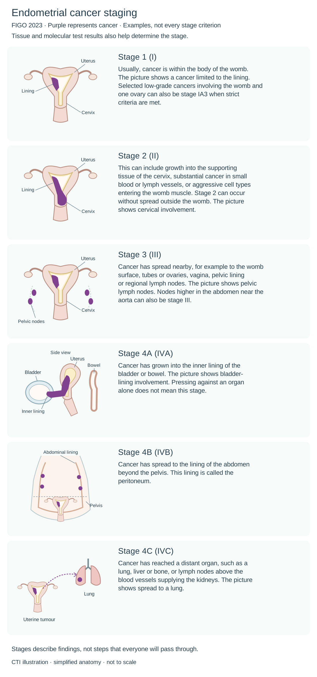
The International Federation of Gynecology and Obstetrics (FIGO) staging system combines where cancer is found with features seen in tissue and molecular tests. The pictures show examples of endometrial cancer, which starts in the womb lining. They do not cover every way a stage can occur.
Lymph nodes are small immune-system structures. Regional nodes are those near or draining the cancer’s starting area; distant spread means cancer has reached a site outside that regional area. Stages describe findings, not a sequence that everyone will pass through.
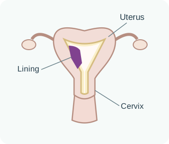
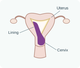
Purple represents cancer. Each picture shows one example.
Stage 1 (I)
Usually, cancer is within the body of the womb. The picture shows a cancer limited to the lining. Selected low-grade cancers involving the womb and one ovary can also be stage IA3 when strict criteria are met.
Stage 2 (II)
This can include growth into the supporting tissue of the cervix, substantial cancer in small blood or lymph vessels, or aggressive cell types entering the womb muscle. Stage 2 can occur without spread outside the womb. The picture shows cervical involvement.
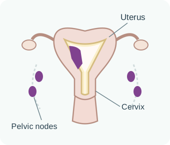
Stage 3 (III)
Cancer has spread nearby, for example to the womb surface, tubes or ovaries, vagina, pelvic lining or regional lymph nodes. The picture shows pelvic lymph nodes. Nodes higher in the abdomen near the aorta can also be stage III.
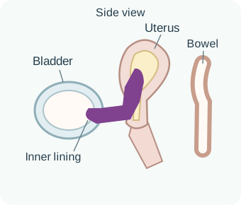
Stage 4A (IVA)
Cancer has grown into the inner lining of the bladder or bowel. The picture shows bladder-lining involvement. Pressing against an organ alone does not mean this stage.
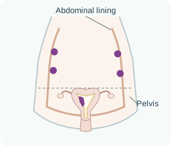
Stage 4B (IVB)
Cancer has spread to the lining of the abdomen beyond the pelvis. This lining is called the peritoneum.
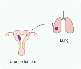
Stage 4C (IVC)
Cancer has reached a distant organ, such as a lung, liver or bone, or lymph nodes above the blood vessels supplying the kidneys. The picture shows spread to a lung.
The first stage estimate comes from examination, biopsies and scans. Surgery can provide more information and change that assessment; this does not necessarily mean cancer grew while you were waiting.
Stage and grade are different: stage describes the cancer’s extent and selected tumour features, while grade describes how abnormal the cells look.
Stage includes extent and tumour features
Stage records where cancer was found; FIGO 2023 also incorporates selected histological and molecular features. Your team combines these findings to estimate risk and plan treatment.
Your results are considered together
The team combines cancer type, grade, growth into small vessels, lymph-node findings and molecular results with stage to estimate the risk of cancer returning.
Planning for a future pregnancy
Tell your team before treatment if you hope to carry a pregnancy. For carefully selected small, low-risk cancers, hormone treatment can delay removal of the womb. This approach requires specialist review, repeated biopsies and a clear plan for surgery if needed.
Who may be considered
- A wish to carry a pregnancy, after discussing the risks and alternatives.
- Grade 1 endometrioid carcinoma confirmed by expert pathology review.
- Cancer limited to the womb lining, with no growth into the muscle or spread outside the womb on specialist-reviewed scans.
- No high-risk cancer type; review of molecular results and inherited risk, including what Lynch syndrome would mean for this choice.
- Ability to attend repeated biopsies and proceed to removal of the womb if cancer remains, grows or returns.
Step | Typical approach |
|---|---|
Treatment | A device placed inside the womb that releases levonorgestrel, a progesterone-like hormone (progestin), is often preferred. Progestin tablets may be used alone or with the device. |
Response check | Repeat endometrial sampling every 3 to 6 months. Imaging and symptoms are reviewed; ultrasound alone is not enough. |
Pregnancy | Once repeat tissue tests show no remaining cancer (a complete response), fertility treatment may help shorten the time to pregnancy. Work with a fertility specialist. |
Definitive surgery | Removal of the womb is advised after childbearing, if cancer grows, or if tests do not show a complete response within an agreed 6 to 12 months. |
How your treatment plan is chosen
Your specialist team considers where the cancer is, its tissue and molecular results, your overall health and your priorities. Each result is interpreted alongside the others.
Question | What guides the answer | Typical direction |
|---|---|---|
Can it be removed safely? | Scans, examination, fitness for an anaesthetic and whether all cancer can be removed | Surgery through small cuts for most cancers limited to the womb; a larger operation or treatment in steps for selected advanced cancers. |
How likely is the cancer to return? | FIGO stage, histology, grade, invasion, LVSI, nodes, margins, POLE, MMR, p53 and ER | Monitoring alone, internal radiation to the upper vagina (vaginal brachytherapy), radiation to the pelvis, cancer medicines or a combination. |
Is fertility preservation safe? | Grade 1 endometrioid cancer, no growth into womb muscle or spread elsewhere, review of molecular and inherited risk, and reliable follow-up | Temporary treatment with a progesterone-like hormone (progestin), with repeat biopsies, then surgery after childbearing or if treatment does not work well enough. |
If cancer is advanced or recurrent? | Symptoms and current fitness, how quickly and where the cancer is growing, previous treatment, and molecular markers such as MMR status, ER and HER2 | Treatment directed at a limited area of returning cancer, or medicines that work throughout the body (systemic therapy), chosen using molecular results. Symptom support starts early. |
Team
A gynaecological oncologist, pathologist, radiologist, radiation oncologist, medical oncologist and genetic counsellor, with nursing and supportive-care specialists as needed.
Your priorities
Chance of cure or control, treatment burden, fertility, menopause, sexuality, work, travel, caregiving, cost and access all belong in the decision.
Treatment options
Topics in this section
- Surgery to remove the cancer
- Sentinel nodes and recovery after surgery
- Treatment after surgery
- Radiotherapy: treating a specific area
- Chemotherapy and combined treatment
- When cancer is advanced or returns
- Medicines for advanced or recurrent cancer
- Options after previous chemotherapy
- Hormone, HER2 and rare-target treatment
- Checking treatment benefit and discussing trials
Surgery to remove the cancer
For most cancers that can be operated on, surgery removes the tumour and provides more accurate stage and risk information. The sections below explain surgery and any treatment needed afterwards, followed by options for cancer that is advanced or has returned.
What the operation involves
Total hysterectomy removes the womb and cervix. Bilateral salpingo-oophorectomy removes both fallopian tubes and ovaries. Depending on risk, the surgeon may map the first lymph nodes that drain the womb (sentinel nodes) or use other checks for spread.
Surgery through small cuts
Keyhole (laparoscopic) or robot-assisted surgery is preferred for most cancers limited to the womb. When suitable, it offers similar cancer control with a usually faster recovery.
Open surgery
A larger abdominal cut may be safer for a very large womb, extensive cancer outside it, previous complex surgery, or when wider access is needed to remove the cancer.
Ovarian preservation
Some people who have not reached menopause and have low-risk endometrioid cancer may keep their ovaries after expert review. The fallopian tubes are usually removed.
Advanced disease
Surgery to remove as much cancer as possible is considered only when all visible cancer is likely to be removed with acceptable risks and effects on quality of life.
Sentinel nodes and recovery after surgery
Good staging should answer the lymph-node question while avoiding unnecessary long-term harm.
Sentinel lymph-node mapping
A tracer, often a dye called indocyanine green, is injected into the cervix to find the first lymph nodes that drain the womb, called sentinel nodes. These are checked in extra detail, called ultrastaging. If the nodes cannot be mapped on one side, the next step depends on cancer risk and the surgical plan.
Why it helps
It can find small cancer deposits while removing fewer nodes than a full lymph-node dissection, reducing the risk of lymphoedema.
When more nodes are needed
Suspicious nodes should be removed. If mapping does not work in a higher-risk cancer, more nodes may need to be removed on that side.
Recovery phase | What often happens | What helps |
|---|---|---|
First day | Pain control, clot prevention, urine catheter as needed, fluids, breathing exercises and early walking. | Tell the team if pain, nausea or dizziness prevents movement. |
Going home | Wound and vaginal-discharge advice, bowel plan, activity restrictions and emergency contacts. | Walk regularly; avoid heavy lifting and driving until advised. |
Next weeks | Tiredness usually improves gradually. The team reviews the final tissue report and decides whether further treatment is needed. | Ask when work, exercise and sexual activity can resume. |
Treatment after surgery
Treatment given after surgery to lower the risk of cancer returning is called adjuvant treatment. Your team weighs its likely benefit against short- and long-term side effects.
Overall risk group | Common direction | What can change the choice |
|---|---|---|
Low risk | No further cancer treatment; follow-up appointments and support with recovery. | Certain POLE changes and low-grade cancers limited to the womb often fall in this group. |
Intermediate | Internal radiation to the upper vagina may be considered. Monitoring without further treatment is also reasonable for selected lower-risk patients. | Age, grade, LVSI, node staging, molecular group and patient preference. |
High-intermediate | Radiation from a machine outside the body is often recommended to reduce the risk of cancer returning in the pelvis. Internal vaginal radiation or monitoring may suit selected people whose lymph nodes are clear. | Substantial lymphovascular space invasion (LVSI), cervical involvement, grade, nodes, MMR and the full risk assessment. |
High risk | Chemotherapy and pelvic radiotherapy, given sequentially or in a combined programme; chemotherapy with or without brachytherapy is an alternative. | Stage, any cancer remaining after surgery, p53 or other high-risk findings, overall health and ability to manage treatment side effects. |
Selected stage III–IVA MMR-deficient cancers after surgery | After complete removal of the cancer, chemotherapy with an immune checkpoint inhibitor may be considered, with radiotherapy according to the risk assessment. | This is a specific treatment-after-surgery recommendation. Options for advanced or recurrent cancer in both MMR groups are described below. |
How much could treatment help?
Ask how many people out of 100 with similar cancer would avoid a recurrence because of this treatment. This helps put percentage benefits into context.
Side effects to weigh up
Discuss effects on the bowel, bladder, vagina, nerves, blood counts and heart, especially when treatments are combined.
Radiotherapy: treating a specific area
Radiotherapy uses radiation to treat a specific area. This may be the top of the vagina after womb removal (the vaginal cuff), pelvic lymph nodes and tissues, or an area where cancer has returned. The treatment area and dose match the risk.
Vaginal brachytherapy
A smooth applicator places a radioactive source close to the upper vagina for a short time. It is commonly outpatient and limits dose to other organs.
Adjuvant pelvic external-beam radiotherapy
Given after surgery to reduce the risk of cancer returning in the pelvis, this treatment uses a machine outside the body. It is usually delivered over several weeks. A planning CT scan helps protect nearby bowel, bladder and bone marrow.
Definitive radiotherapy
If an operation is unsafe or cannot remove the pelvic cancer, external-beam radiation with internal radiation guided by scans may aim for long-term control or cure.
Palliative radiotherapy
Shorter courses can relieve bleeding, pain or pressure from cancer in the pelvis or elsewhere in the body.
During treatment | Later effects to discuss |
|---|---|
Tiredness, diarrhoea, cramps, frequent or burning urination, skin irritation and temporarily reduced blood cell production. | Vaginal narrowing or dryness, bowel or bladder changes, fractures in weakened pelvic bones, lymphoedema and, rarely, tissue injury. |
Hydration, symptom medicines, skin care and nutrition support can keep treatment on schedule. | Vaginal moisturisers, a dilator to help keep the vagina open if advised, physiotherapy for pelvic muscles, bone-health care and prompt review of bleeding or pain. |
Chemotherapy and combined treatment
Carboplatin and paclitaxel are the main chemotherapy combination for high-risk and stage III–IV endometrial cancers, and many cancers that have returned.
What the programme may include
- Chemotherapy every three weeks for a planned number of cycles, commonly six, adjusted for fitness, organ function and side effects.
- Pelvic radiotherapy before, after or between chemotherapy courses. In selected high-risk cancers, chemotherapy is given during radiation to make the cancer more sensitive to it.
- After surgery, an immune checkpoint inhibitor may be added to chemotherapy for selected stage III–IVA MMR-deficient cancers, as described above.
Treatment | Common issues to monitor |
|---|---|
Carboplatin | Low blood counts, infection, bruising, kidney function, nausea and allergy. Effects on blood cell production may build up over several cycles. |
Paclitaxel | Nerve damage causing tingling or numbness in the hands or feet (peripheral neuropathy), hair loss, muscle or joint pain, low blood counts and reactions during an infusion. |
Checkpoint inhibitor | The immune system can inflame healthy organs, including the bowel, lungs, liver, kidneys, skin and hormone-producing glands. See “Managing side effects” for symptoms that need a prompt call. |
Chemoradiotherapy | Chemotherapy and radiotherapy may be given together or in a planned sequence. Bowel, bladder, blood-count and fatigue effects can overlap; the team plans monitoring and symptom support. |
When cancer is advanced or returns
Cancer returning in one small area may still be treatable with the aim of cure. Cancer in many parts of the body usually needs medicines to control it. Your team will explain which situation applies to you.
Isolated vaginal recurrence
If the pelvis has not had radiotherapy, external beam treatment plus image-guided brachytherapy can offer a strong chance of durable control. Surgery is selected carefully.
Previously irradiated pelvis
A major operation, cancer medicines or another course of radiation at a specialist centre may be considered if the expected benefit outweighs the risks.
A few areas of spread (oligometastatic disease)
One to five cancer deposits in limited areas may be treated with surgery, precisely focused radiation (stereotactic radiotherapy) or a procedure that destroys the deposits (ablation). Medicines may also be needed.
Cancer spread across several areas
Medicines that work throughout the body are central. Surgery is used only if all visible cancer can be removed safely, or to relieve bleeding, a blockage or an abnormal connection between organs (fistula).
Re-check before choosing treatment
- Confirm recurrence with biopsy when feasible, especially if the pattern is unusual or a new primary cancer is possible.
- Re-test or retrieve MMR/MSI, ER and HER2; consider broad NGS if it can identify a useful target or trial.
- Review previous platinum chemotherapy, such as carboplatin, as well as radiotherapy and immunotherapy. The time since treatment, any ongoing nerve symptoms and organ function also matter.
- Treat urgent bleeding, blockages, pain, clots, infection, fluid build-up in the abdomen (ascites) or difficulty eating and drinking alongside cancer treatment.
Ask what can still be cured
A central pelvic or isolated vaginal recurrence can differ greatly from widespread metastatic disease.
Ask what success means
Define the hoped-for change in scan, bleeding, pain, daily function or treatment-free time before starting.
Medicines for advanced or recurrent cancer
For cancer that is advanced when first diagnosed or has returned, immunotherapy with chemotherapy is an option whether MMR repair is deficient or working (proficient). The benefit is generally greater in MMR-deficient cancer.
Symptoms, previous treatment and test results guide the choice.
Tumour situation | Evidence-based direction | Key qualification |
|---|---|---|
MMR-deficient / MSI-high | Carboplatin-paclitaxel plus dostarlimab, pembrolizumab or durvalumab, followed by checkpoint-inhibitor maintenance. | Benefit is particularly large in dMMR cancer. The suitable medicine, length of treatment and availability vary. |
MMR-proficient, fit for chemotherapy | Carboplatin and paclitaxel with pembrolizumab or dostarlimab, followed by continued immunotherapy as maintenance. | MMR-proficient disease can benefit, although the benefit is generally smaller than in dMMR disease. Discuss the expected benefit and side effects for you. |
Another option for MMR-proficient cancer | Durvalumab with carboplatin-paclitaxel followed by durvalumab plus olaparib maintenance. | For pMMR cancer that has not grown during initial chemotherapy with durvalumab. Maintenance includes both durvalumab and olaparib; this differs from the dMMR regimen. |
HER2 3+ serous or carcinosarcoma | Carboplatin and paclitaxel with trastuzumab is an option for selected advanced or recurrent cancers. | Heart-function monitoring and checks for infusion reactions are needed. The team considers whether this or an immunotherapy-based combination is more suitable. |
HER2 IHC 3+ cancer after previous treatment | Trastuzumab deruxtecan (T-DXd; Enhertu) is an option for adults whose cancer cannot be removed by surgery or has spread, after previous systemic treatment, when no satisfactory alternative is available. | The HER2 tissue-staining score must be IHC 3+. T-DXd is an antibody–drug conjugate that carries chemotherapy to target cells. It is different from trastuzumab and is not a routine treatment after curative surgery. Lung, heart and blood-count monitoring are needed. |
Small amount of slow-growing, ER-positive cancer | Hormone treatment may be preferred when cancer is slow growing or causes no symptoms. | Improvement is usually slower, so check carefully for symptoms or signs that cancer is growing. |
Options after previous chemotherapy
After platinum chemotherapy such as carboplatin, the next choice depends on molecular results, whether immunotherapy was already used and any ongoing side effects.
Situation | Common direction | Main cautions |
|---|---|---|
dMMR/MSI-H; no previous checkpoint immunotherapy | Dostarlimab or pembrolizumab given on its own is a preferred option after platinum chemotherapy. | Immune-related side effects. A previous organ transplant or an illness in which the immune system attacks the body may affect suitability. |
pMMR/MSS; no previous checkpoint immunotherapy | Pembrolizumab plus lenvatinib is an established post-platinum option. | High blood pressure, diarrhoea, tiredness, weight loss, thyroid changes, protein leaking into the urine and immune-related side effects. Dose changes are common. |
Checkpoint already used | Options may include another course of platinum chemotherapy, doxorubicin, weekly paclitaxel, hormone treatment, HER2-targeted treatment or a clinical trial, depending on previous treatment and test results. | The chance of shrinking the cancer is often modest. Discuss whether the likely benefit is worth the treatment burden, alongside care focused on symptoms and comfort. |
Local or limited recurrence | Surgery, radiotherapy, stereotactic treatment or ablation can be considered by a specialist MDT. | The team needs to assess all important cancer sites and the risk of lasting treatment effects. |
Hormone, HER2 and rare-target treatment
Hormone treatment or medicines directed at a specific cancer feature may suit some slower-growing cancers or cancers with a matching test result. Each has its own benefits and risks.
Approach | Who may benefit | Distinctive issues |
|---|---|---|
Progestin therapy | Low-grade, ER/PR-positive, low-volume endometrioid cancer; also used in fertility-sparing care. | Appetite or weight change, fluid retention and clot risk; response monitoring is essential. |
Aromatase inhibitor | Slow-growing, ER-positive cancer after menopause; sometimes chosen because its side effects may be easier to manage. | Joint pain, bone loss, hot flushes and usually modest response rates. |
Tamoxifen-based sequence | Selected hormone-sensitive disease after discussion of alternatives. | Blood clots and effects on the womb lining. The treatment schedule differs from its use in breast cancer. |
Trastuzumab | HER2-positive serous carcinoma or carcinosarcoma, commonly with carboplatin-paclitaxel. | Heart-function monitoring and infusion reactions. |
Trastuzumab deruxtecan (Enhertu) | Previously treated HER2 IHC 3+ advanced cancer, with the eligibility criteria described above. | Lung inflammation (interstitial lung disease) can be serious or fatal. New cough, breathlessness or fever needs urgent review. Heart function and blood counts are monitored. Nausea and infusion reactions can also occur. |
Rare fusion target | A confirmed NTRK or other treatable gene fusion may allow a medicine selected by the gene change rather than where the cancer started, or a clinical trial. | These findings are very uncommon. Confirm the gene change and that the treatment’s eligibility criteria are met. |
Checking treatment benefit and discussing trials
Agree how the team will check whether treatment is helping, which side effects would mean changing it, and when to discuss the next option.
Check | What the team is looking for |
|---|---|
Every visit | Bleeding, pain, bowel and bladder symptoms, intake, weight, activity, neuropathy, immune symptoms, blood pressure and treatment goals. |
Blood tests | Counts, kidney and liver function, electrolytes; thyroid and endocrine tests with immunotherapy; urine protein with lenvatinib. |
Imaging | Usually CT at agreed intervals, or sooner if symptoms worsen. The team compares the same cancer areas over time, including changes that cannot be measured precisely. |
Cancer antigen 125 (CA-125) | Useful only when elevated and informative at baseline. A trend cannot replace scans and clinical assessment. |
At progression | Confirm that cancer has grown, review the treatment taken and its side effects, and re-check biomarkers when useful. Discuss further options and trials while you are well enough to consider them. |
Questions to ask about clinical trials
- What is the study testing, what is already known about benefits and risks, and what treatment could I have outside the trial?
- Is treatment assigned by chance? Could I receive a placebo, a treatment with no active drug, and what standard care would still be given? Can I switch groups? Which extra biopsies, visits, travel and costs are covered?
- What happens if cancer grows or I have serious side effects? Can I leave the study and receive standard treatment?
- Will the trial accept previous checkpoint treatment, MMR status and my current medicines or illnesses?
Managing side effects
Immunotherapy side effects: when to call
Immunotherapy can cause the immune system to attack healthy organs. Side effects can begin during treatment or months later. Reporting symptoms early can prevent a mild problem becoming dangerous.
Possible immune problem | Symptoms that need a prompt call |
|---|---|
Hormone glands: thyroid, pituitary or adrenal | New severe fatigue, dizziness, headache, confusion, low blood pressure, marked weight change, heat or cold intolerance. |
Lung inflammation | New cough, breathlessness, chest pain or falling oxygen level. |
Bowel inflammation | Persistent diarrhoea, blood or mucus, severe cramps, fever or inability to keep fluids down. |
Liver or pancreas | Yellow eyes, dark urine, severe upper-abdominal pain, vomiting or major liver-test changes. |
Kidney | Reduced urine, swelling, blood-test change or unexplained dehydration. |
Skin, nerves or heart | Widespread rash or blistering, weakness, numbness, drooping eyelids, chest pain, palpitations or fainting. |
Carry a treatment card
Tell every doctor, including emergency teams, that you receive or have received checkpoint immunotherapy. Keep the medicine name and treatment dates with you.
Monitoring continues
Blood counts, kidney and liver tests, thyroid and symptoms are reviewed during maintenance even when chemotherapy has stopped.
Managing treatment side effects
Supportive medicines, rehabilitation and dose changes are part of safe cancer treatment, not signs that care is failing.
Problem | Practical response | Call urgently for |
|---|---|---|
Infection or low counts | Check temperature as instructed; hand hygiene; do not take leftover antibiotics. | Fever at or above the centre's threshold, chills, confusion or feeling suddenly very unwell. |
Nausea or poor intake | Take anti-sickness medicines on schedule; small frequent food and fluids; dietitian review. | Persistent vomiting, dehydration, very little urine or inability to take medicines. |
Nerve symptoms (neuropathy) | Report tingling, numbness, pain, poor balance or difficulty with tasks such as fastening buttons before the next dose. | Rapid weakness, falls or loss of bladder/bowel control. |
Clot | Stay mobile as able; use prescribed prevention after surgery; know personal risk. | One swollen painful leg, chest pain, coughing blood or sudden breathlessness. |
Bleeding | Track amount and pattern; check blood count and treat the source. | Soaking pads rapidly, large clots, dizziness, fainting or shortness of breath. |
Bowel or urinary change | Drink as advised, follow your bowel-care plan and ask about pelvic-muscle support. The team may need to check for infection or a blockage. | Severe pain, blood, inability to pass urine or stool, or a swollen abdomen with vomiting. |
Live well
Supportive and palliative care
Palliative care is specialist symptom and quality-of-life care. It can be given alongside active cancer treatment at any stage.
Symptoms
Help with pain, bleeding, nausea, constipation, breathlessness, tiredness, sleep, appetite, lymphoedema and treatment side effects.
Emotional health
Fear, low mood, anxiety, body-image change, sexual concerns, uncertainty and support for children or family.
Practical needs
Help with work, travel, caregiving, home equipment, getting medicines and coordinating care between teams. Caregivers can ask what support is available for them too.
Future planning
Understanding likely outcomes, naming a decision-maker, documenting priorities and planning for crises without giving up hope.
Symptom | Possible approaches |
|---|---|
Bleeding | Radiotherapy, cancer medicines, a procedure to block bleeding blood vessels (embolisation), surgery or other local treatment, depending on the cause and severity. |
Pain or pressure | Pain medicines, radiotherapy, cancer medicines, procedures to reduce nerve pain, and treatment of constipation or a blockage. |
Bowel or urinary obstruction | A tube to hold a passage open (stent), an operation to create another route for urine or stool, or symptom medicines. Discuss the goal and how long the benefit may last. |
Ascites or fluid | Draining the fluid, medicines for symptoms, nutrition support and cancer treatment when suitable. |
Menopause, sexual health and lymphoedema
Recovery includes problems that may feel difficult to mention. They deserve direct, repeated assessment.
Issue | What may help |
|---|---|
Surgical menopause | Removal of both ovaries before natural menopause causes menopause straight away. Discuss hot flushes, sleep, mood, bone and heart health, and sexual wellbeing. Hormone therapy needs an individual review of your cancer risk. |
Vaginal dryness | Regular vaginal moisturiser, lubricant for sexual activity and review for local oestrogen or non-hormonal prescription options when appropriate. |
Vaginal narrowing after radiotherapy | Dilator programme when advised, moisturiser, pelvic-floor therapy and gradual return to comfortable sexual activity. |
Pelvic-floor symptoms | Specialist physiotherapy for urinary urgency or leakage, bowel control, pelvic pain and sexual function. |
Lymphoedema | Early assessment for persistent leg, groin or genital swelling; skin care, compression, exercise and specialist therapy. |
Fertility loss and grief | Fertility counselling, psychological support and space to discuss identity, relationships and family-building options. |
Protect long-term health
Physical activity, smoking cessation, healthy weight, diabetes and blood-pressure control, vaccination and routine cancer screening remain important.
Ask for rehabilitation
Support with exercise and daily activities can help tiredness, loss of strength, nerve symptoms, pelvic-muscle problems and returning to work.
Follow-up after treatment
Follow-up supports recovery and investigates symptoms. Routine scans are not automatically useful after every early cancer.
Many programmes use visits every 3 to 6 months for the first 2 to 3 years, then every 6 to 12 months until year 5, and yearly thereafter.
Follow-up is personalised to molecular risk, treatment effects, other illnesses and your needs. New symptoms should not wait for a scheduled visit.
Questions worth asking
- What is my exact cancer type, grade and stage? What do the molecular results mean for the risk of it returning?
- Are my MMR and p53 results available? Is POLE testing or any further tumour or inherited-risk testing needed?
- What is the treatment goal, how much might it help me, and which lasting side effects matter most?
- Which symptoms should make me call before the next visit, and what is the 24-hour number?
- What help is available for menopause, sexual health, bone or heart health, pelvic-muscle symptoms or lymphoedema?
- When is the next review or scan, and what question is that test intended to answer?
Sources and further reading
For more information, explore these resources from the National Cancer Institute (NCI) and other medical organisations. The linked organisations have not reviewed or endorsed this CTI guide. Your care team can explain how the information applies to you.
- NCI: Endometrial Cancer Treatment
- Cancer Council Australia: uterine cancer
- ESGO–ESTRO–ESP: endometrial carcinoma guidelines (2025)
- ESGO–ESTRO–ESP: treatment algorithms and evidence appendix (2025)
- FDA: pembrolizumab with chemotherapy for advanced or recurrent endometrial cancer
- FDA: trastuzumab deruxtecan for previously treated HER2-positive solid tumours
- ACR: imaging before treatment and during follow-up of endometrial cancer
Staging and risk-group references
FIGO 2023 endometrial cancer staging, including tissue and molecular features.
Medicine approval and prescribing references
These official records describe licensed uses and prescribing information for the medicines named in this guide. Approval depends on the cancer type, biomarkers, previous treatment, combination and formulation. Some uses discussed in the guide are off-label: outside the medicine’s licence, with evidence from clinical guidelines or studies. FDA or EMA approval does not establish approval or availability in India.
- Carboplatin — FDA prescribing information · Approval history
- Dostarlimab — FDA prescribing information · Approval history
- Doxorubicin — FDA prescribing information · Approval history
- Durvalumab — FDA prescribing information · Approval history
- Lenvatinib — FDA prescribing information · Approval history
- Olaparib — EMA approval record. EU maintenance indication with durvalumab in eligible mismatch-repair-proficient advanced or recurrent endometrial cancer.
- Paclitaxel — FDA prescribing information · Approval history
- Pembrolizumab — FDA prescribing information · Approval history
- Tamoxifen — FDA prescribing information · Approval history. Use in selected endometrial cancer is outside the FDA-labelled cancer indications.
- Trastuzumab — FDA prescribing information · Approval history. Use with chemotherapy in HER2-positive uterine serous cancer is outside the single-antibody FDA label.
- Trastuzumab deruxtecan — FDA prescribing information · Approval history
Additional medicine approval notices
These records explain the specific medicine uses and eligibility requirements discussed in this guide.
Related care
Meet our doctors



Where to get care