
Share
Start here
Testicular cancer at a glance
Testicular cancer starts in a testicle, most often in germ cells: the cells that normally develop into sperm. It commonly affects younger adults, although it can occur at other ages. Most germ cell testicular cancers can be cured, including many that have spread. Treatment is chosen carefully to preserve that chance of cure while limiting effects on fertility (the ability to have children), hormone levels and long-term health. Hormones are chemical messengers that help control how the body works. A reassuring overall outlook does not remove the need for prompt assessment and the right treatment.
This guide focuses on germ cell tumours after puberty: seminoma and non-seminoma. Childhood tumours, testicular lymphoma and rare tumours arising from hormone-producing cells need different specialist pathways.
A lump in the scrotum, the skin pouch that holds the testicles, is not always cancer. You cannot reliably tell its cause by touch alone. An ultrasound scan, which uses sound waves to make pictures, and assessment by a urologist, a specialist in the urinary and male reproductive systems, help establish what is happening.
Symptoms and warning signs
Arrange prompt assessment for a new lump, firmness or enlargement in a testicle, a change in its shape, or a persistent heavy feeling in the scrotum. Some people have a dull ache in the groin or lower abdomen; many have no pain. Less commonly, cancer is discovered during investigation of back pain, a neck lump, breathlessness or breast tenderness. Do not wait for a painless lump to become painful before seeking help.
Sudden severe testicular pain, particularly with feeling sick (nausea) or vomiting, is an emergency because a twisted testicle can lose its blood supply. It needs immediate assessment even when cancer seems unlikely. Severe breathlessness, coughing substantial blood, a new seizure or sudden problems with speech, vision or movement also require urgent care. These symptoms should not be managed through a routine website enquiry.
Making this information useful to you
A cancer diagnosis can arrive before you have had time to understand the tests. You do not need to learn every treatment name at once. Begin with the exact diagnosis, where the cancer is, and what the team recommends next. A relative or friend can join the appointment if you would find that helpful.
This guide explains possibilities, not a prescription for your situation. Your tissue-test results, scan findings, general health, previous treatment and priorities determine which possibilities apply. Two people with the same cancer name may appropriately receive different treatment. If a recommendation differs from something you have read, ask the team to explain the reason and the evidence behind it.
Keep a list of medicines, allergies and other health conditions, including non-prescription products, vitamins and herbal remedies. Tell the team about previous cancer treatment, fertility concerns, and anything that could affect attending appointments. Do not stop an established medicine without advice. Bring your questions in the order that matters most to you, so that the most important concern is discussed first.
Understand
How testicular cancer develops
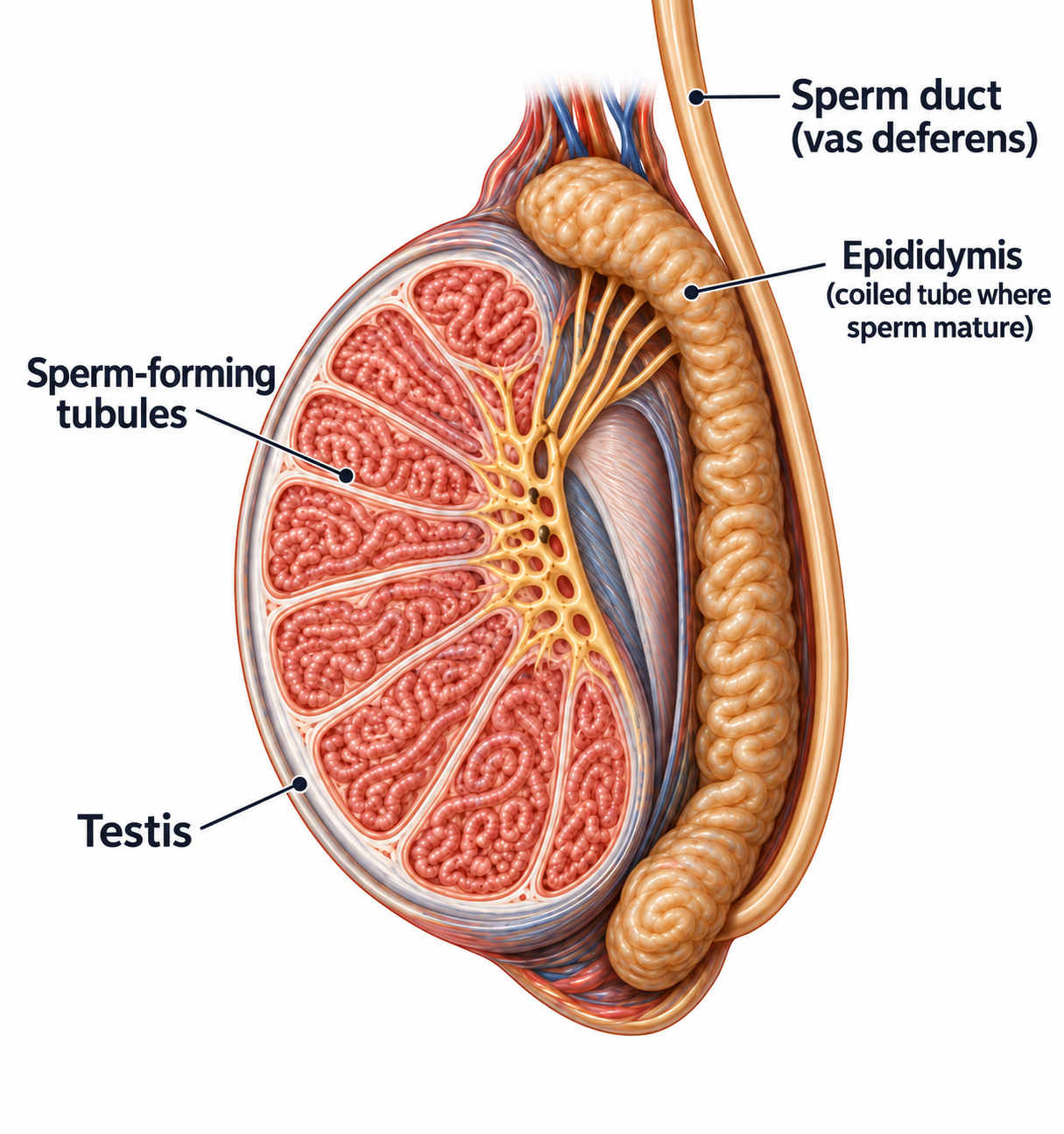
The testicles sit within the scrotum and produce sperm and testosterone, a hormone that helps maintain sex drive, muscle and bone health. The epididymis is a tube behind each testicle that stores and carries sperm; a lump there may have a different explanation from a solid lump inside the testicle. The spermatic cord contains the blood vessels and sperm-carrying tube. Understanding these structures helps explain why the usual cancer operation is performed through the groin.
Lymph nodes are small structures that help the body fight infection. Cancer may spread to nodes at the back of the abdomen (belly), called retroperitoneal nodes, and sometimes to the lungs or other organs. A new cancer can also develop separately in the other testicle. This is different from assuming that the first tumour has simply crossed from one testicle to the other. A new change in the remaining testicle deserves assessment even after successful treatment.
Seminoma and non-seminoma
- Seminoma: one of the two main groups of germ cell cancer. Its treatment options differ from those for non-seminoma. Blood-marker differences are explained below.
- Non-seminoma: a group that includes embryonal carcinoma, yolk sac tumour, choriocarcinoma and teratoma arising after puberty. These names describe different types of germ cell tumour seen under a microscope. A tumour may contain more than one of these components.
- Mixed germ cell tumour: a cancer containing both seminoma and a non-seminoma component is managed using the non-seminoma pathway.
- Teratoma: a tumour that can contain several types of tissue. When it develops after puberty, it can remain after chemotherapy (medicines that kill cancer cells) and may need surgery, even when tumour-marker blood tests are normal. It should not automatically be described as harmless.
The type of tumour found when tissue is examined matters more than age or how fast a lump appeared. Rarely, a different type of cancer can develop within a teratoma and need a different treatment plan. If a report is unusual, uncertain or does not fit the blood results, a pathologist can review it to help clarify the treatment plan. A pathologist is a doctor who diagnoses disease by examining tissue. Experience with germ cell tumours is useful for this review.
Understanding tumour-marker blood tests
Tumour markers are substances measured in the blood that can give clues about germ cell cancer. They help with diagnosis, planning treatment and checking its effect. A result may be described as raised or elevated when it is above the laboratory’s normal range.
- Alpha-fetoprotein (AFP) is a protein that some non-seminomas produce. Pure seminoma does not produce AFP.
- Beta-human chorionic gonadotropin (beta-hCG) is part of a hormone that some germ cell tumours produce. The blood test measures its level. It may be raised in seminoma or non-seminoma. Your report may use the name hCG.
- Lactate dehydrogenase (LDH) is an enzyme, a protein that helps chemical reactions in cells. It is found in many normal body tissues. It can rise in either cancer type and sometimes reflects the amount of cancer. It is less specific because many conditions unrelated to cancer can raise it.
Which tumour types can raise which markers?
These are typical patterns, not a way to diagnose the tumour type from blood tests alone. Mixed tumours can produce markers from more than one component.
| Tumour type | Typical AFP and beta-hCG pattern |
|---|---|
Pure seminoma | Does not produce AFP. Beta-hCG can be raised, but is often normal. |
Yolk sac tumour | Typically produces AFP. |
Choriocarcinoma | Typically produces beta-hCG, sometimes at very high levels. |
Embryonal carcinoma | May raise AFP, beta-hCG, or both. |
Pure teratoma after puberty | Usually does not raise AFP or beta-hCG. Teratoma can remain after chemotherapy even when these markers are normal. |
If AFP is genuinely raised because of the tumour, the team looks for a non-seminoma component even when the tissue report says seminoma. They first consider other explanations, such as liver disease.
What do the results tell us at each step?
- Before surgery: markers support the assessment and provide starting values to compare with later results. Ultrasound and examination of the removed tissue establish the diagnosis.
- After surgery: repeated tests show whether markers are falling as expected. Together with scans, they help determine the stage.
- Just before chemotherapy: the current levels help determine the prognosis group (the expected outlook with treatment), especially in non-seminoma, and guide the treatment plan.
- During treatment: the trend helps show whether treatment is working. A slower-than-expected fall or a rise needs review alongside the scans and clinical assessment.
- After treatment and during follow-up: markers help check the response and can sometimes be the first sign of cancer returning. They are used alongside the planned scans, examinations and symptoms.
How quickly should raised markers fall?
A marker’s half-life is the time the body takes to clear about half of it from the blood once the tumour is no longer producing it. Markers therefore need time to fall after successful surgery or treatment.
- AFP: approximately 5–7 days to fall by half.
- Beta-hCG: approximately 1–3 days to fall by half.
- LDH: there is no similarly reliable clearance timetable for judging cancer response; it is interpreted with the other findings.
A very high starting value may take several weeks to return to normal. A half-life is a guide to the expected trend, not a deadline for an individual patient. Chemotherapy and other changes in the body can complicate the pattern, so the team reviews several results with their dates.
Why one result is not the whole answer
Normal markers do not rule out cancer or prove that all cancer has gone. Some tumours do not produce these markers, and remaining tissue may contain teratoma. Keep the planned follow-up even when the blood results are reassuring.
A raised result does not always mean active cancer. Liver problems can raise AFP; hormone changes or interference with a laboratory test can affect hCG; and illness, tissue injury or damage to blood cells in the sample can raise LDH. A small, stable abnormality may need a repeat test or further checks before it changes the treatment plan.
Markers that remain raised or rise after surgery, beyond the expected fall and without another explanation, can indicate remaining cancer even when scans are normal. This is the reason for the separate stage IS category explained under Planning your treatment.
Factors that increase risk
An undescended testicle (one that did not move down into the scrotum during development), previous testicular cancer, a close family history and some problems with testicular development or fertility are associated with increased risk. Bringing an undescended testicle into the scrotum does not completely remove future risk. Many people have no recognised risk factor. The diagnosis is not evidence of personal fault, poor hygiene or an injury you failed to prevent.
Testicular cancer is not contagious and cannot be passed to a partner through sex. A family history does not mean every relative will develop it. Tell the team about affected relatives and previous testicular problems. Small calcium deposits found on ultrasound, called microlithiasis, do not by themselves establish cancer; further advice depends on the whole clinical picture rather than that word alone.
Tests and diagnosis
How cancer is diagnosed
The initial assessment usually includes examination and ultrasound of both testicles. Ultrasound helps distinguish a solid abnormality within the testicle from fluid or another scrotal problem. Blood is taken for AFP, beta-hCG and LDH before treatment when possible. Kidney function and other routine blood tests help prepare for surgery and any later treatment.
For a suspicious testicular tumour, the usual diagnostic and first treatment step is radical inguinal orchidectomy: removal of the affected testicle and spermatic cord through a cut in the groin. The tissue is then examined to confirm the diagnosis. A biopsy means taking tissue for examination; a needle biopsy through the scrotal skin is generally avoided for a suspected testicular cancer. Very small or uncertain lumps, tumours in both testicles, or a tumour in the only remaining testicle need an individual specialist assessment.
Rarely, when widespread cancer is immediately life-threatening, the team may need to start medicines that work throughout the body (systemic treatment) before removing the testicle. This depends on strong evidence of the diagnosis and the urgency of treatment. A new testicular lump still needs prompt assessment by a urologist.
Understanding your pathology report
The pathology report records what was found when the removed tissue was examined. It describes whether the tumour is seminoma, non-seminoma or mixed, its size and which structures it involves. It also records lymphovascular invasion, meaning tumour cells are seen in small blood vessels or lymph vessels, the channels that drain fluid from tissues. In stage 1 (I) non-seminoma, this helps estimate the chance of cancer returning (relapse or recurrence). It helps the team discuss regular scheduled checks (surveillance) or further treatment.
A report may mention germ cell neoplasia in situ (GCNIS): abnormal germ cells that have not yet grown into surrounding tissue. This is not the same as distant spread. Routine biopsy of the other testicle is not necessary for everyone; selected people with risk factors may discuss it. If surgery to remove the tumour while keeping the rest of the testicle is considered, the possibility of associated GCNIS and the need for further treatment or close monitoring must be explained.
Scans and staging tests
Computed tomography (CT) uses X-rays to make detailed pictures. It commonly checks the abdomen and pelvis, the lower part of the body between the hips. The team also arranges chest imaging as needed to check for spread. Magnetic resonance imaging (MRI) uses magnetic fields rather than X-rays and can sometimes be an alternative for abdominal scans, especially during follow-up.
Brain scans are used when symptoms or other findings suggest they may be needed. Positron emission tomography (PET) uses a small amount of radioactive tracer to show activity in tissues. It is often combined with CT as a PET-CT scan. PET-CT does not replace the usual scans for initial staging, although PET has a role in selected assessments after treatment.
The stage and a separate prognosis group help guide treatment. The next section explains how scans, blood markers and the cancer type contribute to these assessments.
Preparing for your appointment
For your consultation or a second opinion, bring a compact disc (CD) containing your scan images in Digital Imaging and Communications in Medicine (DICOM) format and the software needed to view them, along with the written scan reports. Bring your pathology report. Ask whether the laboratory’s glass slides or stored tissue samples are needed for specialist review or further tests. Keep originals safe and agree how borrowed material will be returned. Having the actual images allows the team to compare changes and plan treatment accurately.
A repeat test is sometimes needed because an earlier sample was too small, a scan did not cover the necessary area, or a new result will change treatment. Ask what question the test will answer, how it may affect the plan, and when you should expect the result. A result described as uncertain does not automatically mean the cancer is worse. Sometimes discussion between the radiologist (the doctor who interprets scans), pathologist and treating specialists resolves the uncertainty without another procedure.
Before a scan or procedure, mention kidney problems, previous reactions to scan contrast dye, implanted devices, diabetes medicines, blood-thinning medicines and any other relevant health conditions. Follow the testing centre's instructions about food, drink and medicines. If you have not received those instructions, contact the centre rather than making changes yourself.
Planning your treatment
Understanding your stage
Tumour, lymph nodes, distant spread and blood markers (TNM + S)
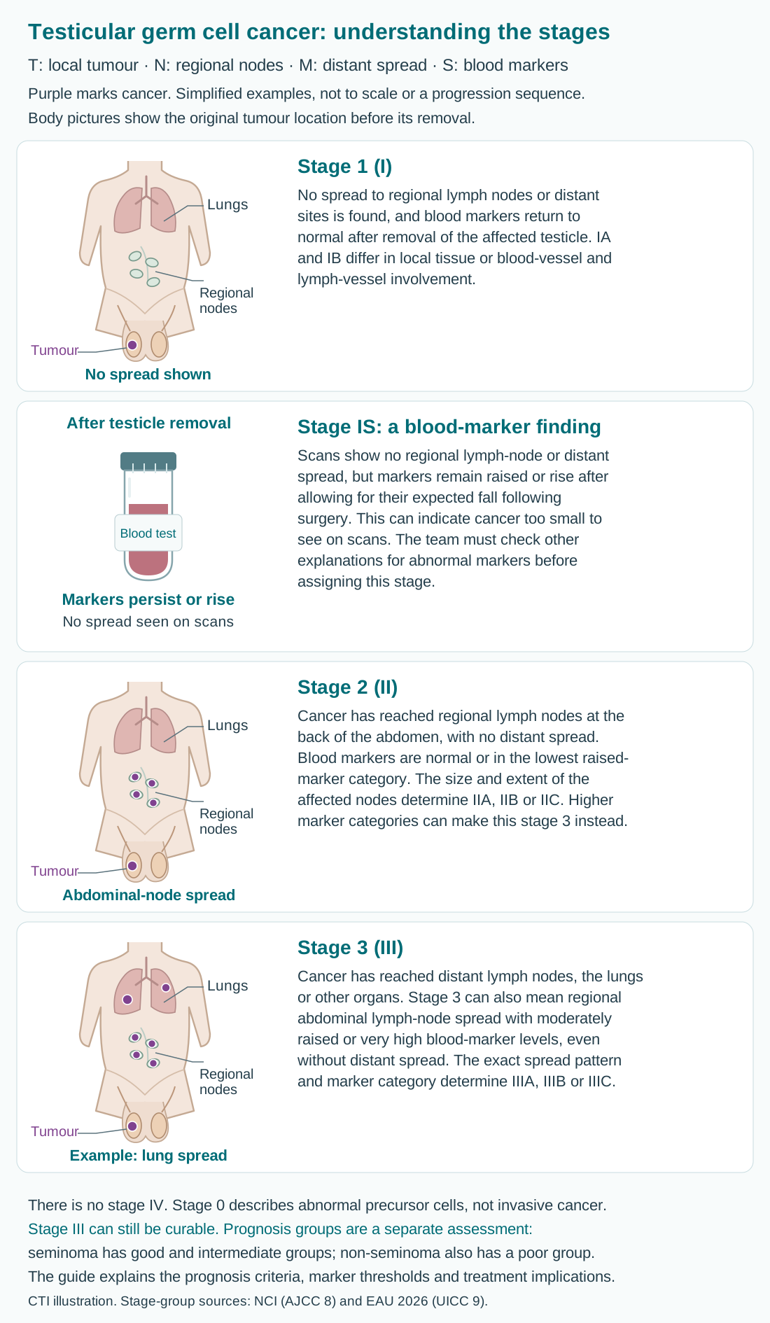
The stage combines the tumour’s local growth (T), spread to regional lymph nodes (N), distant spread or metastasis (M), and serum markers measured in blood (S). Testicular germ cell cancer that has grown into surrounding tissue is called invasive cancer and uses stages 1–3; there is no stage 4. Stage 0 describes abnormal germ cells that have not yet grown into surrounding tissue.
Lymph nodes are small immune-system structures. Regional nodes are those near or draining the cancer’s starting area; distant spread means cancer has reached a site outside that regional area. Stages describe findings, not a sequence that everyone will pass through.
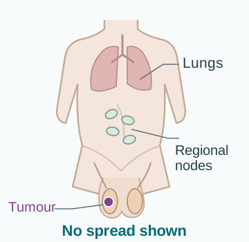
Purple marks cancer in the body pictures. These are simplified examples, not to scale. Pictures with a testicular tumour show its original location before removal. Stages describe findings, not steps that everyone passes through.
Stage 1 (I)
No spread to regional lymph nodes or distant sites is found, and blood markers return to normal after removal of the affected testicle. IA and IB differ in local tissue or blood-vessel and lymph-vessel involvement.
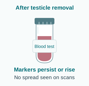
Stage IS: a blood-marker finding
Scans show no regional lymph-node or distant spread, but markers remain raised or rise after allowing for their expected fall following surgery. This can indicate cancer too small to see on scans. The team must check other explanations for abnormal markers before assigning this stage.
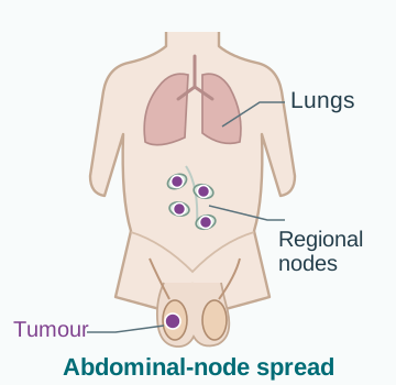
Stage 2 (II)
Cancer has reached regional lymph nodes at the back of the abdomen, with no distant spread. Blood markers are normal or in the lowest raised-marker category. The size and extent of the affected nodes determine IIA, IIB or IIC. Higher marker categories can make this stage 3 instead.
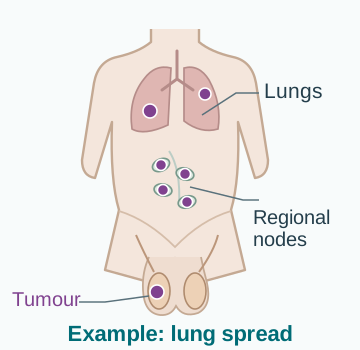
Stage 3 (III)
Cancer has reached distant lymph nodes, the lungs or other organs. Stage 3 can also mean regional abdominal lymph-node spread with moderately raised or very high blood-marker levels, even without distant spread. The exact spread pattern and marker category determine IIIA, IIIB or IIIC.
Even stage 3 cancer can be curable. A separate prognosis group helps guide chemotherapy; it is explained in the next section. A stage number alone does not determine the outlook.
The team combines scans and blood tests with examination of the tissue removed at surgery. Markers are followed over time because they do not disappear immediately after the affected testicle is removed.
Good, intermediate and poor prognosis groups
Prognosis means the expected outlook with treatment. For germ cell cancer that has spread, the International Germ Cell Cancer Collaborative Group (IGCCCG) classification helps the team choose the first chemotherapy plan. It uses the cancer type, where it started, where it has spread and blood-marker results.
These groups are separate from stages 1–3. For example, someone with stage 3 cancer may still be in the good-prognosis group. The groups below apply before the first chemotherapy treatment for advanced disease. They are different from the risk of cancer returning during stage 1 surveillance, or the assessment used when cancer returns after chemotherapy.
Which blood results are used?
The team uses alpha-fetoprotein (AFP), human chorionic gonadotropin (hCG, commonly measured as beta-hCG) and lactate dehydrogenase (LDH). Results should be measured immediately before chemotherapy, ideally on the day it starts. Results taken before the testicle was removed may give a different picture.
LDH is compared with the highest normal value for that laboratory, called the upper limit of normal (ULN). The team checks other possible reasons for raised markers before assigning a group. A mildly abnormal result alone does not prove that cancer remains.
Seminoma: two prognosis groups
Both groups require a normal AFP result and can include a tumour that started at any site. A tumour-related rise in AFP suggests a non-seminoma component, even if the tissue report says seminoma, and needs review.
| Group | What defines it |
|---|---|
Good prognosis | Cancer may involve lymph nodes or the lungs, but has not spread to other organs such as the liver, bones or brain. |
Intermediate prognosis | Cancer has spread to an organ other than the lungs, such as the liver, bones or brain. |
Seminoma has no poor-prognosis group in this classification. Any hCG or LDH level is allowed within either of these two groups. LDH can still add information about the outlook, as explained below.
Non-seminoma: three prognosis groups
Mixed tumours containing any non-seminoma component use this pathway. The highest-risk feature determines the group; the team does not average the results.
- Good prognosis: the tumour started in a testicle or at the back of the abdomen (the retroperitoneum), there is no spread to organs other than the lungs, and all three markers are in the good-prognosis range below.
- Intermediate prognosis: the same starting-site and spread conditions apply, but at least one marker is in the intermediate range and none is in the poor-prognosis range.
- Poor prognosis: any one of the following is enough: the tumour started in the central chest between the lungs (the mediastinum); it has spread to an organ other than the lungs, such as the liver, bones or brain; or at least one marker is in the poor-prognosis range.
Spread to the lungs or to distant lymph nodes does not, by itself, put non-seminoma in the poor-prognosis group. A tumour that starts in the central chest is different from a testicular tumour that spreads to chest lymph nodes.
The table shows the marker ranges used for non-seminoma. The units ng/mL mean nanograms per millilitre; IU/L means international units per litre. An hCG result in milli-international units per millilitre (mIU/mL) has the same numerical value as IU/L.
| Marker | Good range | Intermediate range | Poor range |
|---|---|---|---|
AFP | Below 1,000 ng/mL | 1,000–10,000 ng/mL | Above 10,000 ng/mL |
hCG | Below 5,000 IU/L | 5,000–50,000 IU/L | Above 50,000 IU/L |
LDH | Below 1.5 times the ULN | 1.5–10 times the ULN | Above 10 times the ULN |
The intermediate ranges include both end values. For example, AFP of exactly 1,000 or 10,000 ng/mL falls in the intermediate range. The tumour’s starting site and sites of spread must also be considered.
What do these groups mean for treatment?
The group helps determine the combination of chemotherapy medicines and how many cycles are needed. A cycle is a planned period of treatment followed by time to recover. Good-prognosis cancer usually needs fewer cycles than intermediate- or poor-prognosis cancer; the options are explained under Treatment options.
Poor prognosis does not mean that cure is impossible. Treatment can still aim to cure the cancer. Your team also considers your health, side effects and how the markers fall during treatment.
What do the updated studies show?
Two large international studies published in 2021 updated the expected outcomes with cisplatin-based treatment. Their estimates remain part of current guidance. Five-year overall survival means the proportion of patients alive five years after starting treatment, whether or not the cancer had returned.
| Cancer type and group | Five-year overall survival |
|---|---|
Seminoma — good prognosis | 95% |
Seminoma — intermediate prognosis | 88% |
Non-seminoma — good prognosis | 96% |
Non-seminoma — intermediate prognosis | 89% |
Non-seminoma — poor prognosis | 67% |
These are estimates for groups treated between 1990 and 2013, reported in the 2021 IGCCCG updates. They are not an individual prediction or a direct comparison between treatments. Your response to treatment and general health also matter.
The updates add detail within the groups. In seminoma, LDH above 2.5 times the ULN was linked to a higher risk of the cancer worsening or returning. In non-seminoma, an updated prediction model also considers age, lung spread and an LDH threshold of 2.5 times the ULN. These refinements do not replace the standard group definitions above or create a poor-prognosis seminoma category.
Useful questions for your oncologist: “Which prognosis group am I in, and which finding puts me there? How does it affect the medicines and number of cycles you recommend?”
How your treatment plan is chosen
Your team considers the tissue results, changes in blood markers over time, scans, general health and your wishes about having children. A urologist works with a medical oncologist, a doctor who treats cancer with medicines. A radiation oncologist plans radiotherapy, which uses high-energy rays to treat cancer, when this is needed. A pathologist, fertility team and lymph-node surgeon may also be involved.
If cancer has spread, returned or remains after treatment, advice from a team that regularly treats germ cell cancers can be particularly helpful.
After removal of the affected testicle, some people can choose between surveillance and treatment to reduce the chance of cancer returning. Surveillance is an active plan of scheduled reviews and tests, with treatment if cancer returns. It avoids exposing everyone to treatment they may not need, but depends on reliable attendance. The best choice balances the probability of relapse, the treatment required if relapse occurs, immediate side effects, long-term risks and your preferences.
Sperm banking before treatment
Discuss fertility early, ideally before surgery and certainly before chemotherapy, radiotherapy or an operation that may affect ejaculation. Ejaculation is the release of semen, the fluid that carries sperm, during orgasm. Testicular cancer itself can reduce sperm quality before any treatment. Freezing sperm provides an additional option for future parenthood; it is worth discussing even if you do not currently have a partner or are unsure about having children.
If producing a semen sample is difficult or no sperm are found, a fertility specialist may discuss a procedure to collect sperm directly, sometimes during surgery. Fertility planning should be arranged promptly so urgent cancer treatment is not delayed unsafely. Ask about storage, consent, later use and costs. Having stored sperm is not a guarantee of pregnancy, but it can preserve choices.
Hormones, appearance and personal priorities
One healthy remaining testicle often provides enough testosterone and sperm, but this is not guaranteed. An artificial testicle, called a testicular prosthesis, can be inserted at the time of surgery or later if you want it; choosing no implant is equally reasonable. It changes appearance and feel but does not produce hormones or sperm. Discuss size, expectations and potential discomfort or infection before deciding.
Tell the team about work, study, relationships and any concerns about discussing genital symptoms. You can ask for privacy, a trained chaperone to be present, and clear explanations before examination. Losing a testicle does not define masculinity, sexual worth or the ability to have a satisfying relationship. Practical and psychological support can be part of treatment from the beginning.
Questions to ask your care team
- What is the exact cancer type and stage, and are any important results still pending?
- Is the aim to cure the cancer, reduce the chance of recurrence, control it, or relieve a particular symptom?
- What benefit do you expect from this option, and what are the reasonable alternatives for me?
- How will treatment affect everyday activities, fertility, sexual health and the things I most value?
- How will we know whether it is working, and when will we review the plan?
- Who should I contact between visits, including at night or over a weekend?
Considering a clinical trial
A clinical trial is a research study that tests a treatment approach. It may be worth discussing at diagnosis or when treatment needs to change. Trials ask a defined question; some compare established treatments and others study a newer approach. Eligibility depends on the cancer, previous treatment, test results and health. Taking part is voluntary, and choosing not to participate does not remove your right to standard care. Ask about extra visits, whether treatment groups are assigned by chance, costs, travel, side effects and what happens when the study ends.
Your priorities and supportive care
Tell the team what you are hoping treatment will make possible: returning to work, caring for family, keeping independence, reducing pain or attending an important event. These priorities can influence a choice between reasonable options. Ask how each option balances the chance of cure or cancer control with side effects and the effect on daily life. Decisions can be revisited when your health or circumstances change.
Supportive and palliative care help with symptoms, sleep, mood, family concerns and planning. They can be provided alongside treatment intended to cure or control cancer. A referral does not mean that active treatment is ending. If the cancer cannot be cured, ask what treatments may help you live longer or more comfortably and how their burdens compare with their likely benefits.
Treatment options
Surgery to remove the affected testicle
As described under Tests and diagnosis, radical inguinal orchidectomy removes the affected testicle through the groin. It both treats the tumour and provides tissue to confirm the diagnosis. The other testicle is not routinely removed. The tissue results and blood markers after surgery help determine whether further treatment is needed.
Ask about pain relief, supportive underwear for the scrotum, wound care, lifting, driving and returning to work. Mild bruising or swelling may occur, but increasing pain, redness, discharge, fever or a rapidly enlarging swelling needs review. Removing one testicle does not usually prevent erections or orgasm. Recovery and confidence can nevertheless take time, particularly if further treatment follows soon afterwards.
Stage I seminoma
Surveillance is preferred for many people after surgery when markers and staging support stage I disease. Most will not need further treatment. If relapse occurs, it is usually still highly treatable, but treatment may involve more medicines or more cycles than treatment given straight after surgery to reduce the risk of recurrence (adjuvant treatment). Follow-up visits and scans must remain a priority even when you feel completely well.
A single course of the chemotherapy medicine carboplatin can be considered when surveillance is unsuitable or after discussing the benefits and risks. It reduces the chance of recurrence but does not remove it or replace follow-up.
Radiotherapy is reserved for selected situations because side effects that develop years later matter in people likely to live for many decades. Tumour size and other tissue findings help the discussion but should not automatically lead to extra treatment on their own.
Stage I non-seminoma
Surveillance is a common choice, particularly when lymphovascular invasion is absent and follow-up can be maintained. When relapse risk is higher, one cycle of bleomycin, etoposide and cisplatin (BEP) chemotherapy is an important alternative. A cycle is a planned period of treatment followed by time to recover. The choice should include discussion of both the chance of avoiding all chemotherapy and the possibility of needing several cycles if relapse develops.
Surgery to remove lymph nodes at the back of the abdomen is called retroperitoneal lymph node dissection (RPLND). It may be considered for particular tissue findings or when other approaches are unsuitable. It is not a routine requirement for every stage I cancer. The operation should be performed by an experienced team; its results can sometimes lead to further treatment. Markers that remain raised after surgery need separate assessment before a surveillance plan is chosen.
Seminoma involving abdominal lymph nodes
For stage IIA or selected IIB seminoma, established choices include radiotherapy or cisplatin-based chemotherapy. The size and location of the affected nodes, blood markers, general health and possible long-term side effects help determine which approach fits. More extensive cancer generally favours chemotherapy. A small uncertain node with normal markers may sometimes be reassessed before committing to treatment.
RPLND and other ways to reduce the amount of treatment are being studied for carefully selected patients with a small amount of seminoma in lymph nodes. They need specialist assessment, preferably within a clinical trial, a research study that tests a treatment approach. A reassuring early PET scan alone is not a reason to reduce chemotherapy outside a tested treatment plan.
Non-seminoma involving abdominal lymph nodes
When blood markers are normal and stage IIA non-seminoma involves a small amount of cancer in the abdominal lymph nodes, RPLND may be an option. The surgeon aims to preserve nerves that help control ejaculation when this is safe. This can be particularly important when teratoma is suspected, because teratoma does not respond reliably to chemotherapy. Small nodes whose cause is uncertain can sometimes be checked again before the team decides they truly represent spread.
When markers are clearly raised or disease is more extensive, cisplatin-based chemotherapy is usually the main initial approach, selected according to prognostic group. Surgery may still be needed afterwards. The marker result, tumour type and distribution all matter: a plan for a small amount of cancer with normal markers may differ from a plan for rising markers or more widespread cancer.
Chemotherapy when cancer has spread
BEP chemotherapy is a standard treatment intended to cure germ cell cancer that has spread (metastatic cancer). Good-prognosis disease commonly receives three cycles. Four cycles of etoposide and cisplatin (EP) can be used in suitable patients when bleomycin is inappropriate. The oncology team chooses the combination of medicines, doses and schedule to suit the cancer and your health.
Intermediate-prognosis disease, and poor-prognosis non-seminoma, generally need four cycles of BEP. When bleomycin cannot be used, four cycles of etoposide, ifosfamide and cisplatin (VIP) may be appropriate. VIP requires close monitoring for low blood counts and infection, with medicines and other care to reduce these risks. Carboplatin used after surgery for stage I seminoma is not a routine substitute for cisplatin when treatment aims to cure cancer that has spread.
Blood tests and clinical review are performed through treatment, including tumour-marker trends. Kidney function, hearing, lung health and other factors guide monitoring. Maintaining the intended treatment intensity can be important for cure, but safety decisions belong to the specialist team. Do not skip a treatment or alter doses yourself. If severe illness or side effects develop, contact the team promptly so any change is planned.
What if a lump remains after chemotherapy?
A remaining lump on a scan is called a residual mass. It may contain scar tissue, teratoma or active cancer. Seminoma and non-seminoma are managed differently. For seminoma with a residual mass larger than 3 centimetres (cm), PET assessment may be useful after an adequate interval, generally at least two months after chemotherapy. A positive result alone does not reliably prove that live cancer cells remain and needs expert interpretation before further treatment.
For non-seminoma with markers that have returned to normal, a residual mass larger than 1 cm often needs surgical removal, usually through RPLND when the lump is in abdominal lymph nodes. This operation needs a team experienced in surgery after chemotherapy. PET cannot reliably exclude teratoma and is not used to decide that such surgery is unnecessary. Smaller residual findings require an individual follow-up or surgical decision. Rising markers after treatment need separate assessment and may lead to further drug treatment rather than surgery for the remaining lump.
Lymph-node surgery and recovery
RPLND removes selected deep abdominal lymph nodes and can be technically demanding, especially after chemotherapy. Surgery can be more complex when nearby blood vessels, kidneys or other structures are involved. Ask why surgery is needed, how much tissue is expected to be removed, and whether nerve preservation is possible. The choice between one larger cut (open surgery) and smaller cuts (keyhole surgery) depends on cancer control and safety.
Nerve injury can reduce or stop semen coming out during orgasm, even when erections and the sensation of orgasm remain. Sperm banking should be discussed beforehand. Recovery may involve tiredness (fatigue), bowel changes or leakage of lymph fluid into the abdomen. Increasing abdominal swelling, persistent vomiting, wound problems or difficulty eating should be reported. Dietary restrictions after surgery should follow the treating team's instructions rather than a general internet plan.
Cancer that returns after treatment
Relapse does not automatically mean the cancer is incurable. The team reassesses pathology, markers, scans, previous treatment and how long the cancer remained controlled. A recurrence during stage I surveillance is different from cancer progressing after full cisplatin-based chemotherapy, and the next treatment is chosen accordingly.
After previous chemotherapy, further treatment may include paclitaxel, ifosfamide and cisplatin (TIP), or high-dose chemotherapy with support from the patient's own collected blood-forming stem cells (an autologous stem cell transplant). Stem cells can develop into new blood cells; autologous means using your own cells. The collected cells are returned through a drip after chemotherapy to help restore blood-cell production.
You may also hear the term autologous bone marrow transplant. In testicular cancer, the stem cells are usually collected from the bloodstream rather than directly from the bone marrow, the soft tissue inside bones where blood cells are made.
The choice depends on individual risk and previous treatment. Surgery may be needed for cancer or teratoma remaining after treatment, or for cancer that returns after a long interval. Review at a centre experienced in germ cell cancer is especially valuable before starting further treatment.
Managing side effects
Chemotherapy effects that need early attention
Cisplatin can affect kidney function, hearing and nerves. Fluids and checks of blood salts, called electrolytes, help protect the kidneys; follow the fluid advice tailored to your health. Report ringing in the ears, hearing loss, numbness, balance difficulty or markedly reduced urine. Some effects improve, but others can persist, so early reporting matters.
Bleomycin can cause serious lung inflammation. New cough, breathlessness or fever during or after treatment needs prompt assessment; infection and blood clots are other possible causes. Tell future anaesthetic and emergency teams that you have received bleomycin. Oxygen needed in an emergency should not be refused; clinicians should manage it with awareness of your treatment history. Discuss diving or other activities involving unusual oxygen exposure with a specialist.
Ifosfamide can cause bladder irritation, kidney problems or confusion, and is given with specific protective measures and monitoring. Chemotherapy combinations can lower blood counts, including white blood cells that fight infection. A painful swollen leg, sudden chest pain or new breathlessness may indicate a clot and needs urgent care. Anti-sickness medicines and other supportive treatments should be taken as directed rather than waiting for symptoms to become severe.
Fertility, contraception and sexual function
Chemotherapy and radiotherapy can reduce sperm production temporarily or permanently. Recovery varies and cannot be predicted from erections, ejaculation or feeling well. Ask when a test of the number and quality of sperm in a semen sample (semen analysis) would be useful and when it is safe to try for pregnancy after the particular treatment you received. Reduced fertility does not prevent every pregnancy, so contraception may still be needed, and stored sperm should not be discarded without a considered discussion.
If both testicles are removed, testosterone replacement is generally needed. After removal of one, persistent low energy, reduced sex drive, difficulty getting or keeping an erection, or low mood can prompt a hormone assessment. These symptoms have several possible causes. Testosterone treatment needs specialist guidance, particularly if future fertility matters, because taking testosterone can reduce sperm production.
Radiotherapy and longer-term effects
Abdominal radiotherapy can cause nausea, bowel changes and fatigue; the treatment plan aims to limit exposure of healthy organs and the remaining testicle. Fertility protection and later monitoring should be discussed in advance. Radiotherapy does not make you radioactive. The possibility of later heart or blood-vessel disease, or a second cancer, is part of choosing treatment, especially when surveillance is a reasonable alternative.
After chemotherapy, cold-sensitive fingers, persistent tingling, hearing changes or fatigue may need rehabilitation and symptom treatment. Tell the team about effects on work, driving, sleep or concentration. A cancer-free scan does not mean these difficulties should be dismissed. Recovery support can continue after active cancer treatment has ended.
Keeping treatment manageable
Keep the team's contact details available and know where to go outside clinic hours. Report side effects early rather than waiting until the next appointment. A short record of symptoms, temperature, fluid intake and medicines can help the team decide what is needed. Do not change cancer medicine doses, restart a stopped treatment, or use leftover antibiotics without instructions. A planned delay or dose adjustment can be an important part of safe treatment.
If you receive treatment that can lower white blood cells, a temperature of 38°C (100.4°F) or higher needs urgent advice. Follow any lower threshold or additional instructions your team gives you. Chills, sudden weakness, confusion or feeling very unwell also need urgent assessment, even without a fever. Do not wait for a blood test appointment or try to hide the fever with medicine before speaking to the team.
Severe breathlessness, chest pain, a new seizure, sudden weakness, heavy bleeding or collapse require emergency care. New severe back pain with leg weakness, numbness around the bottom, or loss of bladder or bowel control also needs immediate assessment. Contact the nearest emergency service when necessary; a website contact form or routine messaging service is not an emergency pathway.
Eating, drinking and energy
Treatment may change appetite, taste, bowel habits and energy. Small, frequent meals and foods containing protein can be easier than large meals. Ask for help from a dietitian, a specialist in food and nutrition, if weight is falling, swallowing is difficult, or you are avoiding many foods. There is no single diet that treats cancer, and severe restriction or prolonged fasting can make it harder to tolerate treatment. Check supplements with your team because some interact with medicines.
Take anti-sickness medicine as prescribed and report vomiting that prevents you keeping down fluids or essential medicines. Seek advice for persistent diarrhoea, dizziness, very little urine or increasing weakness. Fluid recommendations may need adjustment if you have heart or kidney disease. Mouth ulcers, pain or infection that interferes with eating should be treated promptly rather than accepted as unavoidable.
Gentle movement, short walks and planned rest may help fatigue, but activity should match your condition and any surgical or bone-safety restrictions. Fatigue can also reflect anaemia (too few healthy red blood cells), infection, hormone changes, pain, poor sleep or low mood. Let the team assess a marked or persistent change. Accepting help with shopping, transport or meals can conserve energy for the activities that matter to you.
Live well
Follow-up after treatment
Follow-up depends on whether the cancer was seminoma or non-seminoma, its stage and the treatment received. Surveillance after surgery alone is different from monitoring after chemotherapy or radiotherapy. Ask for a clear schedule of appointments, blood-marker tests and scans. Visits are generally more frequent early on and less frequent later; late symptoms still need assessment.
A normal marker result does not exclude relapse, and imaging remains important when scheduled. Conversely, a mildly abnormal result should be interpreted in context rather than causing an immediate assumption that cancer has returned. MRI can sometimes be used instead of repeated CT scans to reduce exposure to X-rays. The follow-up team needs experience in using MRI for this purpose. If you miss an appointment, contact the team to rearrange it promptly.
Learn what is normal for the remaining testicle and report a new lump or change. Persistent back or abdominal pain, a neck lump, unexplained weight loss or a persistent cough or breathlessness between visits should also be discussed. Routine scans indefinitely are not necessarily helpful; after the main surveillance period, attention increasingly includes symptoms, late effects and general health.
Protecting long-term health
Long-term care includes checks of blood pressure, cholesterol, blood sugar, kidney health, hearing, hormone symptoms and fertility when relevant. Keep a record of the medicines and radiotherapy you received so future clinicians can recognise treatment-related risks. Avoid tobacco and build up activity gradually, with support for weight, sleep and heart and blood-vessel health.
Returning to education, work, sport or dating can raise concerns that were less pressing during treatment. Ask for help with concentration, anxiety around scans, sexual confidence or explaining your experience to others. You do not need to minimise these concerns because the cancer is often curable. A long-term plan should support the life you want to return to, as well as checking for recurrence.
Emotional and practical support
Feeling frightened, angry, sad or uncertain is understandable. You do not have to appear positive all the time. Ask about counselling, psychological support for people with cancer, support groups or a social worker if worry is affecting sleep, relationships or daily life. Persistent depression, panic or thoughts of self-harm need prompt professional help. Caregivers may need their own support and breaks; one person should not have to manage every appointment and household task alone.
It can help to agree who will update relatives and how much information you want shared. Children usually benefit from honest, age-appropriate explanations and reassurance about who will care for them. Ask permission before discussing private medical information with others. If concentration or memory is difficult, use reminders, a calendar and a trusted companion at important consultations.
Relationships, fertility and sexual health
Cancer and treatment can affect desire, comfort, fertility, body image and confidence. These are valid medical concerns. Raise fertility questions before treatment when possible, because some protective options need to be arranged early. The right approach depends on age, the treatment and how urgently it must start. Reduced fertility does not mean that you cannot father a child. Ask about contraception and when it is safe to try for a pregnancy after your specific treatment.
Explain any pain, difficulty getting or keeping an erection or changes in intimacy to the team. Appropriate treatments, rehabilitation and counselling can help. You can choose how much to involve a partner. Advice about hormone-containing treatments needs to take the cancer type and treatment into account; do not start them without discussing suitability.
Planning ahead while living well
Ask for a clear follow-up plan and know which symptoms should be reported between visits. Continue care for other conditions and discuss vaccinations with your oncology team; the timing and suitability of live vaccines (which contain a weakened germ) can change during treatment. Avoid tobacco and ask for practical help to stop smoking if needed. Activity, food and sleep plans should be realistic for your health rather than another source of pressure.
If treatment is long term, arrange periodic discussions about what is helping, what is difficult and what you want next. Planning for future care can protect your preferences and reduce uncertainty for family. You can discuss who should help with decisions if you become too unwell to speak for yourself, while continuing treatment and everyday life now.
Sources and further reading
For more information, explore these resources from the National Cancer Institute (NCI) and other medical organisations. The linked organisations have not reviewed or endorsed this CTI guide. Your care team can explain how the information applies to you.
- NCI: Testicular Cancer Treatment
- ESMO–EURACAN testicular cancer guideline (2022)
- EAU testicular cancer guidelines (2026)
- ASCO: tumour markers in germ cell tumours
- ASCO: fertility preservation guideline update (2025)
- AUA early-stage testicular cancer guideline amendment (2023)
- Macmillan: testicular cancer
- Cancer Council Australia: testicular cancer
- EAU testicular cancer guidelines (2026): tumour markers and diagnostic evaluation
- American Cancer Society: germ cell tumour types and marker patterns
- ASCO tumour-marker guideline (2010): interpretation, half-lives and false-positive results
- NCI: stem cell and bone marrow transplants, including autologous transplantation
- Cancer Research UK: chemotherapy and autologous stem cell transplant for testicular cancer
Staging and risk-group references
Numbered stages: American Joint Committee on Cancer (AJCC), 8th edition, which remains current for testis in 2026. The 2026 European Association of Urology guidance uses the Union for International Cancer Control (UICC), 9th edition (2025); the broad stage groups shown here are consistent with both. Prognosis groups use the IGCCCG classification with its 2021 outcome updates.
- AJCC: Current staging editions by cancer type (2026)
- NCI: Testicular staging tables and explanation
- EAU testicular cancer guidelines (2026): staging and prognosis groups
- EAU testicular cancer guidelines (2026): treatment by prognosis group
- Beyer et al. (2021), Journal of Clinical Oncology: IGCCCG update for metastatic seminoma
- Gillessen et al. (2021), Journal of Clinical Oncology: IGCCCG update for metastatic non-seminoma
Medicine approval and prescribing references
These official records describe licensed uses and prescribing information for the medicines named in this guide. Approval depends on the cancer type, biomarkers, previous treatment, combination and formulation. Some uses discussed in the guide are off-label: outside the medicine’s licence, with evidence from clinical guidelines or studies. FDA or EMA approval does not establish approval or availability in India.
- Bleomycin — FDA prescribing information · Approval history
- Carboplatin — FDA prescribing information · Approval history
- Cisplatin — FDA prescribing information · Approval history
- Etoposide — FDA prescribing information · Approval history
- Ifosfamide — FDA prescribing information · Approval history
- Paclitaxel — FDA prescribing information · Approval history
Related care
Meet our doctors



Where to get care