
Share
Start here
About this guide
A stomach cancer diagnosis can feel overwhelming. This guide explains tests, treatment choices, recovery and support for you and those caring for you. It focuses on adenocarcinoma, cancer that starts in gland-forming cells in the stomach lining. Other types, including lymphoma, gastrointestinal stromal tumours (GIST) and neuroendocrine tumours, need different treatment plans.
Start with your biopsy result (the examination of a tissue sample), what the scans show and the goal of treatment. You do not need to understand every medicine at once. Tell your team early if eating is difficult or you are losing weight, and ask for dietitian support.
Bring your test reports, medicine and allergy lists, and any questions to your appointment. A family member or caregiver can help you take notes if you wish. Ask who is coordinating your care and which result or appointment comes next.
Stomach cancer at a glance
Most stomach cancers begin in the inner lining. Treatment depends on the cancer’s location, how deeply it has grown, whether it has spread to lymph nodes (small parts of the immune system) or elsewhere, and the results of tests on the cancer cells.
- Early stomach cancer may cause no clear symptoms. Persistent indigestion, feeling full quickly, weight loss, vomiting, swallowing difficulty, anaemia (low red blood cells) or bleeding need assessment.
- Upper gastrointestinal (GI) endoscopy uses a thin, flexible camera tube to examine the upper digestive tract. Several tissue samples are taken to confirm the diagnosis. A scan alone cannot reliably identify the tumour type.
- Scans help establish the stage, or extent, of the cancer. These include a computed tomography (CT) scan with contrast dye of the chest, abdomen and pelvis, and a positron emission tomography combined with CT (PET-CT) scan. Endoscopic ultrasound (EUS), which uses an ultrasound probe on an endoscope, and other tests are added when they answer specific questions.
- Some very early cancers confined to the inner lining (mucosa) can be removed through an endoscope. A specialist may use endoscopic submucosal dissection (ESD), which removes the cancer in one piece.
- For many people well enough for treatment whose stage II–III cancer can be removed, chemotherapy is given before and after gastrectomy (removal of part or all of the stomach). Surgery usually includes D2 lymph-node dissection: removal of defined lymph-node groups around the stomach and its main blood vessels. Durvalumab immunotherapy may be added for suitable patients.
- For advanced cancer, tests on the cancer cells help guide treatment. These biomarker tests are explained under Tests and diagnosis. General health, symptoms and previous treatment also matter.
- Nutrition support and palliative care (help with symptoms and quality of life) can start from diagnosis alongside cancer treatment.
Diagnosis
The exact cancer type, how the cells look under the microscope (grade), and where the tumour began.
Extent
An explanation of the stage, whether surgery is suitable and whether the abdominal lining is involved.
Tumour tests
Biomarker results that may guide treatment, especially when cancer is advanced.
Readiness
Nutrition, strength, organ function and personal priorities.
Bring your scan images
For your consultation or a second opinion, bring a CD containing your scan images in Digital Imaging and Communications in Medicine (DICOM) format, with the software needed to view them, along with the written scan reports.
Understand
Topics in this section
What your stomach does
The stomach is a muscular pouch between the oesophagus (food pipe) and small bowel. It stores and mixes food before passing it into the bowel.
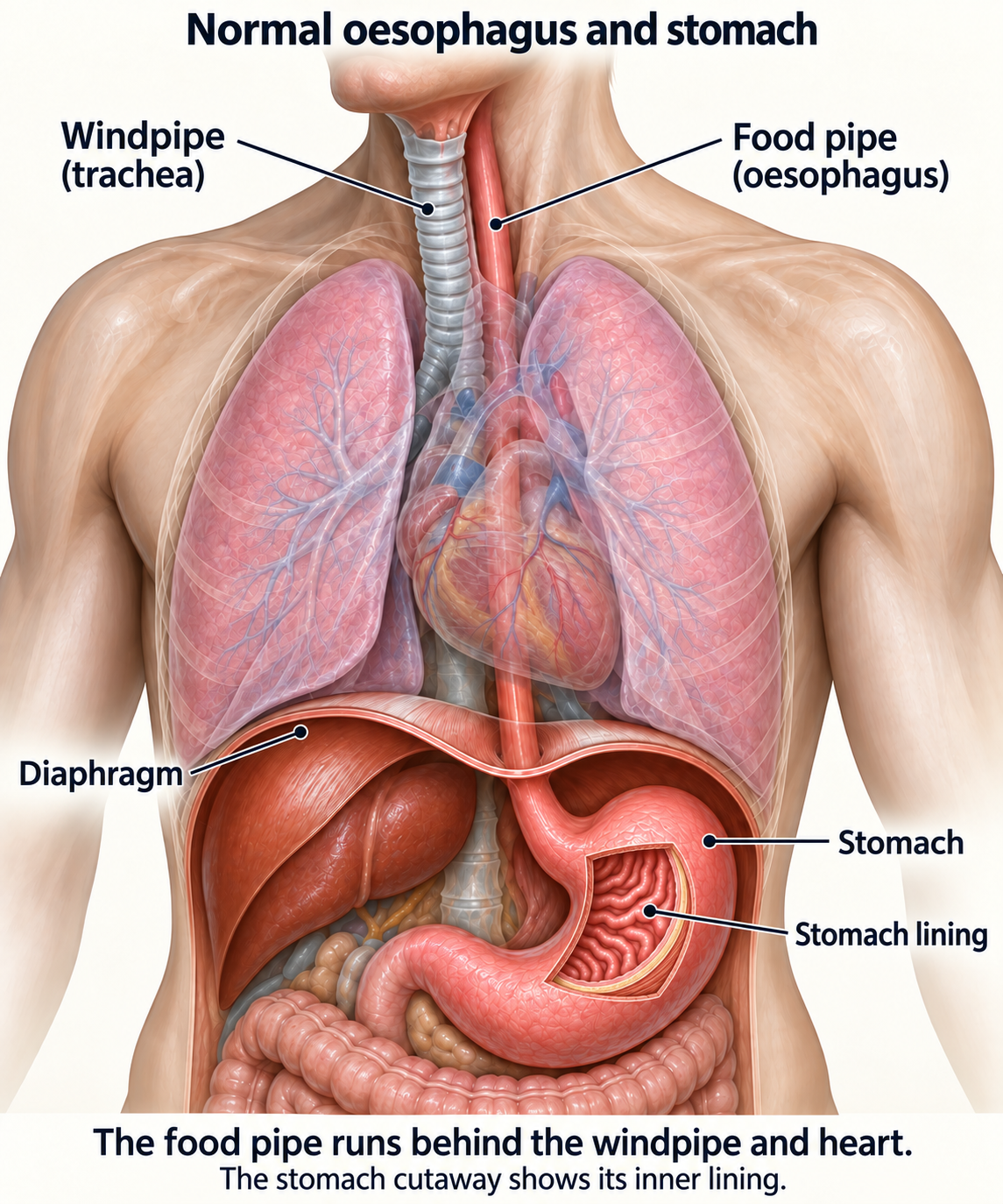
Stores and mixes food
Stomach muscles mix food with acid and digestive enzymes. Food then passes through the stomach outlet (pylorus) into the first part of the small bowel (duodenum).
Supports digestion
Acid begins protein digestion and helps protect against swallowed microbes. Most nutrient absorption occurs later in the small bowel.
Helps absorb vitamin B12
Stomach cells make a protein called intrinsic factor, which helps the body absorb vitamin B12 further along the small bowel.
Signals fullness
Stretch and hormones influence appetite. Cancer and surgery can make fullness arrive after only a few mouthfuls.
The stomach wall and how cancer spreads
As cancer grows deeper into the stomach wall, it can reach blood vessels and lymph channels, which may allow it to spread. The staging section explains how the depth is recorded.
Layer | What it does | Why it matters |
|---|---|---|
Mucosa | Inner glandular lining in contact with food | Very early cancer in this lining may be suitable for endoscopic removal if its size and other features suggest a low risk of spread. |
Submucosa | Support layer containing vessels and lymph channels | Cancer growing into this layer has a greater risk of spreading to lymph nodes; surgery is usually needed. |
Muscularis propria | Thick muscle that churns food | Cancer reaching the muscle layer generally needs a combination of treatments. |
Subserosa and serosa | Outer supporting tissue and surface covering | Cancer reaching these outer layers has a greater risk of spreading to the abdominal lining or nearby organs. |
Four routes of spread
Direct
Into the oesophagus, duodenum, pancreas, colon, spleen or other nearby structures.
Through lymph channels
To lymph nodes near the stomach and, sometimes, to more distant lymph nodes.
Bloodstream
Most often to the liver, and sometimes lung, bone or other organs.
Along the abdominal lining
Cancer cells can spread along the abdominal lining (peritoneum), forming small deposits or causing a build-up of fluid called ascites.
The exact tumour type matters
This guide focuses on gastric adenocarcinoma. A similarly named tumour may need a completely different treatment plan.
Pathology label | What it means for care |
|---|---|
Adenocarcinoma — intestinal type | The cancer cells often form glands. It may develop after long-term inflammation of the stomach lining (gastritis), thinning of the lining (atrophy), and changes that make the lining resemble bowel tissue (intestinal metaplasia). |
Adenocarcinoma — diffuse or signet-ring type | Cancer cells can spread through the stomach wall without forming a clear lump. They may be less visible on PET-CT. The team also assesses the abdominal lining and whether an inherited change in the CDH1 gene could be relevant. |
Gastro-oesophageal junction adenocarcinoma | The gastro-oesophageal junction (GEJ) is where the food pipe meets the stomach. Cancers near this junction may follow a GEJ or oesophageal treatment plan. A multidisciplinary team (MDT), a group of specialists who plan care together, will identify where the tumour began. |
GIST | A tumour arising from cells in the digestive-tract wall. Many have changes in the KIT or PDGFRA genes. Surgery and targeted medicines differ from treatment for adenocarcinoma. |
Gastric lymphoma | A cancer of immune cells. Treatment may include treating Helicobacter pylori (H. pylori), a bacterium that can infect the stomach, or chemotherapy, antibody medicines or radiotherapy. Stomach removal is uncommon. |
Neuroendocrine neoplasm | This tumour follows a separate treatment plan. The grade, Ki-67 result (a measure of how many cells are dividing) and whether the tumour makes hormones help guide care. |
Exact wording
Keep the report’s exact description of the cancer type (histology) and how normal or abnormal the cells look (differentiation).
Exact location
Ask whether the cancer is near the stomach entrance, in the upper or middle stomach, near the outlet, or spread through the wall.
Pattern
The report may describe intestinal, diffuse, mixed, poorly cohesive or signet-ring patterns. Your team can explain how the pattern affects care.
Review
An unusual result or a very small tissue sample may need specialist review.
Symptoms and warning signs
Many symptoms are caused by common non-cancer conditions. Persistence, progression, bleeding or weight loss are reasons to investigate rather than self-treat indefinitely.
Eating-related
Early fullness, reduced appetite, bloating, nausea, vomiting or food sticking.
Pain or discomfort
Persistent upper-abdominal pain, burning or indigestion that does not settle.
Blood loss
Black tarry stools, vomiting blood, tiredness, breathlessness or iron-deficiency anaemia.
Whole-body symptoms
Unintentional weight loss, weakness, loss of muscle or a persistent decline in function.
When to seek urgent help
- vomiting fresh blood or material that looks like coffee grounds; black stools with dizziness or fainting
- inability to keep fluids down, very little urine, confusion or severe weakness
- sudden severe abdominal pain, a rigid swollen abdomen, fever or collapse
- new chest pain, severe breathlessness, one-sided leg swelling or coughing blood
Intake
Record liquids, meals and vomiting over 24 hours when intake falls.
Weight
Track weekly—not several times a day—and report a continuing fall.
Bleeding
Report black stools, blood in vomit or dizziness. Tell the team if you take blood-thinning medicines.
Function
A sudden drop in walking, self-care or alertness can be an urgent clue.
Risk factors
Usually there is no single cause. Risk factors change probability; they do not prove why one person developed cancer.
Factor | What is known | Practical response |
|---|---|---|
H. pylori infection | Can cause long-term inflammation, thinning of the stomach lining and cancer away from the stomach entrance. | Have testing and treatment when advised, followed by a test to check that the infection has cleared. |
Tobacco | Smoking raises risk and also worsens surgical and cardiovascular outcomes. | Stopping helps at any stage; ask for medication and behavioural support. |
Food and salt | High intake of salt-preserved or smoked foods is associated with risk; fruit and vegetables are protective dietary patterns. | Use refrigeration, reduce very salty preserved foods and avoid extreme diets. |
Previous stomach disease or surgery | Risk can be higher with thinning or abnormal changes in the stomach lining, pernicious anaemia (vitamin B12 deficiency caused by poor absorption), some stomach polyps, or a stomach remnant many years after surgery. | Follow a specialist surveillance plan when one is recommended. |
Family or inherited predisposition | A small proportion relate to inherited changes in the CDH1 or CTNNA1 genes, Lynch syndrome, or other inherited conditions. | A genetic counsellor can assess your family history and help choose and interpret a suitable test. |
H. pylori
Ask whether testing or eradication is relevant for you and household members.
Tobacco
Medication plus counselling is more effective than willpower alone.
Diet
Aim for a sustainable varied pattern; avoid fear-driven restrictions.
Surveillance
If the stomach lining has abnormal changes or part of the stomach remains after surgery, ask whether regular specialist checks are needed.
Family history and genetic counselling
Most stomach cancers are not inherited. The types of cancer in the family and ages at diagnosis help determine whether germline testing, which looks for inherited gene changes, may be useful.
Ask for genetic assessment when there is
- diffuse or signet-ring gastric cancer at a young age, especially under 50
- two or more close relatives with gastric cancer, particularly diffuse type
- diffuse gastric cancer plus lobular breast cancer in the person or family
- a known inherited gene change linked to CDH1, CTNNA1, Lynch syndrome or another condition that raises cancer risk
- multiple cancers, numerous polyps, or a family pattern suggesting an inherited syndrome
Possible syndrome | Why it matters |
|---|---|
Hereditary diffuse gastric cancer | Usually involves CDH1 and sometimes CTNNA1. Expert surveillance and, for selected proven carriers, risk-reducing total gastrectomy may be discussed. |
Lynch syndrome | Inherited changes in genes that repair genetic material increase bowel and several other cancer risks. Results help guide screening for the person and their relatives. |
Other rare syndromes | Familial adenomatous polyposis (FAP), gastric adenocarcinoma and proximal polyposis of the stomach (GAPPS), Peutz–Jeghers syndrome and juvenile polyposis may require regular checks of the upper digestive tract. |
Who had what?
Record the exact cancer type and age at diagnosis for both sides of the family.
Which test?
A multigene panel may be better than testing one gene without context.
Before testing
Discuss insurance/privacy, uncertain variants and who receives results.
After testing
If a harmful inherited change is found, the genetics team can help you and your relatives understand the next steps.
Tests and diagnosis
How cancer is diagnosed
You may not need every possible test. Each step should answer a question that changes treatment.
- The specialist examines the tumour with upper GI endoscopy and takes enough tissue samples. The report records its position in relation to the GEJ and stomach outlet.
- A pathologist confirms the cancer type and grade. H. pylori testing may also be relevant. Unusual findings may need specialist pathology review.
- Stage with a contrast CT scan of the chest, abdomen and pelvis and a PET-CT scan. Add EUS or magnetic resonance imaging (MRI) when they answer specific questions.
- Staging laparoscopy and abdominal washings check for spread that scans may miss. They are particularly useful when the tumour reaches deeper wall layers, involves lymph nodes or has a diffuse growth pattern. When indicated, complete these checks before starting neoadjuvant chemotherapy.
- Assess fitness, nutrition, blood count, kidney/liver function, heart-lung health and medicines before a plan is finalised.
- The upper-GI MDT brings together the tissue and scan results, stage, biomarker tests and your priorities to recommend a plan.
Endoscopy complete?
Location photographed and enough representative biopsies obtained.
Staging complete?
Contrast CT and PET-CT scans, with EUS or MRI for specific questions and staging laparoscopy when indicated.
Fitness complete?
The team checks nutrition and organ function and helps you prepare with appropriate food and activity before treatment.
MDT complete?
Talk through the stage, treatment aim, sequence and alternatives with your team.
Upper GI endoscopy and biopsy
During upper GI endoscopy, a thin, flexible camera tube passes through the mouth to examine the food pipe, stomach and duodenum. Small tools passed through the tube take tissue samples.
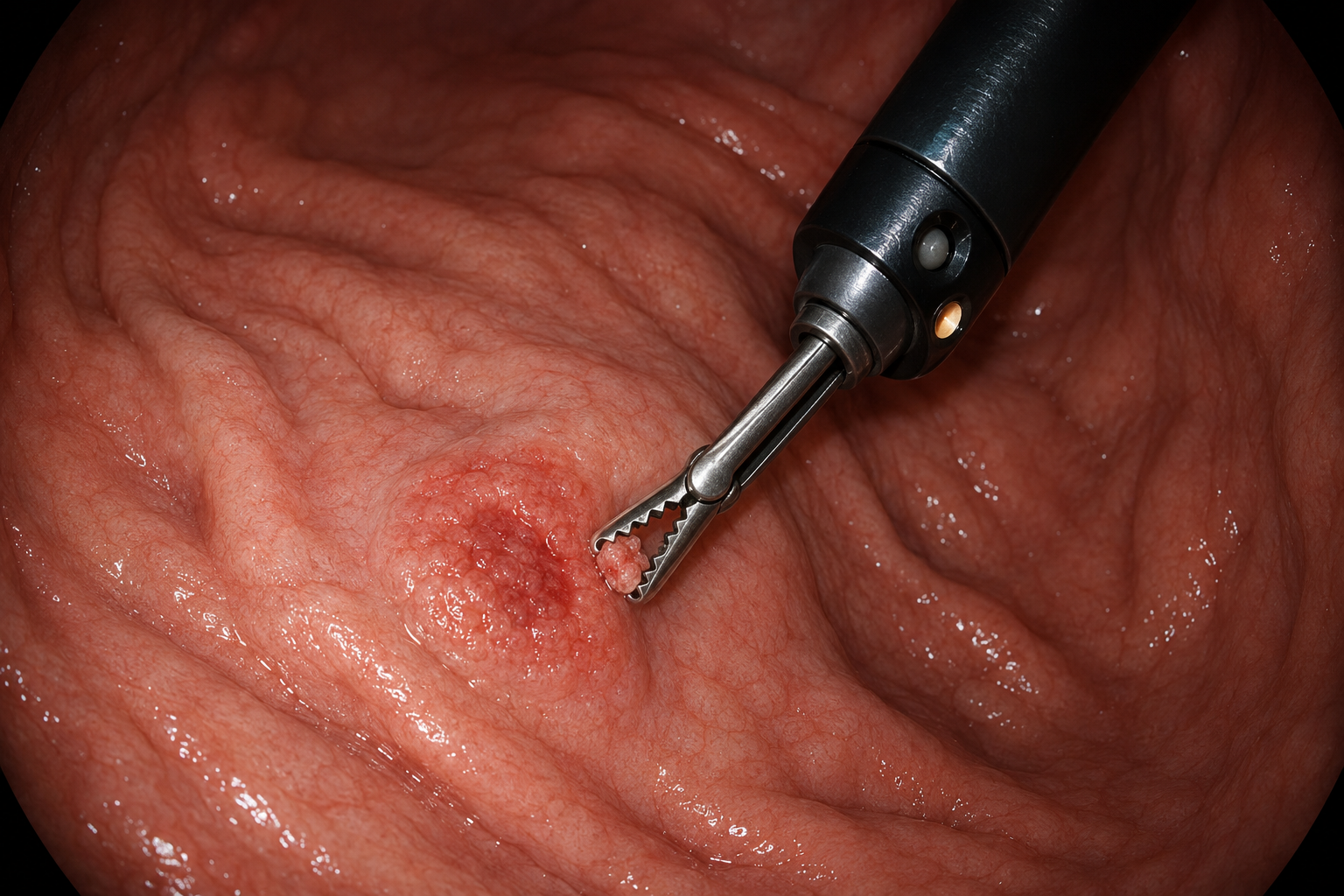
Before
Follow the fasting instructions. Tell the team about blood-thinning and diabetes medicines, allergies, sleep apnoea (breathing pauses during sleep) and previous problems with sedation. Do not stop medicines unless the team tells you to.
During
A throat-numbing spray and/or sedation to help you relax may be used. Several samples are usually taken because different parts of a tumour can have different features.
After
Arrange an escort if sedated. Mild bloating or sore throat can occur. Severe pain, fever, breathlessness, vomiting blood or black stools need urgent advice.
If the biopsy is negative
An initial negative biopsy may need further checking if the area still looks suspicious. A thick, rigid stomach wall (linitis plastica) or a tumour beneath the lining may need deeper repeat biopsies, sampling guided by EUS or specialist review.
Enough tissue for testing
The team checks whether there is enough usable cancer tissue to identify the type and carry out the planned biomarker tests.
Report detail
The report should describe the tumour’s site, extent, any ulceration and its distance from the GEJ and stomach outlet.
Scans and staging laparoscopy
The best staging plan combines tests; no single scan sees every form of spread.
Test | What it adds | Important limitation |
|---|---|---|
Contrast CT | Main map of stomach wall thickening, lymph nodes, liver, lungs and peritoneum. | Small peritoneal deposits and microscopic disease may be missed. |
Endoscopic ultrasound | Estimates depth and nearby nodes; especially helpful when considering endoscopic resection. | Less useful if the scope cannot pass or inflammation distorts layers. |
Fluorodeoxyglucose (FDG) PET-CT | Uses a small amount of radioactive sugar (FDG) to help find spread to distant sites that other tests may miss. | Diffuse, signet-ring and mucinous cancers can show low uptake; not a substitute for quality CT. |
MRI | Problem-solving for liver lesions, pelvis or when CT contrast is unsuitable. | Not routinely required for every patient. |
Staging laparoscopy + washings | Lets the surgeon inspect the abdominal lining and take samples of small deposits. Fluid used to wash the area is tested for cancer cells. | A procedure under anaesthesia; negative findings cannot guarantee that microscopic disease is absent. |
Understanding your pathology report
The report should answer what the cancer is, how aggressive it looks and—after resection—how completely it was removed.
Report item | Plain-language meaning |
|---|---|
Histology | Usually adenocarcinoma; intestinal, diffuse, mixed, poorly cohesive or signet-ring features may be described. |
Grade/differentiation | How closely cancer resembles normal glands. Poorly differentiated cells usually behave more aggressively, but stage remains crucial. |
Tumour depth after surgery (pT) | Deepest layer or nearby structure invaded by the tumour. |
Lymph-node findings after surgery (pN) | Number examined and number containing cancer. A D2 operation and adequate node count improve staging quality. |
Margins (R status) | R0 means all visible cancer is removed and the cut edges are clear under the microscope. R1 means cancer cells are found at a cut edge; R2 means visible cancer remains. |
Lymphovascular/perineural invasion | Cancer seen in lymph/blood vessels or around nerves; these are recurrence-risk features. |
Treatment response | After pre-operative therapy, the pathologist estimates how much viable tumour remains. |
Your biopsy report
The cancer type, grade and whether there is enough tissue for molecular tests.
Resection report
The stage found in tissue removed at surgery uses the prefix p. If treatment was given before surgery, it uses yp. The report also records margins, treatment response and other risk features.
Lymph-node results
Numbers examined and positive should both be visible.
Mismatch
Ask for review when pathology does not fit endoscopy, imaging or clinical behaviour.
Biomarkers that can change treatment
Biomarkers are features of cancer cells that help the team choose treatment. For cancer that cannot be removed by surgery (unresectable) or has spread to distant sites (metastatic), obtain the key results before the first drug treatment whenever possible.
Biomarker | How reported | Why it matters |
|---|---|---|
Human epidermal growth factor receptor 2 (HER2) | Immunohistochemistry (IHC) stains tissue to measure HER2 protein. Scores are 0, 1+, 2+ or 3+. A 2+ result needs in situ hybridisation (ISH), a test for extra copies of the HER2 gene. HER2-positive means IHC 3+, or IHC 2+ with a positive ISH result. | HER2-positive disease can receive trastuzumab-based first-line therapy and, after trastuzumab, trastuzumab deruxtecan in suitable patients. |
Programmed death-ligand 1 (PD-L1) | Combined positive score (CPS): a measure of PD-L1 staining in cancer cells and nearby immune cells. | Helps assess the likely benefit from immunotherapy that blocks programmed cell death protein 1 (PD-1), a signal that can limit the immune response. The required result depends on the medicine and treatment setting. |
Mismatch repair (MMR) / microsatellite instability (MSI) | These tests assess how well cells repair errors in genetic material. Results may show proficient repair (pMMR) and microsatellite stability (MSS), or deficient repair (dMMR) and high microsatellite instability (MSI-H). | MSI-H/dMMR cancers may respond particularly well to immunotherapy. Results can also suggest Lynch syndrome. Testing matters in early as well as advanced disease. |
Claudin 18.2 (CLDN18.2) | A validated IHC tissue test measures this protein on cancer cells. The result must meet the treatment’s required amount and strength of staining. | Positive HER2-negative advanced cancers may receive zolbetuximab with platinum/fluoropyrimidine chemotherapy where available. |
Broad genomic testing | Next-generation sequencing (NGS) tests many genes at once, using tumour tissue or sometimes blood. | May find rare fusions involving neurotrophic tyrosine receptor kinase (NTRK) genes, a high tumour mutational burden (TMB, the number of gene changes in the cancer), or clinical trial options. A negative blood test does not rule out a change in the tumour. |
Planning your treatment
Understanding your stage
TNM staging for stomach adenocarcinoma
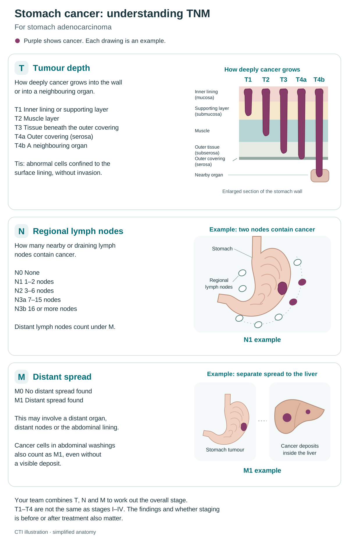
TNM stands for tumour (T), regional lymph nodes (N) and distant spread, or metastasis (M). Stomach staging combines how deeply cancer enters the stomach wall, the number of regional nodes involved and whether there is distant spread. Several combinations can produce the same overall stage.
Lymph nodes are small immune-system structures. Regional nodes are those near or draining the cancer’s starting area; distant spread means cancer has reached a site outside that regional area. Stages describe findings, not a sequence that everyone will pass through.
T — Tumour depth
T describes how deeply cancer grows. T1a is within the inner lining; T1b reaches the supporting layer beneath it. T2 reaches muscle; T3 reaches tissue beneath the outer covering; T4a reaches the outer covering (serosa); T4b enters a neighbouring organ. Tis, or carcinoma in situ, means abnormal cells remain in the surface lining without invading deeper tissue.
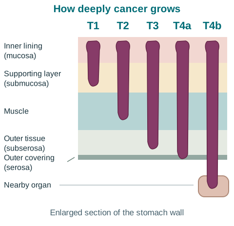
N — Regional lymph nodes
N records how many nearby or draining lymph nodes contain cancer: N0 means none; N1 means 1–2; N2 means 3–6; N3a means 7–15; N3b means 16 or more. Distant lymph nodes count under M.
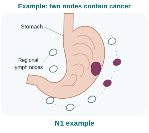
M — Distant spread
M0 means no distant spread was found. M1 means cancer has spread to a distant organ, distant nodes or the abdominal lining. Cancer cells in abdominal washings also count as M1, even without a visible deposit.
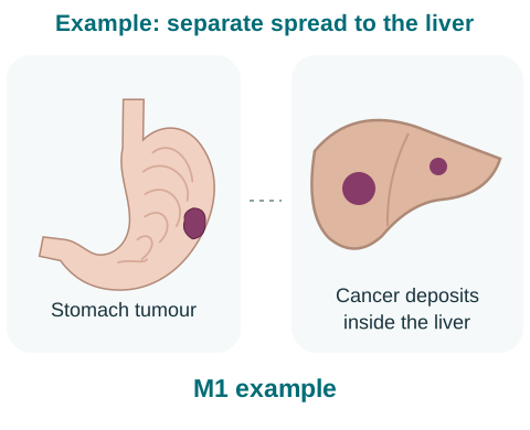
CTI illustration. Enlarge the staging overview.
Stage 0
Cancerous cells stay within the surface lining and have not invaded the supporting tissue beneath it.
Stage 1 (I)
Cancer is relatively shallow: it may reach the supporting layer or muscle. A shallow tumour with a small number of positive regional nodes can also be stage 1.
Stage 2 (II)
There is a greater combination of wall depth and regional node involvement. A deeper tumour without positive nodes, or a shallower tumour with more positive nodes, can fall here. No distant spread is found.
Stage 3 (III)
More extensive growth through the wall, into nearby structures, or a larger burden in regional nodes places the cancer in this group. It has not spread to distant sites.
Stage 4 (IV)
Cancer has reached distant organs, distant nodes or the abdominal lining. Cancer cells found in abdominal washings count as metastatic disease even when no visible deposit is seen.
Scans may miss tiny deposits on the abdominal lining; staging laparoscopy and washings are helpful in selected patients. Clinical stage before treatment and pathological stage after surgery can differ because they use different information.
Clinical stage comes from examination, biopsies and scans. Pathological stage can add information from surgery; a prefix such as yp means the tissue was assessed after treatment. A revised assessment does not necessarily mean the cancer grew while you were waiting. Grade describes the cells’ behaviour and is different from stage.
How stage and the possibility of surgery guide treatment
The team considers the stage, whether all cancer can be removed safely, your general health and your priorities. The table shows the main treatment pathways.
Situation | Typical treatment direction | Main questions |
|---|---|---|
Very early cancer, including selected T1a | Endoscopic submucosal dissection, then careful pathology review. | Is the cancer limited to the lining, completely removed and unlikely to have spread to lymph nodes? |
Cancer that can be removed | Stomach surgery with D2 lymph-node removal. Treatment before and after surgery is usually offered when cancer reaches the muscle layer or deeper, or involves lymph nodes. | Can surgery remove all the cancer with clear margins? Is chemotherapy suitable? Have laparoscopy and washings been completed when indicated? |
Grown nearby / surgery may be difficult | Systemic therapy first; reassess response and resectability. Radiotherapy is selective. | Can surgery safely remove the cancer? Is there hidden spread on the abdominal lining, or growth that makes surgery unlikely to help? |
Distant spread / cancer cells in washings | Biomarker-led systemic treatment plus symptom control. Surgery is rarely curative. | Which biomarker results guide the first treatment? Do any symptoms need a procedure for prompt relief? |
Can the cancer be removed safely?
The surgeon assesses whether all cancer can be removed while preserving enough function and keeping the risks acceptable.
Is surgery likely to help?
The team considers whether hidden spread or rapid cancer growth would limit the benefit of surgery.
Treatment options
Topics in this section
- Endoscopic removal for very early cancer
- Surgery offers the main chance of cure
- Which part of the stomach is removed, and how is it rejoined?
- Recovery after gastrectomy
- Treatment before and after surgery
- Treatment when surgery was done first
- When the cancer cannot be removed
- Choosing the first drug treatment using biomarker results
- HER2-positive disease
- During first-line treatment
- When treatment needs to change
- Precision treatment and clinical trials
Endoscopic removal for very early cancer
A carefully selected cancer confined to the mucosa can sometimes be cured without gastrectomy.
ESD
Endoscopic submucosal dissection removes the lesion in one piece with a margin, allowing accurate assessment of depth and risk features.
EMR
Endoscopic mucosal resection (EMR) may suit small lesions but is less often able to remove larger early cancers in one piece.
The pathology decides whether treatment is complete
- the cancer has been removed in one piece, with no cancer cells at the side or deep cut edges
- the depth, size and cell appearance meet the criteria for endoscopic treatment, with no cancer in blood vessels or lymph channels
- no findings that suggest a significant risk of spread to lymph nodes
Surgery offers the main chance of cure
Curative surgery aims to remove the tumour with clear margins and an appropriate lymph-node dissection while preserving function when safely possible.
Core principles
- The plan is agreed after MDT review and staging, with an upper-GI surgical team.
- Remove enough stomach and any directly involved tissue to achieve an R0 margin; more extensive surgery is not automatically better.
- The planned lymph-node removal is usually a D2 dissection. The spleen is removed only when needed to treat the cancer.
- Plan reconstruction so food can pass into the small bowel and bile/pancreatic juices join downstream.
- Your operation and tissue reports record what was removed, the cut edges, lymph nodes, any complications and the final cancer findings.
Approach | What it means |
|---|---|
Open | A larger abdominal incision. May be preferred for bulky disease, adjacent-organ involvement or when safety requires. |
Laparoscopic | Keyhole surgery through small incisions. Appropriate for selected patients in experienced hands. |
Robotic | Keyhole surgery using robotic instruments controlled by the surgeon. The choice depends on the cancer, your health and the planned operation. |
Clear margins (R0 resection)
The aim is an R0 resection: all visible cancer is removed and no cancer cells are found at the cut edges under the microscope. Your team plans how much stomach and surrounding tissue must be removed to achieve this.
Planned D2 lymph-node dissection
A gastrectomy intended to cure the cancer usually includes D2 lymph-node dissection, removing defined groups of lymph nodes around the stomach and its main blood vessels. This is planned before surgery and explained alongside how much stomach will be removed.
Possible change to open surgery
A keyhole or robotic operation may need to become open surgery through a larger cut for safety or to remove the cancer fully. Discuss this possibility beforehand, including how it could change the recovery plan.
What to expect during recovery
Many people spend about 1–2 weeks in hospital and need 6–8 weeks for initial recovery. Regaining strength and adapting to eating can take longer. Your team will explain your likely timetable, eating and activity plan, and support needed at home.
Which part of the stomach is removed, and how is it rejoined?
The operation is chosen by tumour location and safe margins—not by stage number alone.
Operation | What is removed | How the digestive tract is rejoined |
|---|---|---|
Distal subtotal gastrectomy | The lower stomach, nearby lymph nodes and the omentum (a fatty apron of tissue in the abdomen). Nearby tissue may also be removed if involved. | The remaining stomach is joined to duodenum or jejunum, depending on the operation. |
Total gastrectomy | The whole stomach, omentum and nearby lymph nodes. A short section of food pipe or part of a neighbouring organ is removed only when needed. | The oesophagus is joined to a Roux limb of jejunum. Bile and pancreatic juices meet food further downstream. |
Proximal resection | The upper stomach in selected patients, with the lymph nodes and other tissues needed for the cancer operation. | The way the digestive tract is rejoined varies. The team considers eating and reflux (backflow into the food pipe). Many upper-stomach cancers need total gastrectomy or a GEJ treatment plan. |
How food passes after surgery
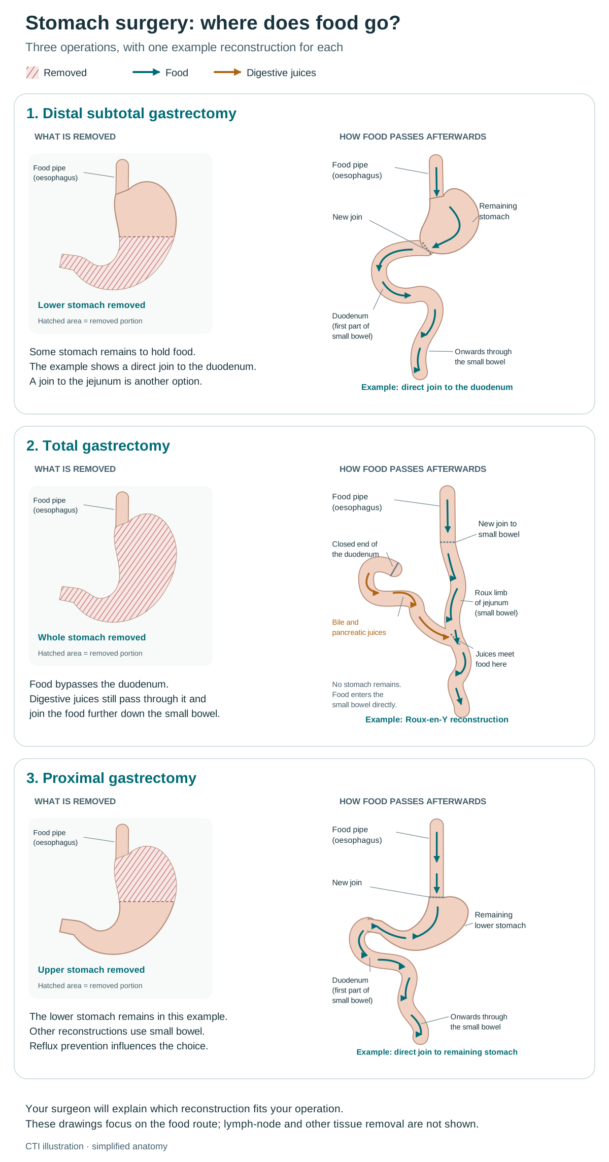
These examples show what is removed and how the digestive tract can be rejoined. Teal arrows show food; orange arrows show digestive juices. The hatched area is the part of the stomach removed.
Distal subtotal gastrectomy
What is removed
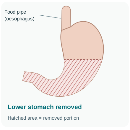
The lower part of the stomach is removed.
How food passes afterwards
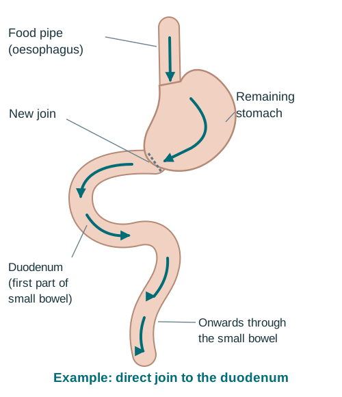
Food pipe → remaining stomach → duodenum → rest of the small bowel.
This example joins the remaining stomach directly to the duodenum, the first part of the small bowel. Other reconstructions join it to the jejunum, a later part of the small bowel.
Total gastrectomy
What is removed
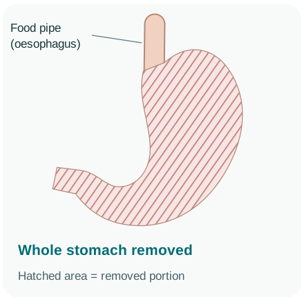
The whole stomach is removed.
How food passes afterwards
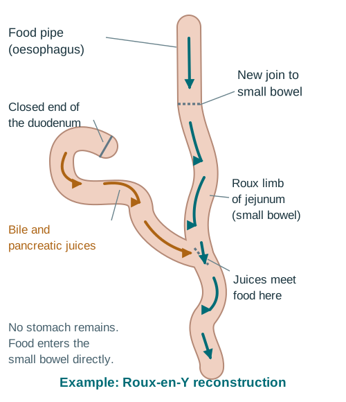
Food pipe → a loop of small bowel → onwards through the bowel.
The food pipe joins a loop of jejunum, called the Roux limb. Bile and pancreatic juices travel through the duodenum and another bowel segment, then meet food further down.
Proximal gastrectomy
What is removed
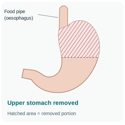
The upper part of the stomach is removed in selected patients.
How food passes afterwards
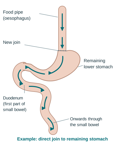
In this example: food pipe → remaining lower stomach → duodenum → rest of the small bowel.
This example joins the food pipe directly to the remaining stomach. Other reconstructions use a segment of small bowel or two routes. The join is planned to help limit reflux; many upper-stomach cancers need a different operation.
Possible complications
- bleeding, infection, blood clot, chest infection or delayed stomach emptying
- leakage or narrowing at a new join, reflux, or bowel slipping through a space inside the abdomen (internal hernia)
- weight loss, dumping syndrome (food moving too quickly into the bowel) and long-term vitamin or mineral deficiency
Recovery after gastrectomy
Preparing for recovery starts before surgery, with nutrition, suitable activity and a plan for pain relief, breathing exercises and getting moving again.
Phase | What often happens | What helps |
|---|---|---|
First 24–48 hours | Monitoring, pain relief, clot prevention, breathing exercises, tubes/drains as needed and early sitting/walking. | Tell the team if pain prevents deep breathing or movement. |
Eating restarts | Small sips and then staged textures, or tube feeding if planned. Timing varies by surgeon and recovery. | Follow the dietitian’s plan; do not force large portions. |
Hospital discharge | You should have wound advice, medicines, eating plan, red flags, pathology appointment and contact numbers. | Arrange support, transport and help with meals. |
First 6–12 weeks | Fatigue, reduced capacity and weight change are common. Food tolerance evolves. | Frequent protein-rich intake, walking, strength recovery and scheduled review. |
Long term | B12, iron, folate, calcium/vitamin D and bone health may need monitoring and replacement. | Keep lifelong nutrition follow-up after total gastrectomy. |
Pain plan
Use enough relief to breathe deeply, cough, walk and sleep.
Clot prevention
Injections, stockings and movement continue for the advised duration.
Eating and feeding plan
Know how much to eat and drink, any tube-feeding instructions and who can adjust the plan.
Where to get urgent help
Know which symptoms require the surgeon, emergency department or ambulance.
Treatment before and after surgery
For many people well enough for treatment whose stomach or GEJ cancer can be removed, drug treatment is given before and after surgery. This is called peri-operative treatment. It is commonly used when cancer reaches the muscle layer or deeper (T2 or above), or has spread to nearby lymph nodes.
Step | Common approach | Purpose |
|---|---|---|
Before surgery | Usually 4 cycles of FLOT: fluorouracil, leucovorin, oxaliplatin and docetaxel. Each cycle includes treatment and a recovery period. If this combination is unsuitable, the team may choose two chemotherapy drugs: oxaliplatin with a fluoropyrimidine such as fluorouracil or capecitabine. | Treat cancer cells too small to see, assess the cancer’s response and improve the chance of removing it completely. |
Restage | Clinical review and CT; repeat laparoscopy in selected high-risk or suspicious situations. | Confirm no progression that would make gastrectomy unhelpful. |
Surgery | Gastrectomy with D2 node dissection at an experienced centre. | Remove visible local and regional disease with clear margins. |
After surgery | Resume the planned chemotherapy when recovery permits: continue FLOT if it was given before surgery, with durvalumab if already included in the treatment plan. Doses and the number of cycles may need adjustment. | Complete the planned drug treatment while monitoring nutrition, nerve symptoms such as tingling or numbness (neuropathy), blood counts and daily activities. |
Adding immunotherapy before and after surgery
Durvalumab is an immunotherapy medicine that helps the immune system attack cancer. It can be given with FLOT before and after surgery, followed by durvalumab alone, for suitable adults with stomach or GEJ adenocarcinoma that can be removed. The MATTERHORN clinical trial supports this approach. Your team considers general health, any autoimmune illness (where the immune system attacks healthy tissue) and the full treatment plan.
Before FLOT
Check general health, existing nerve symptoms, blood counts, nutrition and organ function.
Before surgery
Restage, review laparoscopy status and confirm recovery from chemotherapy.
Treatment when surgery was done first
Sometimes cancer is removed before systemic therapy—for example after an unexpected diagnosis, emergency operation or a surgery-first pathway.
Situation | Possible next step |
|---|---|
Adequate D2 resection, stage II–III | Adjuvant chemotherapy (treatment after surgery) with CAPOX (capecitabine and oxaliplatin) or FOLFOX (fluorouracil, leucovorin and oxaliplatin) can be considered if chemotherapy was not given before surgery. The choice depends on the pathology results, recovery and overall fitness. |
Less-than-D2 nodal surgery or high local-risk features | Chemoradiotherapy (chemotherapy given with radiotherapy) after surgery may be discussed, particularly if no treatment was given before surgery. |
Microscopically involved margin (R1) | Expert pathology confirmation, surgical review and consideration of chemoradiotherapy/systemic therapy. Re-operation is selective. |
Very early, low-risk pathology | Surgery alone may be enough; confirm stage, margins and node assessment. |
Poor recovery or frailty | A lower dose, simpler treatment or monitoring without further treatment may be safer, depending on recovery and overall health. |
Radiotherapy is selective—not routine for everyone
- It can improve local control after inadequate lymph-node dissection or an involved margin in selected patients.
- It may relieve bleeding, pain or obstruction in advanced disease.
- Planning must protect bowel, liver, kidneys and spinal cord; nutrition support is essential.
Pathology first
The team uses the final stage, margins and lymph-node findings to recommend treatment after surgery.
Recovery first
Correct dehydration, infection and major nutrition deficits before chemotherapy.
When the cancer cannot be removed
Systemic treatment aims to shrink or control cancer, relieve symptoms and help people live longer with acceptable quality of life.
Treat the biology
Use HER2, PD-L1, MSI/MMR and CLDN18.2 before first line whenever possible.
Treat urgent problems
Bleeding, obstruction, severe vomiting, pain, ascites or malnutrition may need a procedure before or alongside drug treatment.
Match intensity to fitness
Two chemotherapy drugs are commonly combined. People who are frail may be better suited to one drug, lower doses or symptom-focused care alone.
Plan the next line early
Keep tissue available for testing, monitor nerve symptoms and ability to manage daily activities, and discuss future options while planning current treatment.
The baseline before treatment
- symptoms, weight, muscle loss, eating and drinking, and ability to manage daily activities (performance status)
- blood counts, kidney and liver function, blood salts, albumin (a blood protein), and iron or vitamin B12 when relevant
- HER2, PD-L1 CPS, MMR/MSI and CLDN18.2; consider NGS in selected patients
- scan findings that can be compared later to check how well treatment is working
- treatment goals, likely side effects, travel, costs and what matters most to you
Urgency
Can symptoms safely wait for drug treatment to work?
Biomarkers
Are all first-line results available and technically reliable?
Keeping future options open
How could side effects affect the ability to receive further treatment?
Support
Who is addressing pain, nutrition, finances and family communication now?
Choosing the first drug treatment using biomarker results
First-line treatment means the first drug treatment for advanced cancer. The options below are for adults whose cancer cannot be removed by surgery or has spread to distant sites. Your doctor considers the tumour results, previous treatment and general health.
Tumour result | Treatment options | What to consider |
|---|---|---|
HER2-positive + PD-L1 CPS ≥1 | Trastuzumab + platinum/fluoropyrimidine chemotherapy + pembrolizumab. | Check heart function and monitor immune-related and chemotherapy side effects. |
HER2-positive + CPS <1 | Trastuzumab + platinum/fluoropyrimidine chemotherapy remains a core option. | Pembrolizumab in this setting requires PD-L1 CPS ≥1. |
HER2-negative, PD-L1 positive | Chemotherapy + immunotherapy For adults with HER2-negative stomach or GEJ adenocarcinoma that cannot be removed by surgery or has spread, chemotherapy plus immunotherapy can be considered when PD-L1 CPS is at least 1. Benefit is generally more likely at higher PD-L1 scores. | The exact PD-L1 test and threshold depend on the medicine. Your doctor will also consider your general health and the chemotherapy combination. Both chemotherapy and immune-related side effects are possible; tell the team about autoimmune illness or an organ transplant. |
HER2-negative, CLDN18.2 positive | Zolbetuximab with modified FOLFOX6 (mFOLFOX6, a schedule of the FOLFOX combination) or CAPOX, where available. For adults with HER2-negative stomach or GEJ adenocarcinoma that cannot be removed or has spread. A validated tissue test must show CLDN18.2 positivity, with moderate-to-strong staining in at least 75% of cancer cells. | Nausea and vomiting can be pronounced, especially early. The team plans anti-sickness medicines and monitors appetite and reactions during the infusion. |
MSI-H/dMMR | Immunotherapy is an important option. Some selected patients may receive it without intensive chemotherapy, depending on the treatment setting and previous treatment. | The team checks the exact medicine, previous-treatment requirements and whether assessment for Lynch syndrome is needed. |
No suitable biomarker target or too frail for intensive treatment | Two-drug chemotherapy, an adjusted combination, a single medicine or symptom-focused care, depending on general health. | More treatment is not always better; reassess goals frequently. |
Choosing when both PD-L1 and CLDN18.2 are positive
Discuss the evidence, side effects, availability and clinical trial options with your treating doctor. The discussion should cover:
- the PD-L1 CPS level and the CLDN18.2 tissue-test result
- how much cancer is present, whether it needs to shrink quickly, autoimmune history, nausea and nutrition
- availability, infusion schedule, cost and evidence for the specific treatment
- whether a clinical trial is suitable; there is no established standard combining zolbetuximab with a PD-1 inhibitor and chemotherapy, and combining both antibodies should not be assumed to be safer or more effective
The CPS value matters
CPS 1 and CPS 50 are both ‘positive’ but do not imply equal benefit.
The claudin tissue test
The report should name the validated test and record how much staining is present and how strong it is.
HER2-positive disease
HER2 levels can differ between parts of a stomach tumour and may fall after treatment. A new biopsy may help confirm the current result if the cancer changes or grows.
First-line treatment
As shown in the table above, trastuzumab is combined with chemotherapy. Pembrolizumab may be added when PD-L1 CPS is at least 1 and the other eligibility criteria are met. The team chooses the chemotherapy combination to suit your health; oxaliplatin is often used instead of cisplatin.
Treatment issue | What to monitor |
|---|---|
Trastuzumab | Baseline and periodic heart function; infusion reaction; symptoms of heart failure such as breathlessness, swelling or rapid weight gain. |
Pembrolizumab | The immune system can inflame healthy organs, including the lungs, bowel, liver, skin, kidneys and hormone-producing glands. These effects can occur even after treatment stops. |
Platinum/fluoropyrimidine | Monitor blood counts, infection, nausea, diarrhoea, mouth soreness, painful redness of the hands or feet (hand-foot syndrome), kidney function, and nerve symptoms or cold sensitivity with oxaliplatin. |
After trastuzumab
Trastuzumab deruxtecan (T-DXd) may be used for adults with locally advanced or metastatic HER2-positive stomach or GEJ adenocarcinoma after treatment that included trastuzumab. A recent biopsy may help confirm that the cancer remains HER2-positive.
During first-line treatment
The team checks how well treatment is working, any side effects and your nutrition. Discuss which findings would lead to a dose adjustment or a different treatment.
Check | What the team is looking for |
|---|---|
Every cycle | Symptoms, food and fluid intake, weight, daily activities, nerve symptoms, infection, bowel changes, bleeding and other side effects. |
Blood tests | Blood counts, kidney and liver function, and blood salts. Immunotherapy also needs thyroid and other hormone checks. If ramucirumab is used later, blood pressure and protein in the urine are monitored. |
Imaging | Usually CT after a defined number of cycles or sooner if symptoms worsen. Compare the same target lesions and sites. |
Tumour markers | Carcinoembryonic antigen (CEA) or carbohydrate antigen 19-9 (CA 19-9) blood levels may help track changes if they were raised before treatment. They cannot replace scans and clinical assessment. |
Goals | Is treatment shrinking cancer, stabilising it, relieving symptoms and preserving acceptable life quality? |
Common reasons to adjust treatment
- nerve symptoms or sensitivity to cold that build up with oxaliplatin
- repeated low blood counts, infection, diarrhoea, soreness of the mouth or digestive-tract lining (mucositis), or hand-foot syndrome
- immune-related inflammation, reduced heart function or a severe reaction during an infusion
- ongoing weight/muscle loss, poor intake or a decline in daily function
Response
Use symptoms, function and comparable imaging—not one blood marker alone.
Side effects
Tell the team how severe nerve symptoms, diarrhoea and other side effects are, and whether you have needed hospital care.
Nutrition
Escalate support before loss of weight and muscle becomes severe.
Next line
Discuss likely options while the patient is well enough to receive them.
When treatment needs to change
Second-line treatment is the next treatment if the first stops working or cannot be tolerated. The choice depends on previous medicines, continuing side effects, biomarkers, how quickly the cancer is growing and general health.
Option | When it is considered | Main cautions |
|---|---|---|
Paclitaxel + ramucirumab | A common second treatment for someone well enough to receive it after fluoropyrimidine and platinum chemotherapy. | Nerve symptoms, low blood counts, bleeding, blood clots, high blood pressure, protein in the urine and slower wound healing. |
Ramucirumab alone | When combination chemotherapy is unsuitable but treatment that blocks cancer-related blood-vessel growth remains an option. | Bleeding, clots, high blood pressure, protein in the urine and wound-healing problems remain concerns. Benefit may be smaller than with the combination. |
Irinotecan or FOLFIRI (fluorouracil, leucovorin and irinotecan) | An alternative second chemotherapy treatment, particularly if nerve symptoms limit taxane drugs such as paclitaxel. | Diarrhoea, neutropenia (low infection-fighting white blood cells) and fatigue. |
Trastuzumab deruxtecan | HER2-positive disease after trastuzumab-based therapy. | Interstitial lung disease, nausea, low counts; reassess HER2 when possible. |
Immunotherapy | May suit MSI-H/dMMR cancers or another qualifying biomarker-based use that applies across different cancer types (a tumour-agnostic indication). | Immune-related side effects and the time needed for treatment to work; suitability varies between patients. |
Trifluridine/tipiracil | For adults with metastatic stomach or GEJ adenocarcinoma after at least two previous treatment courses. Earlier treatment must include a fluoropyrimidine, a platinum, a taxane or irinotecan, and HER2-targeted treatment when appropriate. | Low blood counts, infection and fatigue. Take the tablets exactly according to the prescribed schedule. |
Precision treatment and clinical trials
A small number of people have a gene change or other biomarker that allows a treatment used across different cancer types. Others may be eligible for a clinical trial selected by their tumour results.
Finding | Possible relevance |
|---|---|
MSI-H/dMMR | Suggests that PD-1 immunotherapy may help; also prompts assessment for Lynch syndrome. |
NTRK fusion | Rare; may allow an NTRK inhibitor under a tumour-agnostic indication. |
High tumour mutational burden | May allow immunotherapy after previous treatment when the test result and other eligibility requirements are met. Your team interprets it in the context of this cancer. |
Questions to ask about clinical trials
- What phase is it and what is known about benefit and risk?
- Is treatment allocated by chance (randomisation), and what treatment would each study group receive? Is a placebo used, and would standard treatment still be given?
- Which travel, tests, biopsies and costs are covered?
- What happens if the cancer grows or side effects occur?
- Can I withdraw and receive standard treatment later?
Actionable
A test result can guide treatment only when there is suitable evidence, the medicine is available and the person is well enough to receive it.
Tissue limits
If a sample contains too few cancer cells, NGS may miss a gene change that is present.
Managing side effects
Getting help during drug treatment
Keep your treatment team’s daytime and out-of-hours numbers where you and your caregiver can find them. Report side effects early so the team can help and adjust treatment if needed.
Procedures that relieve symptoms
Local procedures can restore eating, control bleeding or drain fluid while systemic treatment addresses disease elsewhere.
Problem | Possible approaches | Key point |
|---|---|---|
Blocked stomach outlet | A stent (a tube placed through an endoscope to hold the passage open), surgery to bypass the blockage, a tube to drain trapped contents, or cancer treatment in selected patients. | Choose according to expected duration of benefit, fitness and ability to eat. |
Bleeding | Treatment through an endoscope, a blood transfusion or iron, radiotherapy, a procedure to block a bleeding blood vessel (embolisation), or surgery in selected emergencies. | Recurrent slow bleeding may first appear as fatigue or anaemia. |
Swallowing difficulty at cardia/GEJ | Stent, radiotherapy or systemic treatment; nutrition access may be required. | A stent can relieve symptoms quickly but may cause reflux, pain or move out of position. |
Ascites | Drainage, medicines that increase urine output in selected patients, other procedures for symptoms, and cancer treatment. | Repeated drainage can worsen protein loss; coordinate nutrition and fluid care. |
Pain or nausea | Pain relief, anti-sickness medicines, acid-reducing medicines, steroids or procedures targeting pain nerves, depending on the cause. | Treat the mechanism—obstruction, constipation, medicines, infection or tumour—not only the symptom. |
Fastest relief
Choose a procedure when symptoms cannot safely wait for systemic response.
Durability
A surgical bypass may provide longer relief than a stent, but involves a bigger procedure and recovery.
Live well
Nutrition before and during treatment
Nutrition care is part of cancer treatment. Waiting for severe weight loss makes recovery harder.
Ask for a dietitian when there is
- unintentional weight loss, early fullness, repeated vomiting, swallowing difficulty or poor intake
- planned gastrectomy, peri-operative FLOT or a prolonged hospital stay
- loss of strength or muscle even when body weight appears stable
- diabetes, kidney disease, food insecurity or a restrictive diet that complicates advice
Priority | Practical approach |
|---|---|
Protein and energy | Use small frequent meals/snacks; add eggs, dairy/curd, pulses, fish, chicken, nut pastes or prescribed supplements according to tolerance. |
Hydration | Sip regularly between meals; use oral rehydration when diarrhoea/vomiting requires it and medical advice supports it. |
Symptoms | Treat nausea, constipation, reflux, pain, oral thrush and obstruction promptly; they are medical barriers to eating. |
Feeding access | A tube into the small bowel, passed through the nose or placed directly, can provide nutrition when eating is not enough. Nutrition through a vein is used only in selected situations. |
Activity | Walking and resistance exercise, adapted to ability, help preserve muscle and function. |
Measure
Track weight, intake and function; consider formal muscle assessment.
Treat causes
Control nausea, pain, constipation, thrush, obstruction and depression.
Eating after gastrectomy
Eating changes after stomach surgery, and what works may change as you recover. Smaller, frequent meals with protein are often helpful. Your dietitian will tailor advice, and long-term checks help identify nutrient deficiencies.
Issue | What may help |
|---|---|
Early fullness and weight loss | Try 6–8 small meals or snacks, include protein and add energy-rich foods as advised. Drink mainly between meals if fluids fill you up. Marked or persistent symptoms need assessment for blockage or poor digestion, including a shortage of pancreatic enzymes. |
Early dumping (10–30 minutes after eating) | Food moving quickly into the bowel may cause cramps, diarrhoea, sweating or dizziness. Smaller meals, fewer sugary foods or drinks, and fluids mainly between meals may help. Include protein, fat and slower-digesting carbohydrates. |
Late dumping (1–3 hours after eating) | Low blood sugar may cause shaking, sweating, hunger or faintness. Try smaller balanced meals. Persistent symptoms need review; blood-sugar monitoring or medicines may help. |
Reflux or bile reflux | Remain upright after eating, use smaller evening meals and take prescribed medicines; persistent symptoms need review of anatomy and narrowing. |
Diarrhoea or pale, greasy stools | The team checks for infection, medicine effects, dumping, excess bile acids in the bowel or too few pancreatic digestive enzymes. Treatment depends on the cause. |
Vitamins and minerals | Lifelong vitamin B12 replacement after total gastrectomy; monitor iron, folate, vitamin D, calcium and bone health. Replace documented deficits. |
Pattern
Small, slow, frequent and protein-led is a starting point—not a rigid rule.
Fluids
Sip between meals if drinking with food worsens fullness or dumping.
Deficiencies
Replacement follows measurements and anatomy; B12 after total gastrectomy is lifelong.
Persistent symptoms
Persistent symptoms may need tests for narrowing, cancer returning, poor digestion from too few pancreatic enzymes, or bile-acid diarrhoea.
Support for you and your caregiver
Agree what help you would find useful, such as preparing small meals, transport, taking notes or keeping track of medicines and symptoms. Share the urgent contact numbers and warning signs. Tell the team about worries, low mood or difficulty coping; support is available for caregivers too.
Follow-up after treatment
Follow-up supports recovery and nutrition, checks for treatment effects and investigates symptoms or possible cancer recurrence. Your schedule is individual. Contact the team about new or worsening symptoms rather than waiting for the next appointment.
At follow-up
Reviews usually cover symptoms, examination, weight and intake, treatment effects, blood tests and imaging when appropriate. After gastrectomy, monitoring may include vitamin B12, iron/folate, vitamin D, calcium and bone health. The exact scan schedule depends on stage, treatment, symptoms and whether a result would change management.
Sources and further reading
For more information, explore these resources from the National Cancer Institute (NCI) and other medical organisations. The linked organisations have not reviewed or endorsed this CTI guide. Your care team can explain how the information applies to you.
- NCI: Stomach Cancer Treatment
- Cancer Council Australia: stomach cancer
- Macmillan: stomach cancer
- NHS: how a gastrectomy is done
- NHS: recovery after gastrectomy
- ESMO: gastric cancer diagnosis, treatment and follow-up guideline
- Cambridge University Hospitals: eating after gastrectomy
- Japanese Gastric Cancer Association: gastrectomy and reconstruction options
- CSCO: gastric cancer clinical practice guideline, including treatment after surgery
- NCI: infection and low white blood cells during cancer treatment
- NCI: inherited stomach cancer risk and genetic testing
Staging and risk-group references
AJCC 8th edition.
Medicine approval and prescribing references
These official records describe licensed uses and prescribing information for the medicines named in this guide. Approval depends on the cancer type, biomarkers, previous treatment, combination and formulation. Some uses discussed in the guide are off-label: outside the medicine’s licence, with evidence from clinical guidelines or studies. FDA or EMA approval does not establish approval or availability in India.
- Capecitabine — FDA prescribing information · Approval history
- Cisplatin — FDA prescribing information · Approval history
- Docetaxel — FDA prescribing information · Approval history
- Durvalumab — FDA prescribing information · Approval history
- Fluorouracil — FDA prescribing information · Approval history
- Irinotecan — FDA prescribing information · Approval history
- Leucovorin (folinic acid) — FDA prescribing information · Approval history
- Nivolumab — FDA prescribing information · Approval history
- Oxaliplatin — FDA prescribing information · Approval history
- Paclitaxel — FDA prescribing information · Approval history
- Pembrolizumab — FDA prescribing information · Approval history
- Ramucirumab — FDA prescribing information · Approval history
- Trastuzumab — FDA prescribing information · Approval history
- Trastuzumab deruxtecan — FDA prescribing information · Approval history
- Zolbetuximab — FDA prescribing information · Approval history
- Trifluridine with tipiracil — FDA prescribing information · Approval history
- Tislelizumab — FDA prescribing information · Approval history
Additional medicine approval notices
These records explain the specific medicine uses and eligibility requirements discussed in this guide.
Related care
Meet our doctors



Where to get care