
Share
Start here
Rectal cancer starts in the rectum, the last part of the large bowel. This guide explains the tests, treatment choices and support you may need. You can read one section at a time and return to the parts that matter to you.
Your plan depends on where the cancer is, how far it has grown or spread, its test results and your general health. Your priorities also matter, including bowel control, fertility, sexual health and daily life.
Rectal cancer at a glance
Magnetic resonance imaging (MRI) gives detailed pictures of the rectum and nearby tissues. Other scans look for cancer elsewhere in the body.
Chemotherapy uses medicines to treat cancer. Radiotherapy uses radiation to destroy cancer cells. These treatments may shrink the tumour and reduce the chance of cancer returning.
A small early cancer may be removed through the anus. Other cancers need an operation to remove part or all of the rectum and nearby tissue.
Immunotherapy helps the immune system attack cancer. For some stage 2 or 3 cancers with a particular fault in cell repair, it may allow surgery to be deferred with close follow-up. The tests and limits of this approach are explained below.
How is rectal cancer different from colon cancer?
The colon and rectum together form the large bowel, so their cancers are often grouped under the word colorectal. The rectum sits within the pelvis, the lower part of the body between the hip bones. It is close to the bladder, reproductive organs and muscles that control the bowel opening. This is why its treatment needs particular attention to bowel, bladder and sexual function.
Surgery is often the first treatment for cancer confined to the colon. Rectal cancer more often needs treatment before surgery. The exact position of the tumour helps decide the plan.
Your first questions
- What do my biopsy and scans show, and are any results still pending?
- What is the aim of treatment, and which treatment comes first?
- How might treatment affect my bowel control and everyday life?
- Who should I contact if symptoms change or I need help?
A biopsy is a small tissue sample checked for cancer. Keep a copy of the biopsy report and your treatment history. Ask for a compact disc (CD) containing your scan images in Digital Imaging and Communications in Medicine (DICOM) format and the software needed to view them, along with the written scan reports. These can help if you seek another opinion.
Understand
Where the rectum sits and what it does
The rectum stores stool before it leaves the body through the anus. Rings of muscle around the anus, called sphincters, help control when you pass stool. Nearby nerves help the bowel, bladder and sexual organs work.
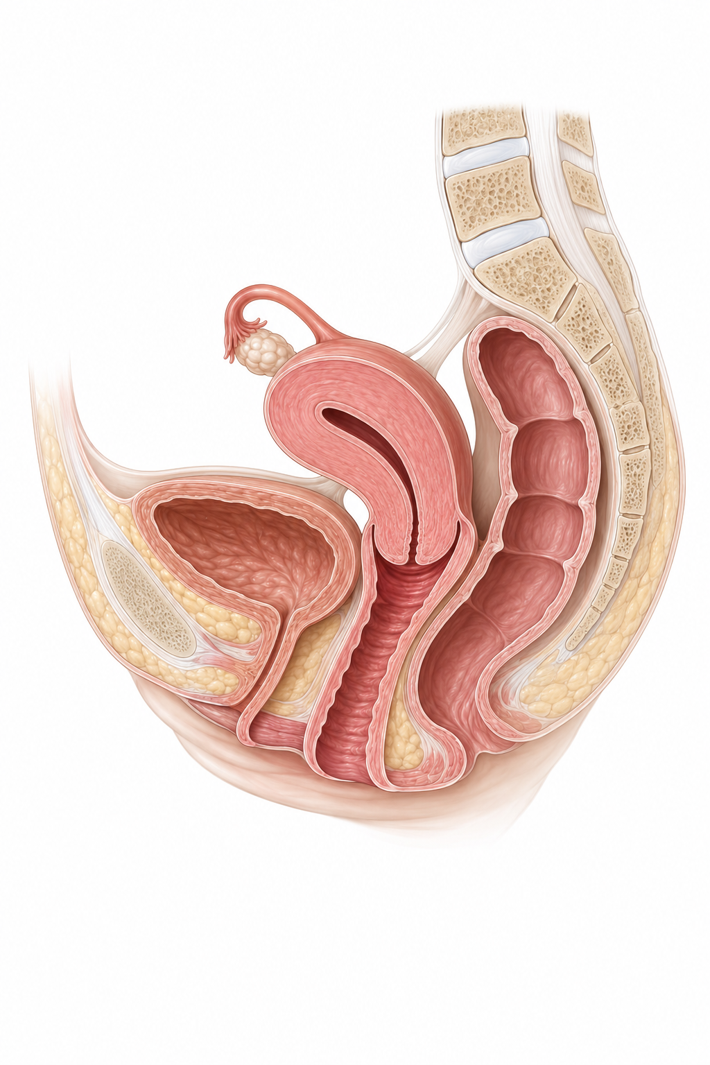
Fatty tissue around the rectum is called the mesorectum. It contains blood vessels and lymph nodes, small structures that are part of the immune system. Its outer covering is called the mesorectal fascia. These tissues matter when planning how to remove the cancer safely.
How rectal cancer can develop
Some rectal cancers develop from a polyp, a growth in the bowel lining. Many polyps never become cancer. Certain types, including adenomas and serrated polyps, can develop abnormal cells over time. Removing these polyps can help prevent cancer.
Dysplasia means abnormal cells that have not yet become an invasive cancer. Invasive means cancer has grown beyond the surface lining into the tissue beneath it. Cancer may grow deeper through the bowel wall, reach nearby lymph nodes or spread to other parts of the body. These findings help determine its stage.
Not every early cancer needs the same operation
A carefully selected small early cancer may be removed through the anus while keeping the rectum. This is called local excision. It leaves the nearby lymph nodes in place. If the removed tissue shows a higher chance of cancer in those nodes, more treatment may be needed.
Symptoms and when to seek help
Possible symptoms include bleeding from the anus, a lasting change in bowel habits, mucus in the stool, or a feeling that you still need to pass stool after going to the toilet. This last symptom is called tenesmus. Unexplained weight loss, tiredness or pain can also occur.
Bleeding may cause anaemia, meaning too few healthy red blood cells to carry oxygen around the body. This can make you tired or breathless. Piles, also called haemorrhoids, and small tears at the anus can cause similar symptoms. They can also occur alongside cancer, so persistent symptoms need checking.
A normal examination with a gloved finger does not rule out rectal cancer. Some tumours are too high to feel. Further tests may still be needed.
Risk, screening and family history
Screening looks for early cancer or polyps before symptoms appear. If you have symptoms, seek assessment even if a previous screening test was normal. Cancer can occur in younger adults as well as older people.
Risk can be higher with previous bowel polyps, a strong family history, or long-standing inflammation of the bowel from ulcerative colitis or Crohn's disease. Smoking, alcohol, excess body weight, little physical activity and some dietary patterns are also linked to risk. A diagnosis does not mean you caused the cancer.
Tests on the tumour and tests for inherited risk
All rectal cancers should have tests for mismatch repair (MMR) and microsatellite instability (MSI). These look for signs that cells cannot properly repair errors in their genetic instructions, called deoxyribonucleic acid (DNA). The results can guide treatment and help identify people who may need tests for inherited cancer risk.
- Deficient mismatch repair (dMMR) means this repair system is not working properly. Microsatellite instability-high (MSI-H) means many changes have built up in short repeated sections of DNA. These results usually identify the same group of cancers and can make immunotherapy an option.
- Proficient mismatch repair (pMMR) means the repair system appears to be working. Microsatellite stable (MSS) means the MSI test does not show this pattern of instability. These cancers usually need a different treatment approach.
A tumour result alone does not prove an inherited condition. An inherited, or germline, test uses blood or saliva to look for a gene change you were born with. A genetic counsellor explains what testing could mean for you and your family.
Inherited conditions include Lynch syndrome and conditions that cause many bowel polyps, such as familial adenomatous polyposis (FAP) and MUTYH-associated polyposis. MUTYH is the name of a DNA-repair gene. Testing may be considered because of your age, family history, multiple cancers or tumour-test results. Having no known family history does not rule out an inherited condition.
If an inherited change is found, close relatives may be offered testing and a screening plan suited to their risk.
Tests and diagnosis
How rectal cancer is diagnosed
Different tests answer different questions. A biopsy confirms the type of cancer. Scans show where it is and help the team plan treatment.
- Talk about symptoms and have an examination
The team asks about bowel habits, bleeding, medicines, family history and how you manage daily activities. A gloved-finger examination may help assess a low rectal tumour.
- Camera examination and biopsy
A thin flexible tube with a camera is passed through the anus to look inside the bowel and take small tissue samples.
- Blood tests
These check for anaemia and assess kidney and liver function. Carcinoembryonic antigen (CEA) is a substance measured in the blood that can help track some bowel cancers.
- Pelvic MRI
This maps the tumour's depth and its relationship to nearby tissues, lymph nodes and the muscles controlling the anus.
- CT scan or PET-CT scan
Computed tomography (CT) uses X-rays to make detailed body pictures. CT of the chest, tummy and pelvis usually checks for distant spread, especially to the liver and lungs. Positron emission tomography combined with CT (PET-CT) may be used for a specific question. It combines pictures of tissue activity with detailed body images.
- Team review
The specialists bring the results together, explain the stage and discuss treatment choices with you.
The camera examination and tissue sample
A sigmoidoscopy examines the rectum and lower colon. A colonoscopy examines the whole colon as well. These camera tests are forms of endoscopy. Checking the rest of the bowel helps find any other polyps or cancers.
The report records the tumour's position, including its distance from the anal verge, the outside edge of the anus. It also describes whether the tumour narrows the bowel. Sometimes a small ink mark, or tattoo, is placed to help locate an area later.
What can a biopsy tell me?
A pathologist, a doctor who examines tissue under a microscope, checks whether cancer is present and what type it is. The biopsy may also describe the grade: how abnormal the cells look and how they may behave. Grade is different from stage, which describes how far the cancer has grown or spread.
A small sample cannot show the full depth of a tumour or all the nearby lymph nodes. Scans and, if surgery takes place, examination of the removed tissue add this information. Sometimes another sample is needed if the first one does not explain a suspicious finding.
What each scan or blood test adds
| Test | What it helps show |
|---|---|
| Pelvic MRI | The tumour and nearby tissues in detail. Scar tissue and inflammation can make results harder to interpret after treatment. |
| CT | Cancer elsewhere in the body. Very small deposits may be too small to see. |
| Ultrasound inside the rectum | An ultrasound probe may help assess the depth of selected small early tumours. It is not needed for everyone. |
| Liver MRI or PET-CT | Further checks when another scan leaves an important question unanswered. Neither test can identify every cancer deposit. |
| CEA blood test | A starting level and, for some patients, changes during treatment or follow-up. Some cancers never raise CEA, and non-cancer conditions can raise it. It is interpreted alongside scans and symptoms. |
Understanding the main terms in an MRI report
You do not need to interpret the report alone. These are the findings most likely to affect your treatment plan.
| Report term | What it means for you |
|---|---|
| Tumour height and length | Where the tumour sits and how much of the rectum it involves. A low tumour may be close to the muscles controlling the anus. |
| Depth, or T category | How far the cancer has grown through the bowel wall or into nearby tissue. It is not simply the tumour's width. |
| Mesorectal fascia and the surgical margin | The fascia is the outer covering of the fatty tissue around the rectum. The margin is the cut edge of tissue removed at surgery. If cancer is close to where the surgeon would cut, treatment first may help achieve a clear margin, with no cancer at that edge. |
| Circumferential resection margin (CRM) | The outer, or side, cut edge around the removed rectum. MRI helps predict whether this edge could be close to the cancer. The final margin is checked on the tissue after surgery. |
| Extramural venous invasion (EMVI) | Cancer seen in veins outside the bowel wall. This can increase the chance of cancer spreading and may influence treatment. |
| Lymph nodes | Nodes that look suspicious for cancer. Their location, shape and other features matter as well as their size. |
| Sphincter or levator involvement | Whether cancer reaches the anal control muscles or nearby pelvic-floor muscles. This affects which operation may be possible. |
What the tissue report and repair tests mean
Alongside the cancer type and grade, the report includes the MMR or MSI result described above. This can affect the choice of medicines and whether inherited-risk testing is recommended.
If you have surgery after treatment, the report may use ypT and ypN. The letters yp mean that tissue was examined after treatment; T describes the remaining tumour's depth and N describes nearby lymph nodes. Regression describes how much the tumour has responded.
The report also checks the cut edges for cancer and may describe cancer in small blood vessels, lymph channels or around nerves. These are called lymphovascular invasion and perineural invasion. Small cancer deposits outside the main tumour can also affect staging and treatment decisions.
When immunotherapy may avoid surgery
For selected patients with stage 2 or 3 dMMR/MSI-H rectal cancer, immunotherapy can be an initial treatment. A medicine such as dostarlimab helps the immune system recognise and attack the cancer. The specialist team checks the response using examination, camera tests and MRI.
If no cancer is detectable, this is called a complete clinical response. Surgery may then be deferred within a structured follow-up programme. This does not prove cure: regular checks remain essential, and surgery or other treatment may be needed if cancer remains or returns.
This immunotherapy approach does not apply to tumours without dMMR/MSI-H. The team also considers your other health conditions and whether immunotherapy is suitable for you.
Planning your treatment
Understanding your stage
Tumour, nodes and metastasis (TNM) staging for rectal cancer
TNM brings together the tumour’s depth (T), nearby lymph nodes (N) and metastasis (M), meaning spread to distant parts of the body. Think of the bowel wall as layers: an inner lining, supporting tissue, muscle and outer tissue. Stage depends on where cancer has reached, not simply how wide the tumour is.
Lymph nodes are small immune-system structures. Regional nodes are those near or draining the cancer’s starting area; distant spread means cancer has reached a site outside that regional area. Stages describe findings, not a sequence that everyone will pass through.
Stage 0
Abnormal or cancerous cells remain within the inner lining and have not entered the supporting layer beneath it. This is also called in situ disease: the cells remain at their starting site.
Stage 1 (I)
Cancer has entered the supporting layer or muscle of the rectum wall, but has not grown through the muscle. Nearby nodes and distant sites are not involved.
Stage 2 (II)
Cancer has grown through the muscle into outer tissue, the outer covering or a neighbouring organ. No nearby lymph-node or distant spread is found. Some stage 2 cancers have features that increase the chance of cancer returning.
Stage 3 (III)
Cancer has reached nearby lymph nodes, or there are particular cancer deposits in the tissue supporting the bowel. It has not spread to distant sites. The depth of growth and the affected nodes or deposits determine the subgroup.
Stage 4 (IV)
Cancer has reached a distant site, such as the liver, lung or abdominal lining. Direct growth into an organ next to the bowel is different from a separate distant deposit.
MRI helps map the cancer and its distance from the planned surgical cut edge. The exact position, response to treatment and ability to remove it fully all matter within each stage. Selected people with a small amount of distant spread may still be offered treatment aiming to remove or destroy all visible cancer.
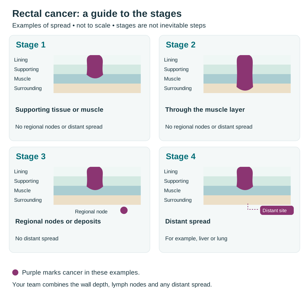
CTI illustration. Enlarge the staging overview.
Clinical stage is the assessment before surgery, using examination, biopsy and scans. Pathological stage adds the findings from tissue removed during an operation. If you had treatment before surgery, the report may use the prefix yp, as explained under Tests and diagnosis. More detailed results can change the stage; this does not necessarily mean the cancer grew while you were waiting.
Treatment choices for stages 0 and 1
Very early cancers may be removed during a camera procedure or through the anus. Others need surgery to remove part or all of the rectum and nearby lymph nodes. The choice depends on the depth, location and tissue-test results, not just the stage number.
| Finding | What may be discussed |
|---|---|
| Stage 0: cells remain in the lining | Removal during endoscopy or through the anus, followed by checks of the removed tissue and follow-up. |
| A small T1 cancer with favourable features | T1 means cancer has reached the supporting layer beneath the lining but not the muscle. Local excision may be possible if there is no evidence of involved nodes and the tumour can be removed fully. |
| An early cancer with higher-risk features | More surgery may be advised to remove nearby lymph nodes and reduce the chance of cancer returning. |
| A stage 1 cancer needing a larger operation | The surgeon explains how much rectum needs removing, whether the bowel can be joined again and whether a stoma is needed. A stoma is an opening on the tummy through which stool passes into a bag. |
For local excision, the team checks that the tumour is small, accessible and removable in one piece with clear cut edges. The removed tissue can reveal deeper growth or features missed by the first biopsy.
Higher-risk features include very abnormal-looking cells, cancer in small vessels, deep growth into the supporting layer, or tumour budding: single cancer cells or small groups breaking away at the tumour edge. Cancer close to a cut edge, or tissue removed in fragments that cannot be assessed reliably, may also lead to a recommendation for more treatment.
Treatment choices for stages 2 and 3
These cancers have grown through the bowel muscle or reached nearby lymph nodes, without distant spread. Treatment often combines medicines, radiotherapy and surgery. MRI helps show which combination and order are most suitable.
The team pays particular attention to cancer near the planned cut edge, in veins outside the bowel wall, or close to the muscles controlling the anus. Your general health and the MMR/MSI result also affect the choice.
Why treatment may come before surgery
Treatment given before an operation is called neoadjuvant treatment. Total neoadjuvant therapy (TNT) means giving the planned chemotherapy and radiotherapy before surgery. Chemotherapy may come before or after the radiotherapy.
This can shrink the tumour, treat cancer cells too small to see on scans and help deliver the planned treatment before recovery from an operation. For selected dMMR/MSI-H cancers, immunotherapy may be the initial treatment instead.
Some lower-risk or upper rectal cancers may be treated with surgery first, or chemotherapy with radiotherapy added only if needed. Ask why the proposed sequence fits your scan and tissue results.
Cancer that has spread or come back
Stage 4, or metastatic, cancer has spread to a distant part of the body. Recurrence means cancer has returned after treatment. A recurrence may be in the pelvis, elsewhere in the body, or both.
| Situation | Possible approach |
|---|---|
| A small amount of spread that can all be treated | Surgery or ablation, which destroys a tumour in place, may be combined with medicines. For selected people, treatment can still aim for a cure. |
| Spread that might become removable | Medicines may shrink the cancer. Repeat scans show whether an operation or another local treatment has become possible. |
| Spread that cannot all be removed or destroyed | Medicines aim to control cancer and help you live as well as possible. Radiotherapy or other local treatments may help bleeding, pain or blockage. |
| Cancer returning in the pelvis | Selected patients may still be offered treatment aiming for a cure. Options depend on earlier treatment and may include further surgery, carefully planned radiation or medicines. |
Symptoms may need treatment first
A blocked bowel may need a stoma or, in selected situations, a stent: a tube placed inside a narrowed section to hold it open. Bleeding and pain may need radiotherapy or other treatment. An infection or fistula, an abnormal connection between the bowel and another organ or the skin, may need drainage, antibiotics or surgery.
Your treatment plan at a glance
- Confirm the diagnosis and extent. Bring together the biopsy, repair tests, scans and your health needs.
- Agree the aim and first treatment. This may be local removal, a larger operation, treatment before surgery, immunotherapy for a suitable tumour or medicines to control spread.
- Check the response. The team explains when repeat examinations and scans will take place and what the results may change.
- Agree the next step. Surgery is often needed. Carefully selected people with a complete clinical response may discuss close monitoring while deferring surgery.
- Plan recovery and follow-up. Arrange bowel or stoma support, rehabilitation and the tests needed after treatment.
Treatment options
Topics in this section
- Removing an early tumour through the anus
- Operations to remove part or all of the rectum
- Understanding and preparing for a stoma
- What treatment before surgery aims to do
- Two ways of giving radiotherapy
- Chemotherapy before surgery
- Checking the response and deciding what comes next
- What watch-and-wait means
- What the operation results mean
- Medicines when rectal cancer has spread
- Treatment directed at liver, lung or pelvic cancer
Removing an early tumour through the anus
Local excision removes a selected early tumour while keeping the rectum. Transanal means through the anus. Techniques include transanal endoscopic microsurgery (TEM, also called TEMS) and transanal minimally invasive surgery (TAMIS). Both use special viewing equipment and surgical instruments through this opening.
This usually means a smaller operation and faster initial recovery than major rectal surgery. It leaves the nearby lymph nodes in place, so careful selection and follow-up are essential. The surgeon aims to remove the tumour in one piece, often with the full thickness of the involved bowel wall, so it can be examined properly.
Risks include bleeding and infection. If the removed tissue shows higher-risk features or cancer at the cut edge, a larger operation or other treatment may be needed. Ask what findings would change the plan and when you will receive the results.
Operations to remove part or all of the rectum
Total mesorectal excision (TME) removes the affected rectum with the surrounding fatty tissue and lymph nodes, keeping their outer covering intact. The aim is to remove the cancer with clear margins. The amount removed depends on the tumour's position and extent.
| Operation | What it involves |
|---|---|
| Low anterior resection (LAR) | The affected rectum is removed and the bowel ends are joined. A temporary stoma is often used when the join is low, to divert stool while it heals. |
| Abdominoperineal resection (APR) | The rectum, anus and muscles controlling the anus are removed. Stool then leaves through a permanent colostomy, a stoma made from the colon. |
| A very low join or intersphincteric resection | Some low tumours can be removed while keeping the anus, sometimes by removing part of the inner control muscle. Bowel control afterwards may be difficult, so expected function matters as well as whether a join is possible. |
| Pelvic exenteration | A larger operation removes the cancer together with affected nearby organs. It is considered for selected cancers and requires detailed planning of recovery and future function. |
Surgery may use one larger cut, called open surgery, or several small cuts, called laparoscopic surgery. Robotic surgery is a form of surgery using instruments controlled by the surgeon. The approach should support safe cancer removal; the technique's name alone does not determine the result.
Possible problems include a leak from the bowel join, infection, bleeding or another operation. Longer-term effects may include changes in bowel control, bladder emptying and sexual function. Keeping the anus does not guarantee normal bowel function. Ask about the likely outcome with each suitable operation.
Understanding and preparing for a stoma
A stoma brings a part of the bowel to an opening on the tummy. Stool collects in a pouch attached to the skin. An ileostomy is made from the small bowel; a colostomy is made from the colon. A stoma may be temporary or permanent.
A temporary ileostomy diverts stool away from a new bowel join while it heals. It reduces the impact of a leak but cannot prevent every leak. Reversal means closing the stoma and restoring the bowel route. It is considered only after healing and recovery; it is not guaranteed and has its own risks.
Meet a stoma nurse before surgery where possible. They can help choose the site, show you a pouch and discuss clothing, bathing, supplies and help at home. The team also checks nutrition, anaemia, medicines, exercise and smoking before the operation.
An ileostomy usually produces more liquid stool than a colostomy. Losing too much fluid can cause dehydration and affect the kidneys. Your nurse will explain how much output is expected and what to drink. Persistent pouch leakage or sore skin needs review; changing the fit or treating a nearby skin or bowel problem can help.
What treatment before surgery aims to do
Treatment before surgery can shrink cancer away from the planned cut edge, reduce the chance of it returning in the pelvis and treat cancer cells elsewhere that are too small to see. It also shows how the tumour responds. For selected patients, a complete clinical response may allow discussion of close monitoring instead of immediate surgery, within an experienced specialist programme.
Treatment and response checks take place before surgery, so the operation happens later. Cancer can sometimes grow despite treatment, and response tests have limits. The team monitors you and reviews the plan if symptoms or results change.
Two ways of giving radiotherapy
The choice depends on the tumour's position, how much it needs to shrink, your health and the rest of the treatment plan.
| Approach | What to expect |
|---|---|
| Long-course chemoradiation | Usually about five to five-and-a-half weeks of radiotherapy with capecitabine tablets or fluorouracil (5-FU) through a vein. The medicine helps radiation work against the cancer. |
| Short-course radiotherapy | Usually five daily treatments, without chemotherapy on the same days. Chemotherapy and surgery may follow according to the plan. |
Radiotherapy may cause loose stools, urgency, tiredness, sore skin or irritation when passing urine. Effects can continue or temporarily worsen after the course ends. Some bowel, bladder and sexual changes can last longer. Careful positioning and planning limit exposure of healthy tissue but cannot remove every risk.
Before pelvic treatment, discuss any wish to have children. Options may include storing sperm, eggs or embryos. For some people, moving the ovaries away from the radiation area may be considered. Radiation can affect fertility, cause an early menopause, or contribute to vaginal dryness or narrowing and difficulty with erections.
Chemotherapy before surgery
Chemotherapy travels around the body through the bloodstream. A cycle is a period of treatment followed by time to recover. Ask which medicines you will receive, how many cycles are planned and when the response will be checked.
| Combination name | Medicines and practical points |
|---|---|
| CAPOX | Capecitabine tablets with oxaliplatin through a vein, commonly in three-week cycles. Follow the tablet schedule carefully and report diarrhoea or sore hands and feet. Kidney function affects dosing. |
| FOLFOX | Folinic acid, also called leucovorin, with fluorouracil and oxaliplatin, commonly in two-week cycles. Part of the treatment is often given through a pump connected to a longer-term tube in a vein. |
| FOLFIRINOX-type treatment | Folinic acid, fluorouracil, irinotecan and oxaliplatin. This is more intensive and is considered for selected fit patients with higher-risk cancer. |
Ask about testing for low levels of dihydropyrimidine dehydrogenase (DPD), an enzyme that helps break down fluorouracil and capecitabine. This may include testing the DPYD gene, which provides instructions for making the enzyme. Low activity can cause severe side effects and may require a different dose or treatment.
Oxaliplatin can cause tingling, numbness or sensitivity to cold, called neuropathy. Report symptoms before they interfere with walking, buttons or other everyday tasks. Reducing the dose or stopping one medicine may protect long-term function while the rest of treatment continues.
Checking the response and deciding what comes next
Restaging means reassessing the cancer after treatment. The team brings together symptoms, a gloved-finger rectal examination, a camera examination, pelvic MRI and checks for cancer elsewhere. CEA may help if it was raised before treatment.
No single test can show whether every cancer cell has gone. Scar tissue can still look or feel abnormal. A surface biopsy may miss deeper cancer, and small deposits may be invisible on scans.
| Result | What may happen next |
|---|---|
| Incomplete response: cancer is still detectable | Surgery is usually the next step if the cancer can be removed. |
| Near-complete response: substantial improvement, but uncertainty remains | A repeat assessment after a defined short interval may clarify the result. Continuing to wait without a clear review plan is unsafe. |
| Complete clinical response: no cancer is detectable on the combined assessments | Discuss surgery and whether a specialist watch-and-wait programme is suitable. This result is not proof that every cancer cell has gone. |
| Progression: cancer has grown or spread | Further assessment is needed promptly so the treatment plan can be changed. |
What watch-and-wait means
For carefully selected patients with a confirmed complete clinical response, a specialist team may discuss watch-and-wait: regular, planned checks while deferring surgery. It is not a recommendation for everyone to avoid surgery. The decision includes the benefits and risks of surgery, your priorities and whether intensive follow-up is practical for you.
This can avoid a major operation and a possible permanent stoma, but cancer too small to detect may remain. Cancer can grow back in the rectum, most often within the first two years. Surgery is often possible if regrowth is found early, but a successful later operation cannot be guaranteed. Some patients then need an operation with a permanent stoma.
Follow-up usually includes frequent rectal and camera examinations, repeated pelvic MRI, CEA tests and CT scans. Checks for distant spread are still needed. The team should give you a written schedule and a route for prompt assessment of new bleeding, pain or bowel changes.
What the operation results mean
The pathologist examines the removed rectum and nodes to check how much cancer remains and whether the cut edges are clear. The report also assesses the quality of the removed surrounding tissue. These findings help estimate the chance of cancer returning.
A complete pathological response means no cancer is found in the tissue examined after treatment and surgery. This is encouraging, but follow-up remains necessary. It is different from a complete clinical response, which is judged using examinations and scans without removing the rectum.
More chemotherapy after surgery is not automatic after completed TNT. The decision depends on treatment already given, the operation results, recovery and side effects. If you have a temporary stoma, discuss the checks and timing needed before reversal.
Medicines when rectal cancer has spread
Biomarkers are features of cancer cells that can help select treatment. These include gene changes and proteins measured in the tumour. Your earlier medicines, current symptoms, general health and other test results also matter.
Another chemotherapy combination is folinic acid, fluorouracil and irinotecan (FOLFIRI). It does not contain oxaliplatin. The team chooses a combination and may add a targeted medicine, which acts on a particular cancer growth signal.
| Test result or situation | What it may mean for treatment |
|---|---|
| MSI-H/dMMR | Immunotherapy can be an option. Nivolumab with ipilimumab is one option for eligible patients aged 12 years or older when the cancer cannot be removed by surgery or has spread. These medicines help release the brakes on an immune response. |
| RAS/BRAF wild-type | RAS and BRAF are genes involved in cell-growth signals. “Wild-type” means the tested cancer-causing changes were not found. FOLFOX or FOLFIRI with cetuximab or panitumumab can be a first treatment option. These added medicines block a growth signal at the cell surface. |
| BRAF V600E | V600E names a specific BRAF gene change. Encorafenib with cetuximab and fluorouracil-based chemotherapy is a first treatment option for eligible adults. |
| KRAS G12C | KRAS is one of the RAS genes; G12C is a particular change in it. Sotorasib with panitumumab or adagrasib with cetuximab may be used in eligible adults after chemotherapy including fluorouracil or a related medicine, oxaliplatin and irinotecan. |
| HER2-positive, RAS wild-type | Human epidermal growth factor receptor 2 (HER2) is a protein involved in cell growth. Too much of it, or extra copies of its gene, may allow HER2-directed treatment. Options for eligible adults include tucatinib with trastuzumab after chemotherapy including fluorouracil or a related medicine, oxaliplatin and irinotecan. |
| A strongly positive HER2 protein test | Immunohistochemistry (IHC) is a tissue-staining test. A score of 3+ means strongly positive. Trastuzumab deruxtecan may be an option for eligible adults with an IHC 3+ tumour that cannot be removed or has spread, after previous treatment when there is no satisfactory alternative. |
| No suitable targeted option, or further treatment is needed | Options may include chemotherapy with a medicine that limits the tumour's blood supply, such as bevacizumab. Later options include trifluridine-tipiracil with bevacizumab, fruquintinib, regorafenib or a clinical trial, depending on previous treatment and health. |
Treatment directed at liver, lung or pelvic cancer
If cancer has spread to a limited number of sites, specialists assess whether all visible cancer can be removed or destroyed. Options may include surgery, ablation using heat or cold, or precisely targeted radiation. Medicines may come before or after these treatments.
For cancer returning in the pelvis, another operation may involve nearby organs, such as the bladder, reproductive organs or part of the lower spine. Radiation to a previously treated area needs especially careful planning. These choices require a clear discussion of cancer control, recovery and lasting effects on daily life.
Ask the team to explain the plan for both the rectal tumour and every area of spread, when scans will be repeated, and what would make the treatment plan change.
Managing side effects
Bowel changes after rectal surgery
After part of the rectum is removed and the bowel is joined again, it may not store stool or control emptying as before. A group of symptoms called low anterior resection syndrome (LARS) can include urgency, frequent trips to the toilet, leakage and difficulty emptying fully. Several bowel movements close together are called clustering.
These symptoms can affect travel, sleep and confidence. They often improve over time, but you do not have to manage them alone. Ask for a bowel-function assessment and a treatment plan.
| Problem | What may help |
|---|---|
| Frequent or loose stools and urgency | A food-and-bowel diary, changes to meal timing and advice about fibre. The team may recommend medicine to slow the bowel. Avoid making major dietary restrictions without advice. |
| Several toilet visits close together | A planned toilet routine and a review of diet and medicines. Selected patients may be taught transanal irrigation, which uses water through the anus to help empty the bowel at a planned time. |
| Leakage or sore skin | Skin protection, continence products and pelvic-floor exercises. Biofeedback uses measurements to help you learn how to coordinate these muscles. |
| Difficulty emptying or a feeling of blockage | An assessment for a narrowed bowel join, constipation with liquid stool leaking around it, or muscles that are not relaxing together. |
| Severe symptoms despite these measures | Specialist options may include irrigation, treatment that uses small electrical signals to influence bowel-control nerves, or a stoma. |
The team also checks for treatable causes, including infection, inflammation, medicine effects or a return of the cancer. Sometimes bile salts irritate the bowel and cause diarrhoea; treatment can help. Persistent symptoms should not automatically be accepted as an unavoidable result of surgery.
Bladder, sexual health and fertility
Pelvic surgery and radiotherapy can affect nearby nerves, blood vessels and organs. Mention changes early, even if they feel difficult to discuss. Help is available.
- Bladder problems: report difficulty emptying, leakage or repeated infections. A bladder specialist or pelvic-floor therapist may help. Being unable to pass urine needs urgent assessment.
- Erections and ejaculation: treatment can affect both. Medicines, devices and specialist support may help with erections; ejaculation and fertility need separate discussion.
- Vaginal dryness or discomfort: moisturisers, lubricants and, when suitable, hormone treatment may help. The team may recommend vaginal dilators, smooth devices used gently to help prevent or manage narrowing.
- Fertility and menopause: ask about storing sperm, eggs or embryos before treatment if relevant. Treatment can cause an early menopause, and support is available for its symptoms.
A caregiver or partner can join these discussions if you want, but you can also ask to speak privately. Recovery is individual, and support may still help months or years after treatment.
Live well
Eating, activity and emotional support
A dietitian can help if you are losing weight, struggling to eat, avoiding food because of bowel symptoms or managing a stoma. Food and fluid advice may change during treatment and recovery. An ileostomy that produces a lot of liquid needs an individual fluid and salt plan.
Build up activity gradually, following the surgical team's advice about lifting and wound healing. Small amounts of regular movement may be easier than a single long session. Ask for help with fatigue, sleep or pain that limits activity.
Worry about recurrence, changes in body image and difficulties with intimacy are common. A specialist nurse, counsellor or support group may help. Caregivers also need rest, information and support; sharing practical tasks can make treatment easier to manage.
Follow-up after treatment
Follow-up checks recovery, helps manage lasting effects and looks for signs that cancer has returned. Surveillance is another word for these planned checks. Your schedule may include appointments, CEA blood tests, scans and colonoscopy. The timing depends on your stage, treatment and individual risk.
Watch-and-wait requires especially close follow-up with rectal examinations, camera tests and MRI, as well as checks elsewhere in the body. Keep appointments even if you feel well. If attending becomes difficult, contact the team so they can help you keep to the plan.
Report new bleeding, persistent pain, a lasting change in bowel habits or unexplained weight loss between appointments. Do not wait for the next scheduled scan. Keep your treatment summary and the team's contact details somewhere you and your caregiver can find them.
Sources and further reading
For more information, explore these resources from the National Cancer Institute (NCI) and other medical organisations. The linked organisations have not reviewed or endorsed this CTI guide. Your care team can explain how the information applies to you.
- NCI: Rectal Cancer Treatment (PDQ®)–Patient Version
- Cancer Council Australia: bowel cancer
- Macmillan: bowel cancer
- ASCO: Management of Locally Advanced Rectal Cancer guideline (2024)
- Cercek and colleagues: Nonoperative Management of Mismatch Repair–Deficient Tumors (NEJM, 2025)
- OPRA trial: long-term results of organ preservation after treatment for rectal cancer (JCO, 2024)
- American College of Radiology: Staging and Disease Monitoring of Rectal Cancer (2025)
- North Tees and Hartlepool NHS: transanal endoscopic microsurgery (TEMS)
- East Kent Hospitals NHS: transanal minimally invasive surgery (TAMIS)
- Macmillan: managing bowel changes after treatment
Staging and risk-group references
AJCC 8th edition.
Medicine approval and prescribing references
These official records describe licensed uses and prescribing information for the medicines named in this guide. Approval depends on the cancer type, biomarkers, previous treatment, combination and formulation. Some uses discussed in the guide are off-label: outside the medicine’s licence, with evidence from clinical guidelines or studies. FDA or EMA approval does not establish approval or availability in India.
- Adagrasib — FDA prescribing information · Approval history
- Bevacizumab — FDA prescribing information · Approval history
- Capecitabine — FDA prescribing information · Approval history
- Cetuximab — FDA prescribing information · Approval history
- Encorafenib — FDA prescribing information · Approval history
- Fluorouracil — FDA prescribing information · Approval history
- Fruquintinib — FDA prescribing information · Approval history
- Ipilimumab — FDA prescribing information · Approval history
- Irinotecan — FDA prescribing information · Approval history
- Leucovorin (folinic acid) — FDA prescribing information · Approval history
- Nivolumab — FDA prescribing information · Approval history
- Oxaliplatin — FDA prescribing information · Approval history
- Panitumumab — FDA prescribing information · Approval history
- Regorafenib — FDA prescribing information · Approval history
- Sotorasib — FDA prescribing information · Approval history
- Trastuzumab — FDA prescribing information · Approval history. The colorectal combination approval is recorded in the tucatinib reference.
- Trastuzumab deruxtecan — FDA prescribing information · Approval history. The tumour-agnostic FDA indication requires HER2 IHC 3+, prior systemic treatment and no satisfactory alternative.
- Trifluridine with tipiracil — FDA prescribing information · Approval history
- Tucatinib — FDA prescribing information · Approval history
Additional medicine approval notices
These records explain the specific medicine uses and eligibility requirements discussed in this guide.
Related care
Meet our doctors



Where to get care