
Share
Start here
Pancreatic cancer at a glance
- The pancreas sits deep behind the stomach. It makes enzymes that help digest food and hormones, including insulin, that help control blood sugar.
- Most pancreatic cancers start in the small tubes (ducts) that carry digestive juices. This type is called pancreatic ductal adenocarcinoma (PDAC) and is the focus of this guide. Pancreatic neuroendocrine tumours start in hormone-producing cells and need a different treatment plan.
- Early pancreatic cancer may cause no clear symptoms. Yellowing of the skin or eyes (jaundice), upper tummy (abdominal) or back pain, weight loss, appetite change, pale oily stools and newly diagnosed diabetes should be assessed.
- A computed tomography (CT) scan uses X-rays to make detailed pictures. A scan specially timed to show the pancreas and nearby blood vessels, using contrast dye, is called a pancreatic-protocol CT. It is usually the main first test to assess the extent of cancer (its stage). Other scans and procedures are chosen when needed and explained below.
- A biopsy takes a small tissue sample to confirm cancer. It is needed before chemotherapy (cancer-killing medicines), radiotherapy (radiation treatment) or most clinical trials (research studies). Some people whose tumour can clearly be removed safely may go straight to surgery after specialist review.
- Treatment depends on whether the cancer can be removed safely by surgery (resectable), is close to blood vessels and may need treatment before surgery (borderline resectable), cannot currently be removed safely but has not spread far (locally advanced), or has spread to distant parts of the body (metastatic). Your health, symptoms and priorities also matter.
- Surgery offers the main chance of cure, but chemotherapy is usually important before or after surgery. Chemotherapy is the main treatment for most advanced cancers.
- Testing for inherited gene changes is recommended for everyone with confirmed PDAC. Separate tests for changes in the cancer cells (tumour profiling) may identify a treatment option for a small minority.
A statistic describes a group, not an individual. Ask your doctor which features of your cancer, treatment response and general health matter most for your outlook.
Bring your scan images
For your consultation or a second opinion, bring a compact disc (CD) containing your scan images in Digital Imaging and Communications in Medicine (DICOM) format and the software needed to view them. DICOM is the standard format for medical scan images. Bring the written scan reports too.
Understand
What your pancreas does
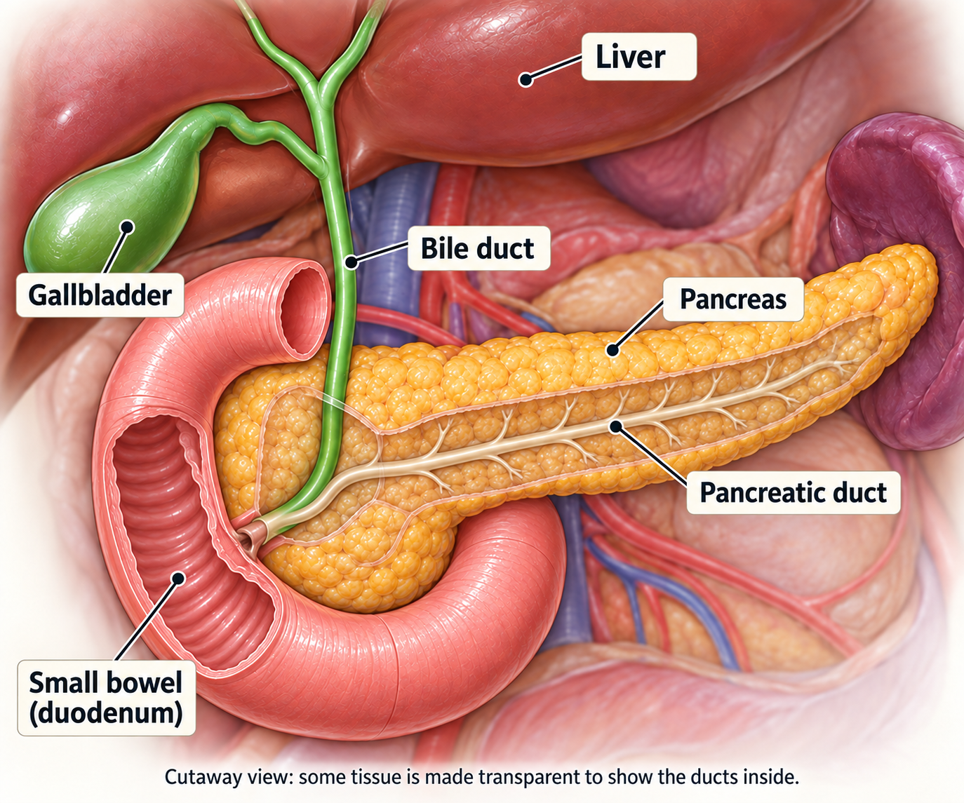
The pancreas is a long, flat gland behind the stomach. Its head lies beside the first part of the small bowel (duodenum) and the bile duct, the tube that carries digestive fluid from the liver to the bowel. Its tail points towards the spleen, an organ that helps fight infection.
Digestion
The part of the pancreas that makes digestive juices (the exocrine pancreas) produces enzymes that travel through the pancreatic duct into the small bowel. They break down fat, protein and carbohydrate so nutrients can be absorbed.
Blood sugar
The hormone-producing part (the endocrine pancreas) releases hormones, chemical messengers that travel in the blood. Insulin lowers blood sugar and glucagon raises it. Cancer or pancreatic surgery can disturb this balance and cause diabetes.
Why anatomy affects symptoms
A tumour in the pancreatic head may block the bile duct and cause jaundice. Loss of enzyme flow can cause pale, oily or floating stools, bloating and weight loss. A body or tail tumour may remain unnoticed for longer.
What is pancreatic cancer?
Cancer begins when abnormal cells keep growing and form a tumour. PDAC usually starts in cells lining the small ducts of the exocrine pancreas.
How it may behave
- It may remain confined to the pancreas at first.
- It can grow into nearby nerves, the duodenum, stomach, bile duct, spleen or important blood vessels.
- Cancer cells can travel through the blood or lymph, the fluid carried by the body’s drainage system. They can form cancer deposits elsewhere (metastases), most often in the liver or the lining of the abdomen (peritoneum), and sometimes in the lungs or other sites.
Some abnormal cells in pancreatic ducts (pancreatic intraepithelial neoplasia) and some mucus-producing growths can develop into cancer. The latter include intraductal papillary mucinous neoplasms (IPMNs), which grow in the ducts, and mucinous cystic neoplasms, which form cysts.
Most pancreatic cysts are not cancer. Some need regular specialist checks or surgery because the risk varies with the type and features of the cyst.
Three labels to record
- Where the tumour began: head, body or tail of the pancreas
- The type of cancer seen under the microscope and, when reported, its grade or differentiation: how abnormal the cells look and how closely they resemble normal cells
- Its extent: resectable, borderline resectable, locally advanced or metastatic
Cancer that started elsewhere and later reached the pancreas is a metastasis from the original cancer. It is not treated as primary pancreatic cancer.
The exact tumour type matters
Histology is the pattern and appearance of cells under a microscope. PDAC is the most common diagnosis, but rarer tumours may need a different plan.
Tumour type | Why the label matters |
|---|---|
Ductal adenocarcinoma | The usual PDAC pathway in this guide. Most evidence for chemotherapy and surgery is based on this type. |
Adenosquamous or undifferentiated | Rarer patterns that often grow quickly. Adenosquamous cancers contain two types of cancer cells; undifferentiated cells look very unlike normal cells. Specialist tissue review can help confirm the diagnosis. |
Acinar cell carcinoma | A rare cancer of the cells that make digestive enzymes. Its behaviour differs from PDAC, and tests for gene changes may be especially useful. |
Solid pseudopapillary neoplasm | An uncommon tumour, often found in younger people, named for its appearance under a microscope. Surgery can be very effective when it can be removed safely. |
Pancreatic neuroendocrine tumour | A separate tumour family. Treatment depends on grade, hormone production and sometimes a scan that detects somatostatin receptors, proteins on the surface of these tumour cells. |
A pathology report may mention
- Tumour type and grade or differentiation
- After surgery: tumour size; any spread to lymph nodes, the small structures that help fight infection; the edges of the removed tissue (margins); growth into vessels or nerves; and response to treatment given before surgery
- On a biopsy: whether there are enough living cancer cells in the sample for molecular testing
Symptoms and warning signs
Early pancreatic cancer may cause no obvious symptoms. Having one of these symptoms does not mean you have cancer, but persistent or worsening symptoms should be assessed.
Possible symptoms
- Yellow skin or eyes, dark urine, pale stools or itching
- Persistent pain in the upper abdomen, side or back, sometimes worse after eating or when lying down
- Unexplained weight loss, poor appetite, early fullness, nausea or vomiting
- Pale, oily, floating or foul-smelling stools; diarrhoea, constipation, bloating or excess wind
- New diabetes, suddenly difficult-to-control diabetes, thirst or frequent urination
- Marked tiredness, weakness, low mood or a blood clot in a leg or lung
There is no routine screening test for people without symptoms who have an average risk of pancreatic cancer. CA 19-9 is not a screening or diagnostic test by itself.
Risk factors
Usually there is no single answer. A risk factor changes probability; it does not prove what caused one person's cancer. This is not your fault.
Factors that increase risk
- Increasing age and smoking tobacco
- Excess body weight and long-standing diabetes
- Long-lasting inflammation of the pancreas (chronic pancreatitis), including an inherited form
- Certain pancreatic cysts, especially selected IPMNs and mucinous cystic neoplasms
- A close family history of pancreatic cancer or an inherited condition that increases the risk of cancer
Screening is only for selected high-risk people
A relative without cancer who has a proven inherited risk or a strong family history may be offered regular checks through a specialist programme.
These usually use magnetic resonance imaging (MRI), which makes pictures using a magnetic field, and endoscopic ultrasound (EUS), which uses an ultrasound probe on a thin camera tube passed through the mouth. Magnetic resonance cholangiopancreatography (MRCP) is a type of MRI that shows the pancreatic and bile ducts. Routine scans or CA 19-9 testing are not recommended for everyone.
Genetic counselling helps you understand test results, arrange testing for relatives and discuss privacy, insurance and questions about having children.
Tests and diagnosis
How cancer is diagnosed
You are unlikely to need every possible test. The team chooses tests that answer a specific question and should avoid delaying treatment without a reason.
- Review symptoms, general health, medicines, weight, nutrition and blood sugar.
- Use detailed scans to show the tumour, nearby blood vessels and any distant spread.
- Treat urgent jaundice, infection, vomiting or pain, and address too little fluid in the body (dehydration) or severe poor nutrition.
- Obtain tissue when treatment requires proof of cancer and keep enough tissue for relevant biomarker tests, which look for features such as gene changes or proteins that may guide treatment.
- Bring together the scan, tissue, blood and genetic test results, your health and your priorities. A multidisciplinary team, with specialists from different areas of care, agrees the plan.
Common blood tests
- Blood-cell counts, kidney function, blood salts (electrolytes) and how well the blood clots
- Liver tests, including bilirubin (a pigment that builds up in jaundice), albumin (a blood protein) and alkaline phosphatase (an enzyme that can rise with bile-duct blockage)
- Blood sugar (glucose) or glycated haemoglobin (HbA1c), which reflects average blood sugar over roughly 2–3 months; CA 19-9 when appropriate
CA 19-9 cannot prove or exclude pancreatic cancer. A blocked or infected bile duct can raise it, and some people do not make the marker. If there is a blockage or infection, a repeat measurement after it has been treated can give a clearer picture. Changes during cancer treatment are interpreted alongside scans and symptoms.
Scans and diagnostic procedures
Test | Typical purpose |
|---|---|
Pancreatic-protocol CT | Usually the main scan to assess the stage. Pictures taken at carefully timed intervals after contrast dye show the tumour, blood vessels, liver, abdominal lining and other organs. |
Chest CT | Checks the lungs and chest lymph nodes for distant spread. |
MRI / MRCP | Shows small abnormal areas in the liver, the pancreatic and bile ducts, or findings that CT has not explained. It can help when CT contrast dye is unsuitable. |
Endoscopic ultrasound | Gives a close view of the pancreas from inside the stomach or bowel. A needle can usually take a small cylinder of tissue (a core biopsy). |
PET-CT | Positron emission tomography combined with CT (PET-CT) uses a small amount of radioactive tracer to highlight areas of activity. It may help when distant spread is strongly suspected or other scans are unclear. It does not replace a pancreatic-protocol CT. |
Staging laparoscopy | A camera is passed through small cuts in the abdomen to look for tiny cancer deposits on the abdominal lining or liver. This may be advised before major surgery for selected patients at higher risk of spread. |
The report should describe tumour-vessel contact, definite or suspected distant spread and any uncertain abnormal area that needs another test before a major treatment decision.
If your condition is stable, scans of the pancreas are ideally done before a bile-duct stent is placed, because the procedure can cause inflammation. Urgent drainage comes first if the bile ducts are infected (cholangitis), the blockage is severe or your condition is worsening.
A radiologist and pancreatic surgeon should review the actual images, not only the written report, before resectability is finalised.
Biopsy and your pathology report
A biopsy removes cells or a small core of tissue so a pathologist can confirm the diagnosis. Endoscopic ultrasound-guided core biopsy is often preferred.
A biopsy is usually needed before
- Chemotherapy, radiotherapy, or chemotherapy given together with radiotherapy (chemoradiation)
- Treatment given before a planned operation (neoadjuvant treatment)
- Most clinical trials or treatment chosen to match a biomarker in the cancer
- Treatment of metastatic disease when the origin or tumour type needs confirmation
What to ask the pathologist or oncologist
- What is the exact histological type and grade?
- Is there enough tissue for the necessary tests, or would another sample help?
- Has tissue been kept for tests of its genetic-material repair system and other molecular tests, if needed?
Endoscopic retrograde cholangiopancreatography (ERCP) uses a flexible camera tube and X-rays. It is mainly used here to open a blocked bile duct. Cells collected with a small brush during ERCP may miss a pancreatic cancer and may not provide enough material for all molecular tests.
Two kinds of genetic testing
Inherited testing and tumour profiling answer different questions. Both can matter even when no relative has had pancreatic cancer.
Germline testing
Uses blood or saliva to look for an inherited gene change present throughout the body. The result may guide the use of platinum-based chemotherapy, a group of medicines that damage cancer-cell genetic material, or medicines called poly(ADP-ribose) polymerase (PARP) inhibitors, which block a repair process in cancer cells.
The result can also guide testing or regular checks for relatives. Germline testing is recommended for everyone with confirmed PDAC.
Tumour profiling
Looks for changes in the cancer cells. Deoxyribonucleic acid (DNA) carries genetic instructions; ribonucleic acid (RNA) helps cells use them. Testing many genes in DNA and, when possible, RNA can be particularly useful in advanced disease or unusual cancer types.
Results may identify a medicine that blocks a specific cancer growth signal (targeted therapy), or one that helps the immune system attack cancer (immunotherapy). Only a small minority have a suitable finding.
Results that may change a discussion
- BRCA1, BRCA2 or PALB2 changes can support use of platinum chemotherapy; an inherited BRCA1 or BRCA2 change can support olaparib in a specific metastatic setting, as explained under Treatment options. This is maintenance treatment: ongoing medicine to help keep the cancer controlled after initial chemotherapy.
- High microsatellite instability (MSI-high) and deficient mismatch repair (dMMR) are signs that cancer cells do not repair certain errors in DNA properly. These cancers may respond to checkpoint inhibitors, medicines that help the immune system recognise and attack cancer.
- Other rare findings may support a matched treatment or trial. Reports may name NTRK, RET or NRG1 gene fusions, where parts of two genes join abnormally, or specific changes called BRAF V600E or KRAS G12C. These genes are involved in signals that control cell growth. Tests may also show high levels of human epidermal growth factor receptor 2 (HER2), a growth-signalling protein, or extra copies of its gene (amplification). Your team can explain whether a finding is useful for treatment.
Testing availability, cost, laboratory quality and drug access vary in India. Ask which result could realistically change your current or later treatment before paying for a panel, a test that checks many genes at once.
Planning your treatment
Understanding your stage
Tumour, node and metastasis (TNM) staging for pancreatic adenocarcinoma
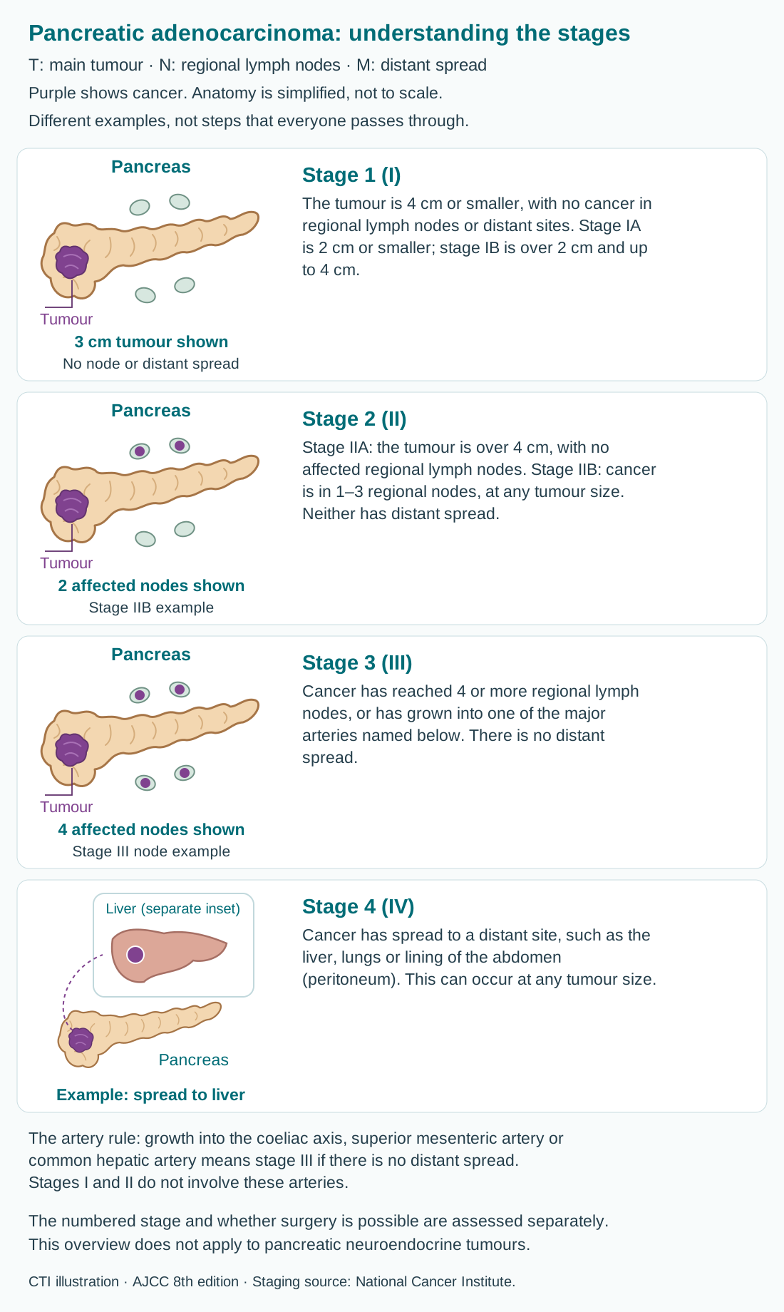
TNM describes the main tumour (T), regional lymph nodes (N) and distant spread, called metastasis (M). These findings give the numbered stage. This overview applies to pancreatic adenocarcinoma; pancreatic neuroendocrine tumours use a different staging system.
Lymph nodes are small immune-system structures. Regional nodes are those near or draining the cancer’s starting area; distant spread means cancer has reached a site outside that regional area. Stages describe findings, not a sequence that everyone will pass through.
Purple shows cancer. These are simplified examples, not to scale. Each picture shows one way a stage can occur; read the text beside it for the other possibilities.
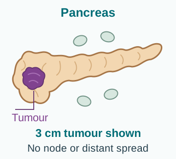
Stage 1 (I)
The tumour is 4 centimetres (cm) or smaller, with no cancer in regional lymph nodes or distant sites. Stage IA is 2 cm or smaller; stage IB is over 2 cm and up to 4 cm.
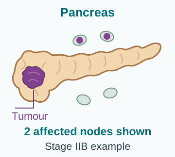
Stage 2 (II)
Stage IIA: the tumour is over 4 cm, with no affected regional lymph nodes. Stage IIB: cancer is in 1–3 regional nodes, at any tumour size. Neither has distant spread.
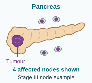
Stage 3 (III)
Cancer has reached 4 or more regional lymph nodes, or has grown into one of the major arteries named below. There is no distant spread.
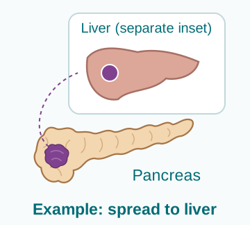
Stage 4 (IV)
Cancer has spread to a distant site, such as the liver, lungs or lining of the abdomen (peritoneum). This can occur at any tumour size.
Which arteries matter? The coeliac axis, superior mesenteric artery and common hepatic artery are major arteries beside the pancreas. Growth into any of these makes the cancer stage 3 if there is no distant spread. Stages 1 and 2 do not involve these arteries.
Whether surgery can safely remove the cancer is assessed separately. Your team may use the terms resectable, borderline resectable or locally advanced. Vein involvement is different from the arterial involvement described above; some veins can be removed and rebuilt during surgery. Ask how your scan findings affect the treatment sequence and whether reassessment for surgery is planned.
Clinical stage comes from examination, biopsies and scans. Pathological stage adds information from surgery. The letters “yp” before a stage mean that tissue removed at surgery was assessed after treatment given before the operation. A revised assessment does not necessarily mean the cancer grew while you were waiting.
Grade describes how abnormal the cells look under the microscope and can suggest how they may behave. It is different from stage, which describes how far cancer has grown or spread.
Whether surgery can remove the cancer safely depends on its contact with blood vessels, the chance of removing the tumour with no cancer at the cut edges (a clear margin), any distant spread, surgical expertise and your health. It should be decided in a pancreatic multidisciplinary meeting.
Your care team
Pancreatic cancer care works best when specialists review the scans, tissue, fitness and goals together and one person coordinates the plan.
Team member | Main role |
|---|---|
Pancreatic surgeon | Assesses whether surgery can remove the cancer safely and which operation is suitable. Operations to remove part or all of the pancreas (pancreatectomy) are explained below. |
Medical oncologist (cancer-medicine specialist) | Plans chemotherapy, biomarker-directed treatment and clinical trials. |
Gastroenterologist (digestive-system specialist) | Performs EUS-guided biopsy and ERCP, and places stents to relieve a blockage. |
Radiation oncologist | Plans radiotherapy, sometimes with chemotherapy. Options may include stereotactic body radiotherapy (SBRT), which delivers precisely focused radiation in a small number of sessions. |
Radiologist | Reads the scans and assesses how the tumour relates to nearby blood vessels. |
Pathologist / molecular laboratory | Examines tissue to confirm the cancer type and performs relevant biomarker tests. |
Dietitian / diabetes team | Provides food and nutrition advice, including help with weight, digestive enzyme medicines, tube feeding and blood sugar. |
Palliative, pain and supportive team | Treats symptoms, distress and practical needs alongside cancer treatment. |
Useful question: Who is my main contact for results, nutrition problems, side effects and urgent concerns? Save the centre's 24-hour number.
How your treatment plan is chosen
Good decisions combine medical evidence with what matters to you. The same stage can lead to different plans when anatomy, fitness or priorities differ.
The team considers
- Stage, resectability, tumour location and relationship to major vessels
- Pathology, germline testing and tumour biomarkers
- Your ability to manage daily activities (performance status), age, nutrition, bilirubin level, kidney and liver function, diabetes and other illnesses
- Symptoms, previous treatment, likely benefit, serious risks and recovery time
- Your priorities, work and family responsibilities, travel, cost and access
Shared decision-making
Ask for the reasonable alternatives, including a less intensive option when appropriate. Ask how many people are likely to benefit, and compare the side effects that matter most, what treatment days look like and what would make the plan change. A second opinion is appropriate if you remain unsure.
Treatment recommendations change. Your oncology team and pharmacist should confirm that the medicine is suitable, its dose, whether other medicines could affect it, and options for managing cost.
Your treatment options at a glance
The first major question is whether treatment should lead towards surgery, control cancer throughout the body, or do both in sequence.
Treatment options
Topics in this section
Two routes towards surgery
For a fit person without distant spread, treatment aims to remove all visible cancer and treat unseen cells that may already be elsewhere.
Surgery first
Some clearly resectable cancers proceed directly to surgery, followed by chemotherapy after recovery. A Whipple operation removes the head of the pancreas and nearby structures; other operations are used for tumours elsewhere in the pancreas. Surgery first remains a standard route for selected patients.
Drug treatment first
Chemotherapy using a combination of drugs may be advised before surgery (neoadjuvant treatment), especially when the tumour has high-risk features or its relationship to nearby blood vessels makes it borderline for surgery. The cancer is reassessed before an operation is planned.
Features that may favour drug treatment first
- Markedly raised CA 19-9 after jaundice has been relieved
- A large tumour in the pancreas, lymph nodes that may contain cancer, severe weight loss or significant pain
- Concern that surgery now might leave cancer at the cut edge (a positive margin), or uncertainty about tiny abnormal areas elsewhere in the body
- A need to see how the cancer responds and give treatment that reaches cells throughout the body (systemic treatment) before a major operation
A biopsy is required before neoadjuvant treatment. If surgery is first, examination of tissue removed during the operation provides the pathological stage.
Whipple, distal or total pancreatectomy
Pancreatic surgery is major treatment. The safest operation is the one that can achieve complete removal while preserving function where possible.
Operation | What is usually removed |
|---|---|
Whipple procedure | For a tumour in the head. Removes the pancreatic head, duodenum, gallbladder, part of the bile duct and nearby nodes; sometimes part of the stomach. The digestive tract is reconnected. |
Distal pancreatectomy | For a body or tail tumour. Removes the left part of the pancreas and usually the spleen. Vaccinations are needed when the spleen is removed. |
Total pancreatectomy | Removes the whole pancreas, usually the spleen and related structures. Lifelong insulin and pancreatic enzyme replacement are then essential. |
Possible complications
- Bleeding, infection, blood clots or injury to nearby structures
- Leakage of pancreatic fluid or bile, pockets of fluid inside the abdomen and slower emptying of the stomach
- Weight loss, poor absorption of nutrients (malabsorption), too few digestive enzymes reaching the bowel (pancreatic exocrine insufficiency) and diabetes
Occasionally an operation cannot be completed because unexpected spread or unsafe vessel involvement is found. The surgeon may instead create a route around the blockage (a bypass) or arrange a stent to hold it open.
Recovery and treatment after surgery
Surgery removes visible disease. Chemotherapy treats microscopic cancer cells that scans and the surgeon cannot see and lowers the risk of recurrence.
The pathology report guides the next conversation
- Tumour size and final tumour (T) category in the staging system
- Number of lymph nodes examined and number involved
- The edges of the removed tissue (resection margins): R0 means no cancer at the assessed edge; R1 means cancer at or very close to it under the microscope, depending on the reporting system
- Grade; cancer in lymphatic channels or blood vessels (lymphovascular invasion) or around nerves (perineural invasion); and response to any treatment before surgery
Common postoperative chemotherapy choices
Modified FOLFIRINOX is often preferred for someone well enough for combination chemotherapy who has recovered well. FOLFIRINOX combines folinic acid (also called leucovorin), fluorouracil, irinotecan and oxaliplatin. “Modified” means the doses or schedule are adjusted. Gemcitabine plus capecitabine is an important alternative.
The aim is commonly about 6 months of drug treatment in total, counting treatment given before surgery when relevant.
Radiation is not routine after every operation that completely removes the visible cancer. It may be discussed if you have a positive surgical margin or are at high risk of the cancer returning near the operation site (local recurrence), usually after chemotherapy and specialist review.
Why might I need drug treatment before surgery?
For borderline resectable cancer, the tumour has not spread to distant parts of the body, but its contact with important blood vessels makes complete and safe removal uncertain at diagnosis. Drug treatment first may improve the chance of surgery.
- Confirm the diagnosis with a biopsy and correct jaundice, infection, dehydration and major nutrition problems.
- Give combination chemotherapy, commonly modified FOLFIRINOX or gemcitabine plus nab-paclitaxel, according to your health and organ function. Nab-paclitaxel is paclitaxel attached to tiny particles of the protein albumin.
- Repeat pancreatic-protocol imaging, clinical assessment and CA 19-9 when informative. A tumour does not always need to shrink visibly for treatment to be useful.
- Consider chemoradiation or SBRT for selected patients; routine use after chemotherapy is not required for everyone.
- Proceed to surgery only if there is no new or worsening distant spread and a specialist team believes complete removal is now realistic.
If the cancer grows or spreads during treatment before surgery, a major operation is unlikely to help. The plan then shifts towards the locally advanced or metastatic pathway, with supportive care throughout.
How is locally advanced pancreatic cancer treated?
Locally advanced pancreatic cancer has not spread to distant parts of the body, but it cannot currently be removed safely with surgery, usually because it involves major blood vessels. Treatment aims to control the cancer, followed by scans and specialist review to decide the next step.
The usual starting point
Chemotherapy that travels through the body is normally given first. The team selects a combination to suit your health, such as modified FOLFIRINOX, NALIRIFOX or gemcitabine plus nab-paclitaxel.
NALIRIFOX combines fluorouracil, leucovorin, liposomal irinotecan and oxaliplatin. “Liposomal” means the irinotecan is enclosed in tiny fat particles; this is a different preparation from the irinotecan in FOLFIRINOX. Scans and a clinical review commonly follow several months of treatment.
If the cancer is controlled
- Continue or simplify chemotherapy, or take a planned break in selected situations
- Consider chemoradiation or SBRT to control cancer in the treated area, relieve pain or address a serious problem that local growth may cause
- Ask for repeat surgical review if the tumour’s relationship to blood vessels, CA 19-9 and overall health improve substantially
Support may include a stent for a blocked bile duct or duodenum, digestive enzyme medicines, nutrition advice, pain relief and medicines that reduce blood clotting (anticoagulation) when needed.
A coeliac plexus block is an injection around a group of nerves near the pancreas to reduce pain. This and other palliative care can be offered alongside cancer treatment.
First treatment for cancer that has spread
When PDAC has spread to a distant site, treatment usually aims to control cancer, prolong life where possible, relieve symptoms and protect day-to-day function. For someone well enough for combination chemotherapy, first-line (initial) options include the following.
First-line chemotherapy options
- Modified FOLFIRINOX: 5-fluorouracil, leucovorin, irinotecan and oxaliplatin
- NALIRIFOX: 5-fluorouracil, leucovorin, liposomal irinotecan and oxaliplatin
- Gemcitabine plus nab-paclitaxel
There is no single best regimen for everyone. The choice considers performance status, bilirubin and organ function, any existing nerve damage causing numbness or tingling (neuropathy), diarrhoea risk, blood counts, diabetes, practicalities of treatment through a drip, previous treatment after surgery, cost and local availability.
When intensive combination treatment is not suitable
A modified schedule, gemcitabine-based treatment, capecitabine or another single drug may offer a better balance for some people. When performance status is very poor because of advanced cancer or other illness, symptom-focused care may help more than chemotherapy. The team checks whether poor health is caused by a treatable problem, such as jaundice or dehydration, or by illness that cannot be reversed.
The team checks the treatment response using symptoms, daily activity, blood tests, CA 19-9 when useful and scans.
When treatment changes - and when a biomarker matters
If another treatment is needed, the choice depends on what you have already received, how long it worked, remaining side effects, your current health and the tumour’s molecular test results.
Changing the chemotherapy combination
- After gemcitabine-based treatment, 5-fluorouracil, leucovorin and liposomal irinotecan may be considered. Evidence is clearest without prior irinotecan; earlier irinotecan needs specialist judgement.
- After FOLFIRINOX or NALIRIFOX, gemcitabine plus nab-paclitaxel or gemcitabine alone may be considered if fitness allows.
Daraxonrasib: a newer option for metastatic pancreatic cancer
Daraxonrasib is an option for adults with metastatic pancreatic adenocarcinoma after at least one previous treatment that acts throughout the body, or when treatment using a combination of such medicines is unsuitable. This does not mean it is appropriate for every pancreatic tumour or disease stage.
How does it work?
Daraxonrasib is taken by mouth. It blocks RAS proteins, a family of proteins that pass on signals telling cells to grow. Your oncologist will consider your diagnosis, previous treatment, fitness and molecular results when discussing it.
Is KRAS mutation testing required?
Under its United States (US) approval, a test for a change (mutation) in the KRAS gene is not mandatory before daraxonrasib, and treatment does not require a positive KRAS result. Your oncologist may still recommend broader tumour testing to identify other treatment options.
Side effects and practical questions
Important risks include skin and mouth problems, diarrhoea, a hole in the bowel wall (perforation), inflammation of the lungs and harm to an unborn baby. Ask about monitoring, effects of other medicines or supplements, pregnancy precautions and access to treatment.
New breathlessness, severe abdominal pain or severe diarrhoea needs urgent medical advice. Do not start or change treatment without your oncology team's guidance.
Finding | What it may change |
|---|---|
Germline BRCA1/2 | For metastatic cancer that has not worsened during at least 16 weeks of first-line platinum-based chemotherapy, maintenance olaparib may be discussed. |
MSI-high / dMMR | A checkpoint inhibitor such as pembrolizumab may be considered. |
NTRK, RET, BRAF V600E or KRAS G12C | A matched targeted drug may be available after prior treatment or through a trial. |
NRG1 fusion | Zenocutuzumab is an option after prior systemic therapy. |
HER2-positive tumour | Selected HER2-directed treatment or a clinical trial may be considered after expert review. |
Managing side effects
Managing treatment side effects
Side effects depend on the drugs, dose, schedule and your health. Reporting problems early allows supportive treatment, a planned pause or a safer dose.
Treatment | Effects to discuss early |
|---|---|
FOLFIRINOX / NALIRIFOX | Low blood counts and infection, nausea, diarrhoea, mouth soreness, tiredness, cold sensitivity and numbness or tingling. |
Gemcitabine + nab-paclitaxel | Low blood counts and infection, tiredness, hair loss, swelling, rash, muscle aches and neuropathy. |
Gemcitabine + capecitabine | Low blood counts, diarrhoea, sore mouth, skin changes and painful red hands or feet. |
Chemoradiation / SBRT | Tiredness, nausea, acid coming back up from the stomach (reflux), appetite loss, diarrhoea and irritation of the stomach or small bowel. |
Your practical safety checklist
- Keep the 24-hour number and know the temperature threshold given by your centre; many teams use 38°C (100.4°F) or higher as an emergency during chemotherapy.
- Take supportive medicines as prescribed. Report diarrhoea, dehydration, mouth ulcers, neuropathy or poor intake early.
- Share every medicine, vitamin, herb and supplement. Ask about food instructions, blood-thinning medicines and whether genetic testing for drug safety is needed. Do not alter a cancer drug without instructions.
When to seek urgent help
Pancreatic cancer, bile-duct stents, surgery and chemotherapy can cause emergencies. Follow your own centre's instructions; use emergency care when symptoms are severe.
Other problems that should not wait for the next routine visit
- More frequent pale or oily stools, rapidly falling weight or inability to eat
- Repeated low or very high blood sugar, severe thirst, drowsiness or sweating and shaking
- Diarrhoea that is frequent, bloody, associated with fever, or not controlled by the plan you were given
- A blocked, leaking or painful treatment line or port (a device used to give medicines into a vein); redness, swelling or discharge from a surgical wound
Carry a current medicine list and details of your chemotherapy, allergies, anticoagulant use, bile-duct stent and recent surgery. Tell every emergency clinician that you are receiving cancer treatment.
Live well
Supportive and palliative care
Supportive care treats the person, not just the scan. Palliative care can be added alongside active cancer treatment at any stage.
Problem | Possible help |
|---|---|
Blocked bile duct | A stent placed during ERCP, a drainage tube passed through the skin, or an operation to bypass the blockage. Infection needs antibiotics and urgent drainage. |
Blocked duodenum | Anti-sickness medicines, a stent placed using an endoscope, or an operation connecting the stomach to the small bowel beyond the blockage (gastrojejunostomy). Other nutrition support may help. |
Pain | Regular pain medicines, including adjusting strong painkillers called opioids; a nerve block or a procedure that interrupts pain signals (neurolysis); chemotherapy; or focused radiotherapy. |
Blood clot | Imaging and anticoagulation when safe; urgent assessment for chest symptoms or leg swelling. |
Fluid build-up in the abdomen (ascites) or breathlessness | Drainage, medicines and treatment of the cause when appropriate. |
Distress and caregiver strain | Counselling, emotional support for people affected by cancer (psycho-oncology), social work, spiritual care, short breaks for caregivers (respite) and planning ahead. |
Tell the team about pain before it becomes severe. A pain or palliative specialist can combine medicines with nerve procedures and address sleep, anxiety, constipation and family concerns.
A special diet or supplement cannot relieve jaundice caused by a blocked bile duct. Fever when you have a bile-duct stent may signal an infection in the ducts (cholangitis) and needs urgent care.
Nutrition and pancreatic enzymes
Weight and muscle can fall quickly in pancreatic cancer. Early help from a dietitian and, when needed, pancreatic enzyme replacement therapy (PERT) can improve nutrition and help you cope with treatment. PERT provides capsules of digestive enzymes when the pancreas is not supplying enough.
Clues that enzymes may be insufficient
- Pale, oily, floating or foul-smelling stools that are hard to flush
- Bloating, cramps, excess wind, urgency or diarrhoea after meals
- Continued weight loss despite eating, or low levels of vitamins absorbed with fat, such as vitamins A, D, E and K
Practical eating principles
- Use small, frequent, energy- and protein-rich meals when appetite is limited.
- Do not cut out fat automatically. Adequate PERT often allows a more normal diet and helps prevent further weight loss.
- Treat nausea, pain, constipation, mouth problems and obstruction because they reduce intake.
- Use nutrition drinks, tube feeding or nutrition given through a vein only after an individual assessment of goals and likely benefit.
No restrictive diet, detox, supplement, fasting plan or pancreatic-enzyme regimen has been proven to cure PDAC. Check any complementary therapy with the oncology team because interactions and malnutrition can cause harm.
Diabetes, follow-up and recurrence
Pancreatic cancer and pancreatic surgery can reduce both insulin and digestive enzymes. Diabetes and malabsorption should be managed together, not as separate problems.
Diabetes after pancreatic disease
Blood sugar may become unstable before or after treatment. Lifelong insulin is needed after total pancreatectomy and may also be needed after partial surgery. Ask for a plan for monitoring, sick days, low sugar and high sugar, with help from a diabetes specialist and dietitian.
Follow-up after curative treatment
Visits usually review symptoms, weight, nutrition, enzymes, diabetes, physical recovery and emotional health. CT or MRI and CA 19-9, when informative, are used at intervals based on stage, treatment and local practice. Ask who orders each test and how results are shared.
If you are receiving treatment for advanced cancer
Scans, blood tests and symptom review are repeated to assess benefit and side effects. A CA 19-9 change is interpreted with imaging and clinical condition, not alone. Small scan changes do not always require an immediate switch if symptoms and the overall pattern remain stable.
If cancer returns
Treatment depends on where and when recurrence appears, previous chemotherapy, molecular findings, current fitness and goals. Options can include a different chemotherapy combination, a medicine matched to a biomarker, radiation for a local problem, a procedure for obstruction, a clinical trial and supportive care.
Emotional and practical support
There is no correct way to feel. Shock, fear, anger, sadness, hope and uncertainty can appear together or change from day to day.
For patients
Tell the team if anxiety, low mood, poor sleep, panic or loss of interest persists or affects daily life. Ask for psycho-oncology, counselling or palliative support. If having children in the future matters to you, ask before treatment about ways to protect your fertility. Accepting help with food, transport or childcare can preserve energy for treatment.
For caregivers
Ask what to monitor, who to call and how to share responsibility. Your own sleep, health, work and emotional needs matter. Request clear instructions rather than trying to interpret every symptom alone.
Sources and further reading
For more information, explore these resources from the National Cancer Institute (NCI) and other medical organisations. The linked organisations have not reviewed or endorsed this CTI guide. Your care team can explain how the information applies to you.
- NCI: Pancreatic Cancer Treatment
- Cancer Council Australia: pancreatic cancer
- Macmillan: pancreatic cancer
Staging and risk-group references
American Joint Committee on Cancer (AJCC), 8th edition.
Medicine approval and prescribing references
These official records describe licensed uses and prescribing information for the medicines named in this guide. Approval depends on the cancer type, biomarkers, previous treatment, combination and formulation. Some uses discussed in the guide are off-label: outside the medicine’s licence, with evidence from clinical guidelines or studies. Approval by the US Food and Drug Administration (FDA) or the European Medicines Agency (EMA) does not establish approval or availability in India.
- Albumin-bound paclitaxel (Abraxane) — FDA prescribing information · Approval history
- Capecitabine — FDA prescribing information · Approval history
- Cisplatin — FDA prescribing information · Approval history
- Daraxonrasib — FDA prescribing information · Approval history
- Fluorouracil — FDA prescribing information · Approval history
- Gemcitabine — FDA prescribing information · Approval history
- Irinotecan — FDA prescribing information · Approval history
- Leucovorin (folinic acid) — FDA prescribing information · Approval history
- Liposomal irinotecan — FDA prescribing information · Approval history
- Olaparib — FDA prescribing information · Approval history
- Oxaliplatin — FDA prescribing information · Approval history
- Pembrolizumab — FDA prescribing information · Approval history
- Zenocutuzumab — FDA prescribing information · Approval history
Additional medicine approval notices
These records explain the specific medicine uses and eligibility requirements discussed in this guide.
Related care
Meet our doctors



Where to get care