
Share
Start here
About this guide
Melanoma is often curable when found early. When it has spread, modern treatment can still give meaningful and sometimes long-lasting control.
This guide explains melanoma of the skin, including the palms, soles and nails. It also covers mucosal melanoma, which starts in the moist lining of areas such as the nose, mouth or lower bowel, and uveal melanoma, which starts inside the eye.
Bring your scan images
For your consultation or a second opinion, bring a compact disc (CD) containing your scan images in Digital Imaging and Communications in Medicine (DICOM) format and the software needed to view them. DICOM is the standard format for medical scan images. Bring the written scan reports too.
Understand
What melanoma is
Melanoma starts in melanocytes, the cells that make melanin, the pigment that gives skin its colour. Most melanomas begin in the skin.
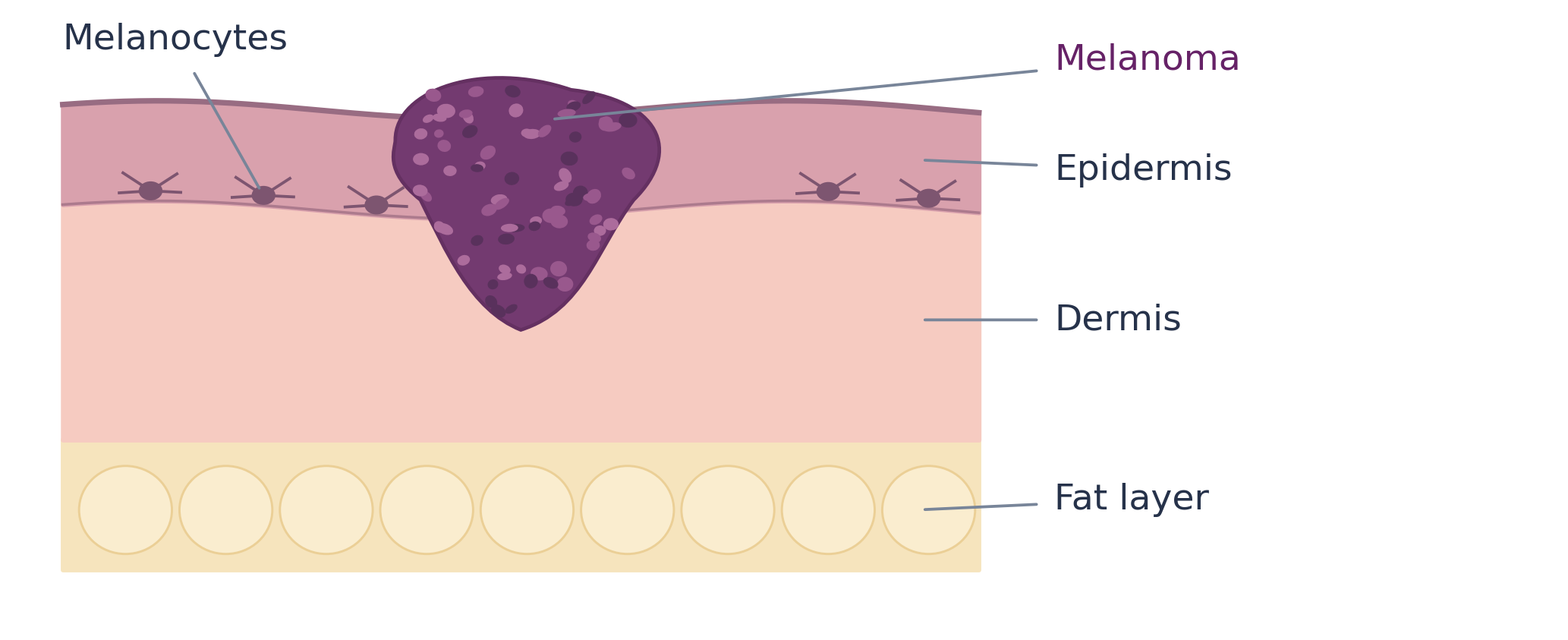
The epidermis is the outer layer. Melanocytes normally sit near its base. The dermis underneath contains supporting tissue, blood vessels and lymphatic vessels, which carry fluid away from tissues. Beneath this is a layer of fat.
Melanoma in situ stays within the epidermis. Invasive melanoma has grown into deeper skin. The depth measured under the microscope is called Breslow thickness. This is not the same as the width of a mole on the skin.
Cancer cells can sometimes travel through lymphatic vessels to nearby lymph nodes, small structures that filter fluid and help fight infection. They can also travel through the bloodstream to other organs. Spread to a distant site is called metastatic melanoma.
Symptoms and warning signs
A new, changing or unusual mark matters more than any single rule. A dermatologist, a doctor specialising in skin conditions, can examine it with a magnifying instrument called a dermatoscope.
Clue | What to look for |
|---|---|
A • Asymmetry | One half looks different from the other. |
B • Border | An uneven, notched or poorly defined edge. |
C • Colour | Uneven colour or a change in colour. |
D • Diameter | Often over 6 millimetres (mm), but melanoma can be smaller. |
E • Evolving | A change in size, shape, colour, surface or symptoms. |
Not all melanomas follow ABCDE
A raised, firm lump that keeps growing can be melanoma even if it is small and evenly coloured. A spot that looks different from your other moles also needs attention. Report unexplained bleeding, persistent itching or a sore that does not heal.
Palms, soles and nails
Look for an enlarging dark patch, a persistent wound, or a new or widening dark nail band, especially in one nail. Colour spreading onto the surrounding skin, nail splitting or a lump under a nail also needs assessment.
Types and risk factors
Melanoma is not one uniform disease. Where it starts and the characteristics of its cells help explain which tests and treatments are useful.
Type | What the name means |
|---|---|
Superficial spreading | Often spreads across the skin before growing more deeply. |
Nodular | Often grows as a raised lump and can become deep quickly. |
Lentigo maligna melanoma | Usually develops on skin damaged by many years of sun exposure, often the face. |
Acral | Starts on palms, soles or under or around a nail. It is not usually caused by ultraviolet radiation from sunlight. |
Other uncommon forms include desmoplastic melanoma, which contains scar-like tissue, and melanoma of the mucosal linings or the eye. Sometimes melanoma is found in a lymph node or elsewhere even though the original skin melanoma cannot be found.
Factors that increase risk
Ultraviolet exposure, sunburns and tanning beds increase the risk of many skin melanomas. Fair skin, many moles or unusual-looking moles, a previous melanoma, a weakened immune system and family history can also increase risk. People without these factors can still develop melanoma.
Tests and diagnosis
Biopsy and your pathology report
A biopsy removes tissue for examination under a microscope to confirm melanoma. A photograph, scan or blood test alone cannot reliably make the diagnosis.
Where possible, the doctor removes the whole suspicious area, a small rim of surrounding skin (the margin) and enough tissue underneath for accurate assessment. A large area or a difficult site, such as a nail, may need a carefully planned biopsy of part of the area. A local anaesthetic medicine usually numbs the skin.
Report item | Why it matters |
|---|---|
Breslow thickness | The depth of an invasive melanoma, in millimetres. It helps guide surgery and node assessment. |
Ulceration | Loss of the surface layer seen under the microscope. It affects the risk category and stage. |
Margins | Whether melanoma reaches an edge of the tissue removed. A wider operation may still be needed. |
Subtype and other features | The report may describe how quickly cells are dividing (mitotic activity), tiny nearby melanoma deposits (microsatellites), or scar-like tissue (desmoplasia). These findings can influence planning. |
The pathology report records what was found when the tissue was examined. Ask for a copy. A skin pathologist, a doctor who diagnoses disease by examining skin tissue, may review an uncertain result or an unusual type. Special laboratory dyes or tests for gene changes may help clarify the diagnosis.
Which tests are needed
Your team examines the whole skin and relevant lymph nodes. Early, lower-risk melanoma often does not need routine body scans. The team chooses scans according to the risk of spread, your symptoms and the planned treatment.
Ultrasound uses sound waves. Computed tomography (CT) uses X-rays to make detailed pictures. Positron emission tomography combined with CT (PET-CT) uses a small amount of radioactive tracer to highlight areas of activity. Magnetic resonance imaging (MRI) uses a magnetic field to produce detailed images and may be used to check the brain.
Blood tests check general health and organ function. Lactate dehydrogenase (LDH), a protein measured in the blood, can help assess advanced disease, but it does not diagnose or exclude melanoma. A normal scan cannot rule out cancer deposits too small to be seen.
Nodes and molecular tests
Sentinel node testing looks for spread to nearby lymph nodes. Tumour biomarker testing looks for features such as gene changes or proteins that may help guide medicine choices. Neither test is needed for every patient.
Sentinel lymph node biopsy
This operation checks the first lymph node or nodes that receive fluid from the skin around the melanoma. It looks for cancer cells too small to be found on examination or scans. It is usually discussed for melanomas 0.8–1.0 mm thick, or thinner melanomas with ulceration, and generally offered when thickness exceeds 1 mm if the result will guide care.
A small amount of radioactive tracer, sometimes with dye, helps locate these nodes. The procedure is often done during the operation to remove a wider rim of skin around the biopsy site (wide local excision). Risks include pain, a fluid collection, infection and, less commonly, persistent swelling. It helps staging; it is not a guarantee that cancer cannot return.
A positive result means melanoma cells were found in the sentinel node. It does not routinely mean that every nearby node must be removed. Regular ultrasound checks and drug treatment may be considered. A clearly enlarged suspicious node usually needs a sample taken with a needle rather than a sentinel node biopsy.
Tests that may guide medicines
BRAF V600 testing looks for a particular type of change in the BRAF gene. It is important when medicines that block specific growth signals in cancer cells (targeted therapy) may be used, especially in stage III or IV disease. A tumour mutation usually is not inherited. Testing for additional gene changes may help selected patients, particularly with acral or mucosal melanoma, or after standard treatments.
Planning your treatment
Understanding your stage
TNM staging for melanoma of the skin
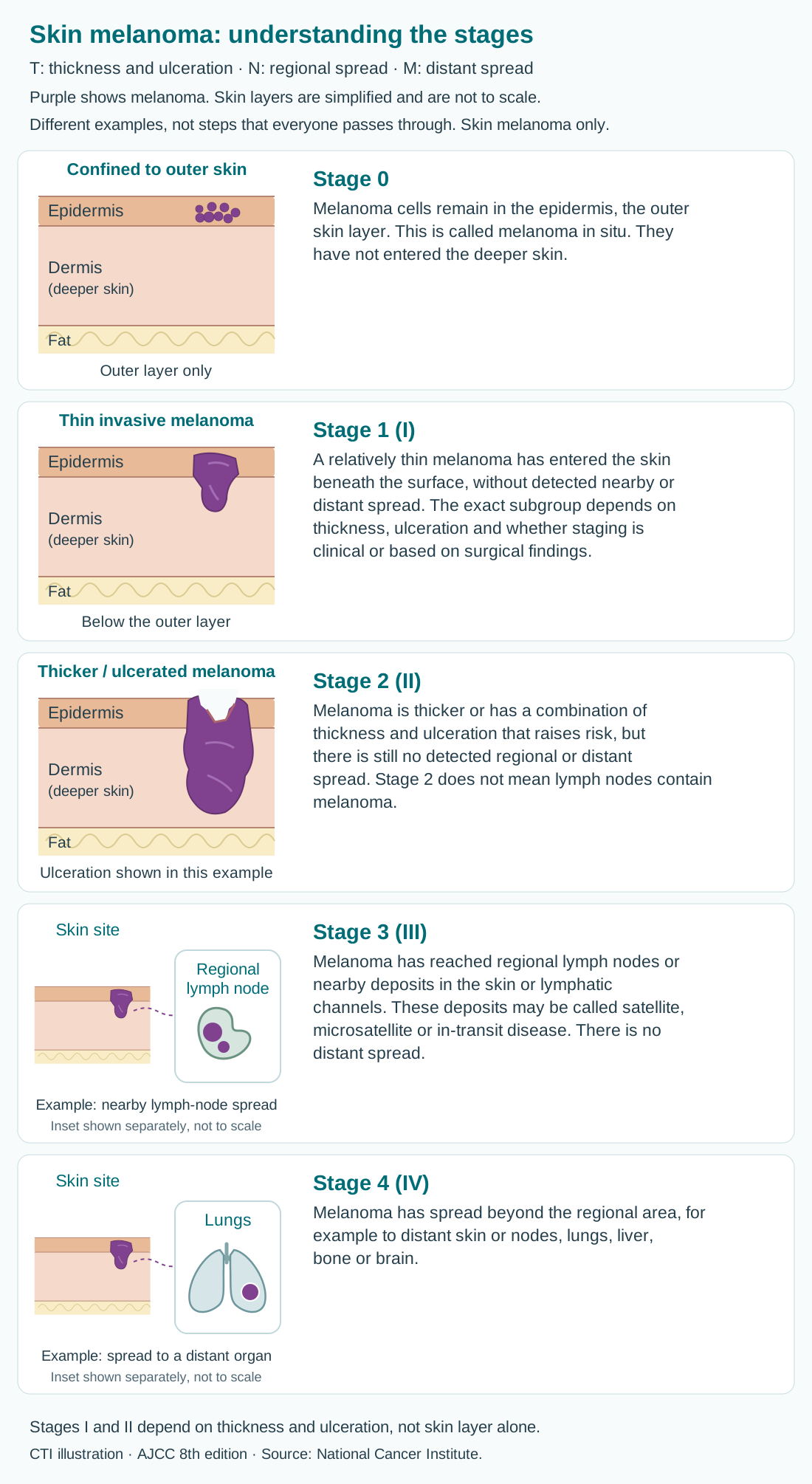
The tumour, node and metastasis (TNM) system describes the main tumour (T), regional spread (N) and distant spread (M). For skin melanoma, tumour thickness in millimetres and a break in the tumour’s surface (ulceration) help determine the stage. The width of a mole is not its depth. The diagram below applies to skin melanoma; mucosal and eye melanoma use different staging approaches.
Lymph nodes are small immune-system structures. Regional nodes are those near or draining the cancer’s starting area; distant spread means cancer has reached a site outside that regional area. Stages describe findings, not a sequence that everyone will pass through.
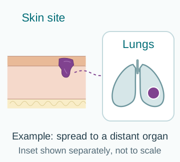
Purple shows melanoma. The skin layers and separate insets are simplified and not to scale. Each picture shows one example; the text explains other ways that stage can occur.
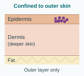
Stage 0
Melanoma cells remain in the epidermis, the outer skin layer. This is called melanoma in situ. They have not entered the deeper skin.
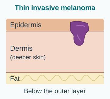
Stage 1 (I)
A relatively thin melanoma has entered the skin beneath the surface, without detected nearby or distant spread. The exact subgroup depends on thickness, ulceration and the findings from examinations, scans and tissue removed during surgery.
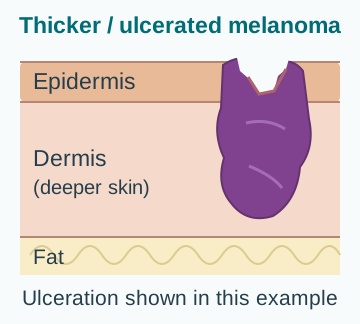
Stage 2 (II)
Melanoma is thicker or has a combination of thickness and ulceration that raises risk, but there is still no detected regional or distant spread. Stage 2 does not mean lymph nodes contain melanoma.
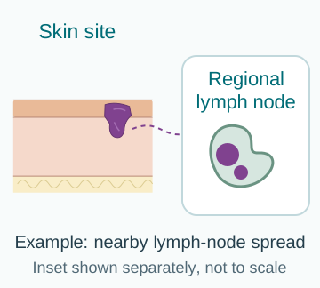
Stage 3 (III)
Melanoma has reached regional lymph nodes or nearby deposits in the skin or lymphatic channels. These deposits may be called satellite, microsatellite or in-transit disease. There is no distant spread.
Stage 4 (IV)
Melanoma has spread beyond the regional area, for example to distant skin or nodes, lungs, liver, bone or brain.
Thickness and ulceration matter: stages 1 and 2 depend on the measured thickness and whether the tumour surface has broken down, not simply which skin layer is reached. A stage 2 melanoma does not always have ulceration.
A sentinel-node biopsy can find tiny deposits in nearby lymph nodes that scans cannot show. Some high-risk stage 2 melanomas and stage 3 melanomas with only small deposits can have similar chances of returning. The team considers the stage, the cancer’s features and your health when discussing further treatment.
Clinical stage is based on examination, biopsies and scans. Pathological stage adds information from tissue examined after surgery. The letters yp before a stage mean the tissue was assessed after treatment given before surgery. More information may refine the stage; this does not necessarily mean the cancer grew while you were waiting.
How your treatment plan is chosen
Your stage, melanoma type, symptoms, BRAF result, general health and priorities all matter. Ask the team to explain the purpose of each treatment.
Situation | Usual discussion |
|---|---|
Stage 0 to IIA | Surgery, selected sentinel node assessment and follow-up. Drug treatment is not routine for everyone. |
Stage IIB or IIC | Surgery followed by a discussion about immunotherapy or regular follow-up checks (surveillance). |
Stage III that can be removed by surgery | A specialist team considers treatment before surgery, surgery and treatment afterwards. |
Cancer that cannot be fully removed by surgery, or stage IV | Medicines that travel around the body (systemic treatment), with surgery, radiotherapy (radiation treatment) or other treatment of specific sites when appropriate. |
Resectable means the cancer can be removed by surgery; unresectable means it cannot be fully removed. A multidisciplinary team brings together doctors specialising in skin, surgery, cancer medicines, scans and tissue diagnosis. Nurses, pharmacists and teams supporting recovery and symptom relief help you manage treatment.
Ask whether the aim is cure, lowering the risk of cancer returning (recurrence), long-term control or symptom relief. Ask for the expected benefit in people like you, the main risks, alternatives and the consequences of waiting or choosing surveillance.
Treatment options
Topics in this section
Surgery for skin melanoma
Wide local excision removes the biopsy site and an appropriate rim of normal-looking skin. The required margin depends mainly on Breslow thickness.
Melanoma thickness | Usual rim of skin removed |
|---|---|
In situ | 0.5–1 centimetre (cm) |
Up to 1 mm | 1 cm |
More than 1 mm to 2 mm | 1–2 cm |
More than 2 mm | 2 cm |
These are usual starting points, not instructions for an individual operation. The surgeon adapts the plan to the site, pathology and function. Some melanomas in situ, especially on the face, may be removed in steps, with the tissue edges checked to make sure the melanoma has been removed.
The wound may close with stitches. A larger wound may need a skin graft, a patch of skin taken from elsewhere, or a flap, nearby tissue moved with its blood supply. Ask about the scar, pain relief, dressings, time off work, movement and when it is safe to drive or exercise. Surgery on a sole or nail needs particular attention to walking and hand function.
If lymph nodes are involved
Removal of affected nodes may be appropriate. When melanoma can be removed by surgery and the involved nodes can be found on examination or scans, the team should discuss treatment before surgery. This differs from tiny deposits found only on sentinel node biopsy.
Treatment after surgery
Adjuvant treatment is given after all visible melanoma has been removed. Its aim is to reduce the chance of recurrence, not to treat a visible tumour.
Higher-risk stage II
For completely removed stage IIB or IIC melanoma, pembrolizumab or nivolumab may reduce recurrence risk. Surveillance is also a reasonable choice for some people. The team should explain your chance of recurrence without additional medicine, how much treatment may lower that chance, and the possibility of lasting side effects from the immune system attacking healthy organs.
Stage III and selected stage IV
Immunotherapy that blocks programmed cell death protein 1 (PD-1) is often considered after surgery has removed all visible cancer. PD-1 acts as a brake on immune cells; blocking it helps them attack cancer. For stage III melanoma with a suitable BRAF V600 mutation, dabrafenib plus trametinib is an alternative. The precise evidence and licensing conditions differ between medicines and settings.
Adjuvant treatment commonly lasts about one year, but the plan may change with side effects, recurrence or the response to treatment already given before surgery. Stage IIIA with only a small amount of cancer in the lymph nodes needs an individual discussion; not everyone benefits enough to justify the risks.
Understanding the trade-off
Some people are already cured by surgery and their cancer would not have returned without medicine. Treatment can lower risk, but cannot eliminate it. Reducing recurrence is not the same as proving that everyone will live longer.
Treatment before surgery
For some resectable stage III melanomas, immunotherapy before surgery is now an important option to discuss with the melanoma specialist team.
Neoadjuvant treatment is given while the tumour is still present. It can treat disease throughout the body and show how the melanoma responds. It is mainly considered when affected nodes or other nearby deposits can be found on examination or scans, not for every early melanoma or tiny sentinel node deposit.
What the evidence supports
The NADINA trial supports nivolumab plus ipilimumab before surgery, followed by treatment guided by how much cancer remains in the tissue removed at surgery, in selected resectable stage III disease. SWOG S1801 supports pembrolizumab before and after surgery in selected resectable stage III or IV disease.
Compared with the surgery-first approaches tested, these studies found a lower risk of events such as cancer progression, recurrence or death. Combination immunotherapy can cause serious side effects. Your surgeon and medical oncologist, the doctor who plans cancer medicines, should agree on the schedule, checks and backup plan together.
What happens next
The team reassesses the cancer, performs the planned operation and examines the removed tissue. The amount of melanoma cells still alive in the removed tissue helps guide further treatment. A good scan response does not automatically mean surgery can be omitted.
Advanced melanoma immunotherapy
Checkpoint immunotherapy releases brakes on immune cells, helping your immune system recognise and attack melanoma. It can work whether or not the tumour has a BRAF mutation.
Option | Main consideration |
|---|---|
Pembrolizumab or nivolumab | A single PD-1-blocking medicine. Often causes fewer serious side effects than combination immunotherapy. |
Nivolumab plus ipilimumab | A combination that can give lasting control, including in selected people whose melanoma has spread to the brain, but with a higher risk of serious immune side effects. |
Nivolumab plus relatlimab | Another combination, supplied as Opdualag. Discuss its benefits, risks and treatment-specific eligibility. |
The choice depends on how much cancer there is, how quickly it is growing, whether it has reached the brain, previous treatment and your ability to tolerate side effects. Transplant recipients and people with significant autoimmune disease need particularly careful advice.
Treatment is usually given at regular hospital visits. Scans and symptoms help assess response. Some responses last after treatment stops, but not everyone responds and no medicine guarantees a cure.
The planned duration varies. Under-the-skin versions of some checkpoint medicines can shorten administration in certain settings. They do not remove immune side-effect risks and are not interchangeable with every combination given through a drip into a vein (intravenously).
BRAF-targeted treatment
BRAF and mitogen-activated protein kinase kinase (MEK) are proteins in a pathway that signals cells to grow. BRAF–MEK inhibitors are medicines that block these signals. They are used only when testing confirms a suitable BRAF V600 mutation.
BRAF medicine | Usual MEK partner |
|---|---|
Dabrafenib | Trametinib |
Encorafenib | Binimetinib |
Vemurafenib | Cobimetinib |
These tablets are generally used in pairs to improve control and reduce some side effects. They can shrink advanced melanoma quickly, but cancer can become resistant, meaning the medicines stop controlling it. Regular assessment is essential.
Which comes first
Immunotherapy is often considered first in suitable patients for its potential for lasting benefit. Targeted treatment can be useful when rapid control is needed, immunotherapy is unsuitable, or disease progresses after immunotherapy.
Selected patients may be offered atezolizumab with vemurafenib and cobimetinib, or a clinical trial. This three-drug approach is not automatically better for every patient and can add side effects.
Taking tablets safely
Follow the exact food, timing and missed-dose instructions for your medicines. Ask whether other medicines, supplements or foods could affect how your treatment works. Do not double doses. Regular blood tests, skin checks and sometimes heart and eye assessment are needed.
If the first treatment stops working
Progression means the cancer is growing or spreading despite treatment. It does not mean that every option has been exhausted. The next choice depends on what you have already received and why it stopped.
The team may review the biopsy, repeat selected tests for gene changes and reassess all disease sites. A different immunotherapy medicine or combination, BRAF–MEK treatment, a local treatment, a clinical trial or symptom-focused care may be appropriate. Chemotherapy uses medicines that kill rapidly dividing cells. It has a more limited role when better-supported options are unsuitable or have stopped working.
Tumour-infiltrating lymphocyte therapy
Tumour-infiltrating lymphocytes are immune cells that have entered a tumour. Lifileucel (Amtagvi) uses these cells: they are collected from a removed tumour sample, multiplied in a laboratory and given back through a drip after preparatory chemotherapy.
Here, chemotherapy prepares the body to receive the immune cells. Interleukin-2, a protein that helps immune cells grow and work, is also given as part of the programme.
Lifileucel is an option for adults with unresectable or metastatic melanoma previously treated with a PD-1 blocker and, if the tumour has a BRAF V600 mutation, a BRAF inhibitor with or without a MEK inhibitor.
It requires a specialist inpatient centre, time for cell manufacture and careful fitness assessment. Serious risks include prolonged low blood counts, infections, bleeding, heart, lung or kidney problems and treatment-related death. It is not suitable for everyone.
Medicines injected into tumours
Some melanoma deposits can be treated directly. A specialist must be able to reach the melanoma deposits safely with an injection, sometimes using scans to guide the needle.
Talimogene laherparepvec
Talimogene laherparepvec (T-VEC, Imlygic) uses a modified herpes virus for selected injectable skin, under-skin or lymph-node deposits. It is unsuitable in pregnancy or with a weakened immune system. It does not replace whole-body treatment for widespread disease.
Tudriqev with nivolumab
Vusolimogene oderparepvec-wtpg (Tudriqev, previously called RP1) with nivolumab is an option for adults with advanced skin (cutaneous) melanoma that cannot be removed by surgery and has grown or spread despite treatment based on a PD-1-blocking antibody.
Tudriqev is injected into tumours; nivolumab is given intravenously. The approval was based on tumour response and its duration. A confirmatory study must verify clinical benefit. This is a later-line option, not a routine replacement for initial treatment or treatment of eye melanoma.
Care at home
Follow dressing, handling and contact precautions carefully. Modified herpes-virus treatments can cause accidental exposure and infection with the herpes virus. Injection procedures and accompanying immunotherapy have additional risks. Report fever, worsening pain, spreading redness or new blisters.
Radiotherapy and brain spread
Local treatment can control a troublesome area even when systemic medicines are also needed.
When radiotherapy may help
Radiotherapy uses focused radiation to treat cancer. It may relieve pain, bleeding or pressure, control selected brain or other metastases, or reduce the risk of local recurrence in particular high-risk situations. It is not routinely needed after every melanoma operation.
The team chooses the technique and number of visits according to the site and goal. Side effects depend on the treated area and may include fatigue, skin changes or temporary swelling. External-beam radiotherapy does not make you radioactive.
If melanoma has reached the brain
A brain MRI helps plan care. Options may include precisely focused radiation (stereotactic radiotherapy), surgery, immunotherapy or BRAF–MEK treatment if the mutation is present. The number, size and location of deposits, symptoms and need for steroids all affect the choice.
Nivolumab plus ipilimumab can be useful in selected patients, particularly when melanoma in the brain is not causing symptoms. Symptoms, swelling or a large cancer deposit may need urgent treatment directed at that area. Do not stop prescribed steroids suddenly; dosing should be coordinated by your team.
For melanoma limited to a limb, specialist regional treatments may occasionally be discussed. Their role depends on specialist expertise, where the cancer is and which whole-body treatments are available.
Acral, mucosal and eye melanoma
These less common melanomas need specialist review. Evidence from common sun-related skin melanoma does not always apply in the same way.
Acral melanoma
Melanoma of the palms, soles and nails uses the skin-melanoma staging system. Surgery aims for cancer control while preserving function. Immunotherapy and, when relevant, mutation-directed treatment may be used. BRAF mutations are less common than in many sun-related melanomas.
Mucosal melanoma
This begins in moist internal linings, such as the nose, mouth, genital area, anus or rectum. Surgery, radiotherapy and whole-body treatment are planned for the individual site.
Tests for gene changes may identify a change that makes the KIT growth-signalling protein overactive. A medicine that blocks KIT may help selected patients. This is often an off-label use, meaning it is outside the medicine’s approved licence; it is not routine treatment for all mucosal melanoma.
Uveal melanoma
This starts inside the eye and is biologically different from skin melanoma. An eye-cancer specialist team may use radiation treatment to the eye or surgery. Follow-up often focuses on the liver as well as the eye.
Tebentafusp (Kimmtrak) is an option for adults whose uveal melanoma cannot be removed by surgery or has spread, if they have a particular human leukocyte antigen (HLA) type called HLA-A*02:01. HLA proteins help the immune system recognise cells. A blood test checks this inherited type.
Treatment needs careful monitoring, especially early on, for cytokine-release reactions. These are strong immune reactions that can cause fever, low blood pressure and other serious symptoms.
Managing side effects
When to seek urgent help
Immunotherapy can inflame healthy organs. Symptoms may start during treatment or months after it finishes. Early reporting can prevent a mild problem becoming serious.
Get emergency help now | Contact the oncology team the same day |
|---|---|
Severe breathing difficulty, chest pain, collapse or marked confusion. | New cough or breathlessness; persistent diarrhoea, blood in stools or abdominal pain. |
A seizure, new weakness, severe headache or trouble speaking. | Yellow eyes, dark urine, much less urine, severe fatigue, dizziness, marked thirst or frequent urination. |
A blistering/peeling rash, a rapidly worsening illness, or inability to keep fluids down. | New rash, eye pain or blurred vision, unusual muscle weakness, fever of 38°C (100.4°F) or above, or shaking chills. |
Use your own team’s fever threshold if it is more cautious. Do not wait for several symptoms to appear. If you cannot reach the team and you feel unwell, use the nearest emergency department.
Tell the emergency clinician the name of your cancer treatment and when you last received it. Bring your alert card and medicine list. A problem may still be treatment-related even if a scan shows the cancer is improving.
Managing everyday side effects
Tell the team early about symptoms that affect eating, sleep, movement or daily life. Treatment can often be adjusted safely.
Problem | Practical first steps |
|---|---|
Tiredness | Pace activity and rest, keep gentle movement, and report a sudden change. Tests may be needed for anaemia (too few healthy red blood cells) or problems with the thyroid gland or other hormone-producing organs. |
Nausea or poor appetite | Use prescribed anti-sickness medicines, eat small frequent meals and ask a dietitian, a food and nutrition specialist, for help if weight is falling. |
Dry or itchy skin | Use a simple moisturiser and gentle washing. Ask before using new creams; report a spreading or blistering rash. |
Swelling after node surgery | Seek lymphoedema assessment. Exercise and properly fitted pressure garments may help when the team advises them. |
Extra checks with targeted tablets
Fever, diarrhoea, rash and sun sensitivity are common. New visual problems, chest symptoms, fainting or unusual breathlessness need urgent assessment. Blood tests and heart checks may be needed even when you feel well.
Keep a simple record
Record symptoms, temperature, bowel changes and missed doses. Keep all prescribed and non-prescribed medicines on one list. Check interactions before taking herbal products, high-dose vitamins or new medicines.
Live well
Follow-up after treatment
Follow-up checks for recurrence, new skin cancers and longer-term effects of treatment. The schedule should match your risk and treatment history.
Your situation | What follow-up often involves |
|---|---|
Lower-risk disease | Skin and lymph-node examinations. Routine scans are often unnecessary without symptoms or concerning findings. |
Higher-risk disease | Closer visits and selected body/brain imaging, especially in the first few years. |
Positive sentinel node without further node surgery | Planned ultrasound assessment of the relevant node region, where appropriate. |
Receiving systemic treatment | Symptom review, blood tests and scans at agreed intervals to assess benefit and safety. |
Ask who coordinates your follow-up and who checks the whole skin. A lifelong habit of skin awareness is useful because another melanoma can develop even after the first has been cured.
Symptoms to report between appointments
Tell the team about a new lump near the scar or in a node region, a persistent cough, unexplained weight loss, ongoing pain, headaches or other persistent new symptoms. Most symptoms have other causes, but checking is safer than waiting.
Skin checks and sun protection
Regular self-checks help you notice change. They complement, but do not replace, professional skin examinations.
A monthly head-to-toe check
- Use good light and a mirror. Ask someone to help with your scalp, back and other hard-to-see areas.
- Check palms, soles, between toes, all nails, and around the operation scar. Ask the team how to check the relevant lymph-node region.
- Compare with earlier photographs where helpful. Report a new or changing spot promptly rather than repeatedly photographing it without seeking advice.
Protect skin of every colour
- Use shade, protective clothing, a broad-brimmed hat and sunglasses. Avoid tanning beds and deliberate tanning.
- Use water-resistant sunscreen with a sun protection factor (SPF) of 50+ on exposed skin. Choose broad-spectrum protection, which covers both main types of ultraviolet rays. Apply generously and reapply every two hours outdoors, and after swimming, sweating or towel drying.
- Sunscreen does not make prolonged sun exposure safe. Be especially careful if treatment increases sun sensitivity.
You do not need deliberate sun exposure for vitamin D. Ask whether diet, a supplement or a blood test is appropriate. Family members may need individual advice about regular skin checks if there is a strong family history.
Living with melanoma
Good supportive care is part of melanoma treatment. It can help with physical symptoms, emotional strain and everyday practical problems.
Food, activity and recovery
Aim for a balanced diet with enough protein and fluids for your needs. Keep moving within your energy and surgical restrictions. A physiotherapist can guide exercises and movement after limb or node surgery. Avoid smoking, and discuss alcohol with your team.
Emotions, relationships and work
Worry before scans, changes in body image and fear of recurrence are common. Tell someone if anxiety, low mood or poor sleep is affecting your life. Counselling, peer support and practical adjustments at home or work can help. Caregivers also need breaks and support.
Sexual health and fertility
If having children in the future matters to you, ask before treatment about ways to protect your fertility. Follow the advice about preventing pregnancy (contraception) for your specific medicines, including after the last dose. Pregnancy and breastfeeding require a specialist discussion; do not assume an oral or immune treatment is safe.
Palliative care and future planning
Palliative care treats pain and other symptoms, and supports you and your family. It can be provided alongside anticancer treatment at any stage. If disease becomes difficult to control, discuss what matters most to you and the care you would want.
Sources and further reading
For more information, explore these resources from the National Cancer Institute (NCI) and other medical organisations. The linked organisations have not reviewed or endorsed this CTI guide. Your care team can explain how the information applies to you.
- NCI: Melanoma Treatment (PDQ®)–Patient Version
- Cancer Council Australia: melanoma
- Macmillan: melanoma
- ESMO: cutaneous melanoma clinical practice guideline (2025)
- NADINA: immunotherapy before surgery in stage III melanoma
- SWOG S1801: pembrolizumab before and after surgery
- FDA: Tudriqev with nivolumab — approval and prescribing information
- FDA: HEPZATO KIT prescribing information (2026)
Staging and risk-group references
AJCC 8th edition.
Medicine approval and prescribing references
These official records describe licensed uses and prescribing information for the medicines named in this guide. Approval depends on the cancer type, biomarkers, previous treatment, combination and formulation. Some uses discussed in the guide are off-label: outside the medicine’s licence, with evidence from clinical guidelines or studies. FDA or EMA approval does not establish approval or availability in India.
- Atezolizumab — FDA prescribing information · Approval history
- Binimetinib — FDA prescribing information · Approval history
- Cobimetinib — FDA prescribing information · Approval history
- Dabrafenib — FDA prescribing information · Approval history
- Encorafenib — FDA prescribing information · Approval history
- Ipilimumab — FDA prescribing information · Approval history
- Lifileucel — FDA approval record
- Nivolumab — FDA prescribing information · Approval history
- Pembrolizumab — FDA prescribing information · Approval history
- Relatlimab with nivolumab — FDA prescribing information · Approval history
- Talimogene laherparepvec — FDA approval record
- Tebentafusp — FDA prescribing information · Approval history
- Trametinib — FDA prescribing information · Approval history
- Vemurafenib — FDA prescribing information · Approval history
Additional medicine approval notices
These records explain the specific medicine uses and eligibility requirements discussed in this guide.
Related care
Meet our doctors



Where to get care