
Share
Start here
Liver cancer at a glance
- Hepatocellular carcinoma (HCC) starts in the main working cells of the liver, called hepatocytes. It often develops in a liver affected by long-term inflammation (chronic hepatitis), severe scarring (cirrhosis) or fatty liver disease linked to conditions such as diabetes.
- Scans can sometimes confirm HCC without a biopsy, which means taking a small tissue sample for testing. Computed tomography (CT) and magnetic resonance imaging (MRI) produce detailed pictures. A multiphasic liver scan takes pictures at different times after an injection of contrast dye to look for the pattern typical of HCC.
- Stage describes how much cancer is present and where it is. It is only part of the treatment decision. The team also checks how well your liver is working, the pressure in the vein carrying blood to the liver, your overall health, whether surgery can remove the cancer, whether a transplant is possible and your goals.
- Early HCC may be curable. Options include surgery to remove the affected part of the liver (resection), replacing the liver with a donated liver (transplant), or destroying the tumour in place (ablation). Treatment with curative intent means treatment aimed at a cure.
- Liver-directed treatment targets cancer within the liver. It includes ablation, blocking the tumour’s blood supply (embolisation), delivering radiation through the liver’s arteries (radioembolisation), and focused radiation from outside the body.
- Systemic treatment uses medicines that travel through the body. Immunotherapy helps the immune system attack cancer; targeted drugs block signals that help cancer grow. The choice depends on liver function, bleeding risk, other illnesses, any previous transplant and your preferences.
- Follow-up matters even when treatment appears to have removed or destroyed all the cancer. HCC can return, or a new cancer can develop in the remaining liver.
Bring your scan images
For your consultation or a second opinion, bring a CD containing your scan images (DICOM format) and the software needed to view them, along with the written scan reports. DICOM stands for Digital Imaging and Communications in Medicine, the standard format used for medical scan files.
Understand
What your liver does
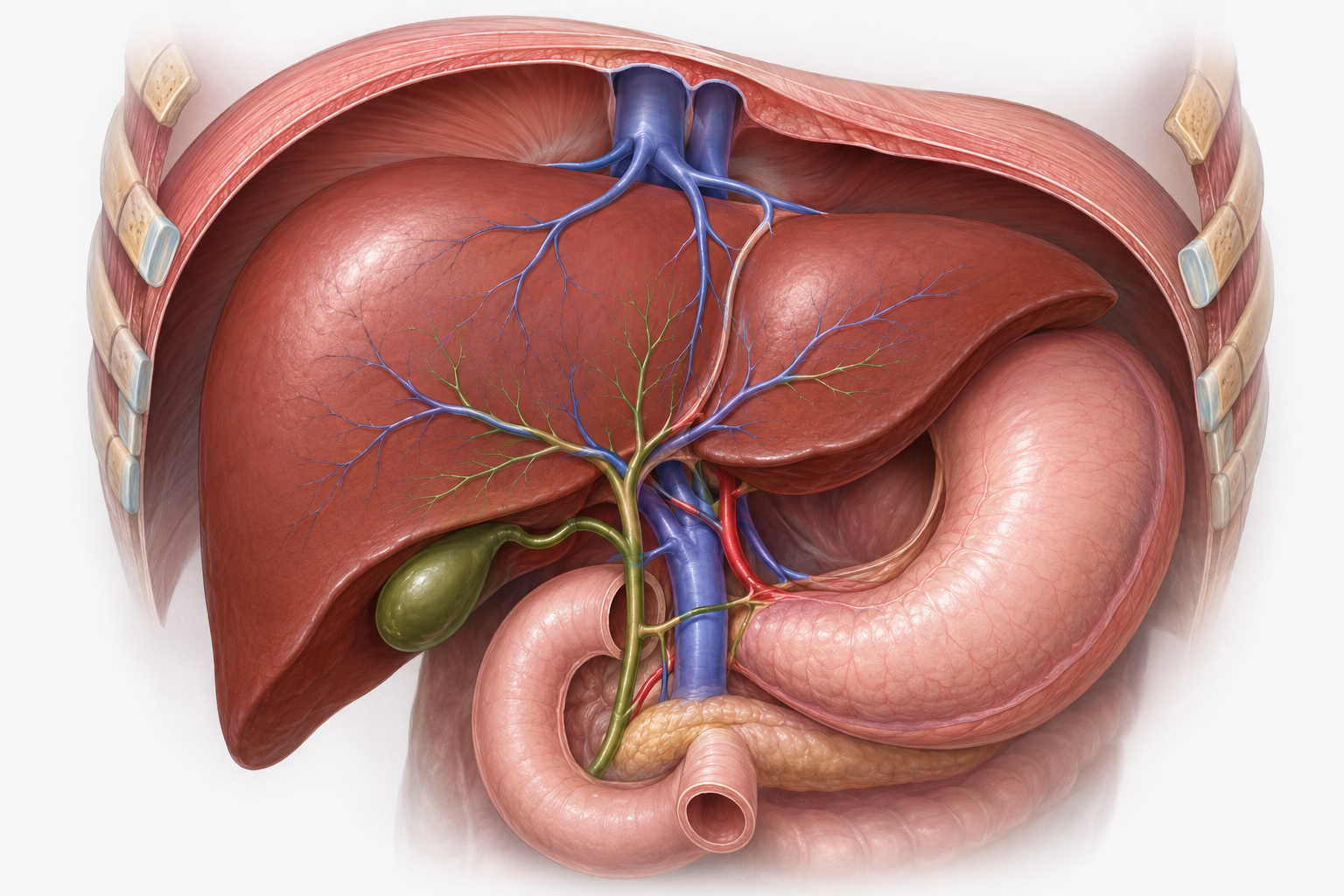
The liver sits under the right ribs. It processes nutrients and medicines, removes waste from the blood, makes proteins that help blood clot, and produces bile to help digest fat. It can keep working despite damage for a long time, so both liver disease and cancer may cause no symptoms at first.
Why this changes cancer care
- Surgery must leave enough functioning liver behind.
- Embolisation or radiation can injure healthy liver as well as the tumour. The team carefully plans the amount of treatment and the area it reaches.
- Many medicines are processed by the liver; dosing and side effects may change as liver function changes.
- Cirrhosis can raise the pressure in the portal vein, which carries blood to the liver. This is called portal hypertension. It can cause a low platelet count (fewer blood cells that help clotting), fluid in the abdomen (ascites) and enlarged veins (varices) in the lower part of the food pipe (oesophagus) that may bleed.
Which type of liver cancer is this?
A mass in the liver is not automatically HCC. The place where a cancer started determines what it is and how it is treated.
Hepatocellular carcinoma
The commonest primary liver cancer in adults. It starts in hepatocytes and is the focus of this guide.
Intrahepatic cholangiocarcinoma
Starts in the bile ducts, the tubes carrying bile inside the liver. It needs its own treatment plan, examination of tissue (pathology) and sometimes tests for changes in cancer genes or proteins (molecular testing).
Fibrolamellar carcinoma
A rare liver cancer, often in younger people without cirrhosis. Management should involve an experienced specialist centre.
Combined HCC-cholangiocarcinoma
Contains features of both HCC and bile duct cancer. A biopsy and review by a specialist who examines tissue are usually important.
Cancer metastatic to the liver
Colon, breast, lung and other cancers can spread to the liver. They remain cancers of their original organ.
Benign liver lesions
Fluid-filled sacs (cysts), growths made of blood vessels (haemangiomas) and certain liver-cell growths (adenomas) are not cancer. Scans usually help identify them; uncertain findings may need further assessment.
Why the distinction changes care
- Scan findings and tumour markers, substances measured in blood that may help assess cancer, mean different things in HCC and bile duct cancer.
- The value of biopsy, molecular testing, surgery, transplantation and drug treatment depends on the exact diagnosis.
- If the diagnosis is uncertain, specialists can review the scans and tissue samples together before treatment is chosen.
Symptoms and warning signs
Early HCC often causes no symptoms. New, persistent or worsening symptoms should still be assessed, especially in someone with chronic liver disease.
Possible symptoms
- Dull pain, pressure or fullness under the right ribs; a new abdominal lump.
- Reduced appetite, unexplained weight loss, nausea or feeling full quickly.
- Tiredness, weakness, fever or a general decline without a clear cause.
- Jaundice, dark urine, pale stools or troublesome itching.
- Abdominal swelling from ascites or swelling of the feet and legs.
Symptoms may come from the tumour, cirrhosis, portal hypertension, infection or another condition. A normal-feeling day does not prove that the cancer is stable, and a bad day does not always mean that it has grown.
Risk factors
Usually there is no single answer. Risk factors increase probability; they do not prove cause, and HCC is not a patient's fault.
Chronic viral hepatitis
Hepatitis B can cause HCC with or without cirrhosis. Hepatitis C raises risk mainly when it has caused substantial liver scarring (advanced fibrosis or cirrhosis).
Cirrhosis
Severe liver scarring is a major risk factor. Causes include viral hepatitis, alcohol, fatty liver disease and autoimmune disease, in which the immune system attacks healthy liver cells.
Metabolic liver disease
Fat build-up linked to conditions such as obesity and type 2 diabetes is called metabolic dysfunction-associated steatotic liver disease (MASLD). When it also causes liver inflammation and injury, it is called metabolic dysfunction-associated steatohepatitis (MASH). Risk rises particularly when significant scarring develops.
Alcohol and tobacco
Long-term heavy alcohol use can cause cirrhosis. Smoking adds a smaller but real risk.
Aflatoxin exposure
Toxins from mould in poorly stored grains or nuts can increase risk, especially with hepatitis B.
Rare conditions
Inherited conditions that damage the liver can raise risk. Examples include haemochromatosis (too much iron), Wilson disease (copper build-up) and alpha-1 antitrypsin deficiency (a problem with a protective protein).
Lowering future risk
- Hepatitis B vaccination; testing and treatment for hepatitis B or C when indicated.
- Avoid alcohol when the liver is damaged; stop tobacco; work gradually on weight, diabetes and blood pressure.
- Some moulds on poorly stored grains and nuts produce harmful substances called aflatoxins, which can increase liver cancer risk. Keep these foods in a cool, dry place and throw away any that look mouldy, are discoloured or smell unusual.
- Attend the surveillance schedule recommended by the liver team.
Surveillance before cancer is diagnosed
Surveillance means regular checks to find HCC early in people at increased risk, even when they feel well. It is not a one-off 'liver cancer test' for everyone.
Who may need it
- Most adults with cirrhosis who could benefit from HCC treatment.
- Selected people with chronic hepatitis B even without cirrhosis, based on age, origin, family history, viral activity and other risk features.
- People previously treated for HCC, using a follow-up schedule rather than general surveillance.
What happens after an abnormal result
- The team checks how clearly the liver was seen, the size of any abnormal area and whether it has changed, and the AFP blood results over time.
- A new abnormal area may need repeat ultrasound or a multiphasic CT or MRI scan, depending on its size and the other findings.
- An abnormal AFP or ultrasound is not itself a cancer diagnosis.
Tests and diagnosis
Your care team
HCC care works best when liver and cancer specialists review your results together. This is called a multidisciplinary team (MDT). A hepatobiliary team specialises in the liver, bile ducts and gallbladder.
Team member | What they add |
|---|---|
Hepatologist / gastroenterologist | A liver or digestive-system specialist who treats cirrhosis, portal hypertension, hepatitis and worsening liver function. |
Hepatobiliary / transplant surgeon | Assesses surgery, how much healthy liver would remain, whether transplant is suitable and possible donor options. |
Interventional radiologist | Uses scans to guide procedures such as biopsy, ablation, embolisation and radioembolisation. |
Diagnostic radiologist | Interprets scans, checks blood vessels and assesses changes after treatment. |
Medical oncologist | Plans cancer medicines, their order and whether a clinical trial may suit you. Trials are research studies of treatments or care. |
Radiation oncologist | Plans focused radiation from a machine outside the body, when suitable. |
Pathologist | Examines tissue to identify the cancer type and features that help guide care, including how the cells look and whether cancer reaches the edges of removed tissue. |
Support team | Nurses, pharmacists, dietitians, physiotherapists and specialists in symptom relief, emotional wellbeing and practical support. |
How cancer is diagnosed
You are unlikely to need every possible test. The sequence below shows the common logic.
A nodule is found
A nodule is a small lump or spot, often found on ultrasound or another scan.
Multiphasic CT or MRI
Shows how the abnormal area takes up contrast dye and whether nearby blood vessels are involved.
Diagnosis is confirmed
Imaging may be sufficient; some people need a tissue sample.
Treatment planning
Combines cancer stage, how well the liver works (liver reserve), your overall health, the options available and your goals.
Four questions the team is answering
- Is this HCC? A typical scan pattern may confirm it in a person at high risk. Otherwise, a biopsy may be needed.
- Where is it? The team checks the number, size and location of tumours, nearby blood vessels and bile ducts, and any spread to lymph nodes or other organs.
- How well is the liver working? Blood results, portal hypertension, ascites, encephalopathy and the amount of healthy liver all matter.
- Which treatment is suitable? The team first considers whether a cure is possible, then discusses other ways to control cancer and symptoms in line with your goals.
Scans and staging tests
Test | Main question | Important limits / preparation |
|---|---|---|
Ultrasound | Is there a new nodule, fluid or change in an at-risk liver? | How clearly the liver can be seen varies. An abnormal finding may need further scans. |
Multiphasic CT | Does the abnormal area brighten soon after contrast dye is given, then look darker than the surrounding liver later (washout)? Are blood vessels involved? | Uses x-rays and contrast dye containing iodine. Tell the team about kidney problems or previous reactions to contrast. |
Contrast MRI | Shows small or uncertain abnormal areas and the surrounding liver in detail. | Takes longer than CT. Tell staff about implants or metal in your body, kidney problems or fear of enclosed spaces (claustrophobia). |
Chest CT | Has cancer spread to the lungs or chest nodes? | May find unrelated small nodules that need observation rather than treatment. |
Positron emission tomography with CT (PET-CT) / bone scan | Selected questions about spread or symptoms. | These tests use a small amount of radioactive tracer. They are not routine for everyone: some HCCs do not show clearly on PET-CT, and a bright area is not always cancer. |
Blood tests and liver reserve
Blood tests help assess liver and kidney function, clotting and tumour markers. They help your team plan treatment safely, but cannot show the full cancer stage on their own.
Test group | What it helps answer |
|---|---|
AFP | A tumour marker that may support assessment and follow-up. Normal AFP does not exclude HCC; a high AFP is not proof by itself. |
PIVKA-II (DCP) | PIVKA-II means protein induced by vitamin K absence or antagonist-II. It is also called des-gamma-carboxy prothrombin (DCP). Some centres use this blood marker alongside AFP to help assess the likely course of HCC or follow treatment. It cannot diagnose HCC alone; low vitamin K or medicines such as warfarin can also raise it. |
Bilirubin, albumin, enzymes | Bilirubin is a waste pigment processed by the liver; albumin is a protein the liver makes. These help assess liver function. Liver enzymes may rise because of inflammation, medicines, cancer or a blocked bile duct. |
International normalised ratio (INR) / prothrombin time | Measures how quickly blood clots and helps assess the liver’s production of clotting proteins. Blood-thinning medicines affect the result. |
Complete blood count (CBC) and platelets | Checks red blood cells, white blood cells and platelets. Low red cells (anaemia) can cause tiredness or breathlessness; other results help assess infection and bleeding risks. |
Creatinine, sodium and electrolytes | Checks kidney function and blood salts. Results guide medicines, fluid management, contrast scans and transplant assessment. |
Hepatitis B virus (HBV) / hepatitis C virus (HCV) tests | Checks for current or past infection and, when needed, the amount of virus in blood. Results guide treatment and prevention of the virus becoming active again. |
How liver reserve is described (4 useful views)
Liver reserve means how well your liver can carry out its normal jobs. It matters alongside the cancer stage because it affects which treatments your liver can safely tolerate. Your team uses the following assessments to help plan your care.
- Child–Pugh score. Combines three blood results (bilirubin, albumin and INR, a blood-clotting measure) with assessments of fluid in the abdomen (ascites) and changes in brain function caused by liver disease (hepatic encephalopathy).Use the Child–Pugh calculator
- Albumin–bilirubin (ALBI) score. Uses the albumin and bilirubin blood results to assess liver function.Use the ALBI calculator
- Model for End-stage Liver Disease (MELD) score. Uses blood test results to assess how severe liver disease is and helps guide priority for a liver transplant.
- Portal hypertension. This means high pressure in the vein carrying blood to the liver. Your team assesses it using your medical history, platelet count, spleen size, scans, endoscopy (a camera test) and sometimes direct pressure measurements.
Biopsy and your pathology report
HCC can sometimes be diagnosed confidently on scans without a biopsy. If a biopsy is recommended, ask how the result could change your care, how bleeding risk is checked and when you will receive the result.
When imaging may be enough
In a person with cirrhosis or another recognised high-risk setting, a lesion with a characteristic multiphasic CT/MRI pattern may meet imaging criteria for HCC. Avoiding biopsy can reduce delay and procedure risk.
When tissue may add value
- Scans do not give a clear answer, or the person does not have the liver conditions in which scans alone can reliably diagnose HCC.
- Cholangiocarcinoma, combined HCC-cholangiocarcinoma, metastasis or another tumour is possible.
- A clinical trial or selected molecular testing requires tissue.
- The result would change the treatment plan.
After surgery, the pathology report describes the tumour and the removed tissue. Grade describes how abnormal the cells look. Vascular invasion means cancer is in blood vessels; margins are the edges of the removed tissue.
The report may also describe a capsule around the tumour or nearby small tumours (satellite nodules). These findings help estimate the chance of cancer returning, but do not automatically mean extra drug treatment is needed.
Planning your treatment
Understanding your stage
HCC staging: TNM and BCLC
Doctors use two main systems. TNM describes the main tumour (T), nearby lymph nodes (N) and spread to other organs, called metastasis (M). Its stages run from 1 to 4. Barcelona Clinic Liver Cancer (BCLC) staging also considers liver function and daily activity. BCLC letters do not translate directly into TNM numbers.
Lymph nodes are small immune-system structures. Regional nodes are those near or draining the cancer’s starting area; distant spread means cancer has reached a site outside that regional area. Stages describe findings, not a sequence that everyone will pass through.
Stage 1 (I), TNM
A single liver tumour, with no regional nodes or distant spread. IA is 2 cm or smaller; IB is larger than 2 cm without blood-vessel invasion.
Stage 2 (II), TNM
A single tumour larger than 2 cm invades blood vessels, or there are several tumours with none larger than 5 cm. No regional nodes or distant spread.
Stage 3 (III), TNM
There are several tumours with at least one larger than 5 cm, or cancer involves a major portal/hepatic vein branch, grows through the liver surface or directly into an adjacent organ other than the gallbladder. No regional nodes or distant spread.
Stage 4 (IV), TNM
IVA means regional lymph-node involvement. IVB means distant metastases. These are different situations.
What the BCLC letters mean
Barcelona Clinic Liver Cancer (BCLC) staging brings together three parts of your health.
BCLC 0: very early
One tumour no larger than 2 cm, preserved liver function and no cancer-related limitation in daily activity.
BCLC A: early
One tumour, or up to three with none larger than 3 cm; no major-vessel invasion or spread outside the liver, with preserved liver function and fitness.
BCLC B: intermediate
Multiple tumours beyond the early-stage pattern, but no major-vessel invasion or spread outside the liver. Liver function and fitness are sufficiently preserved.
BCLC C: advanced
Major-vessel invasion or spread outside the liver in someone with adequate liver function and fitness for treatment.
BCLC D: severely impaired liver function or fitness
Severe cancer-related disability or poor liver function, without a transplant option. Care focuses on comfort and individual priorities.
Simplified diagram.
CTI illustration.
Anatomical stage alone cannot decide whether surgery, transplantation or drug treatment is safe. Severe liver disease can limit treatment even when the tumour is small. BCLC stage D is not simply another name for metastatic cancer.
Clinical stage comes from examination, biopsies and scans. Pathological stage can add information from surgery; a prefix such as yp means the tissue was assessed after treatment. A revised assessment does not necessarily mean the cancer grew while you were waiting. Grade describes the cells’ behaviour and is different from stage.
Five factors shape the treatment plan
For HCC, a stage label alone is never enough.
Tumour extent
The number, size and location of tumours, growth into blood vessels, and spread to lymph nodes or other organs.
Liver reserve
Child–Pugh or ALBI results, complications of liver disease and how much healthy liver would remain after treatment.
Overall fitness
Ability to manage daily activities (performance status), strength, heart and kidney health, infection risk and nutrition.
Treatment feasibility
What can be delivered safely now at an experienced centre, including donor and technology access.
Your priorities
Chance of cure or control, side effects, travel, caregiving, work, fertility, time and cost.
Treatment options
Topics in this section
- Your treatment options at a glance
- How your treatment plan is chosen
- Liver resection
- Liver transplant
- Tumour ablation
- TACE, TAE and arterial treatment
- Radioembolisation and focused radiation
- When systemic treatment is needed
- First-line immunotherapy options
- First-line targeted tablets: lenvatinib and sorafenib
- When treatment changes and when further tests help
Your treatment options at a glance
The first question is whether treatment can aim for a cure. Other goals include lasting cancer control, preserving liver function, helping you live longer and relieving symptoms. A treatment may have several aims.
Potentially removable
Resection may cure when all HCC can be removed and enough well-functioning liver will remain.
Transplant pathway
Transplant may treat both HCC and the damaged liver when the cancer’s behaviour, your health and the transplant centre’s criteria make it suitable.
Liver-limited, not removable
Ablation, treatments delivered through the liver arteries or focused radiation may control the cancer, keep it under control while waiting for transplant, or shrink it enough to make another treatment possible. Some people may be treated with the aim of a cure.
Advanced or spread beyond the liver
Medicines that treat cancer throughout the body are often central. Treatment to a specific area and supportive care may also protect function or relieve symptoms.
Treatment routes can change
- Bridging therapy aims to keep HCC controlled while a patient waits for transplant.
- Downstaging aims to shrink the cancer enough to make transplant or another treatment aimed at cure possible.
- Conversion treatment may make a cancer that cannot currently be removed by surgery (unresectable) removable. This is possible for some people, but is not guaranteed.
- Supportive and palliative care helps with symptoms, emotional wellbeing and practical needs at every stage, including during treatment aimed at a cure.
How your treatment plan is chosen
Good decisions combine evidence with anatomy, liver reserve, feasibility and what matters to you.
Questions the team considers
- Could a liver resection remove all visible HCC while leaving enough healthy liver?
- Would transplant better treat both the cancer and the underlying liver disease?
- Could ablation provide curative local control for a small, accessible tumour?
- If cure is not currently feasible, which liver-directed or systemic treatment best preserves future options?
- What supportive measures are needed now to protect function and quality of life?
A very small spot (under 1 cm) or an uncertain finding may be best checked with another scan after a short interval. Other findings need prompt treatment. Your team should explain the reason for either approach and when the next step will happen.
Liver resection
Resection removes the tumour-bearing part of the liver and can cure selected patients.
Who may be suitable
- All visible HCC can be removed, with no cancer at the cut edges (clear margins).
- Enough working liver with a good blood supply will remain. This is called the future liver remnant.
- Liver function is well preserved and portal hypertension is absent or acceptable.
- The patient is fit enough for a major operation.
The operation may remove a small wedge or one or more sections of liver. It may use a larger incision (open surgery) or smaller incisions with a camera (laparoscopic or robot-assisted surgery). The priority is safe, complete removal while preserving enough healthy liver.
Preparing the liver
If too little healthy liver would remain, a procedure called portal vein embolisation may help. It redirects blood flow to encourage the part that will remain to grow before surgery. Other specialist approaches may also be considered. Nutrition, exercise, diabetes control, smoking cessation and treatment of infection or hepatitis may also reduce risk.
Early risks
Bleeding, infection, bile leak, clots, chest complications and liver failure.
Recovery
Pain control, walking, breathing exercises, blood tests, wound care and gradual return to activity.
Pathology
The cut edges, cell grade and any cancer in small blood vessels help estimate the chance of cancer returning.
Afterwards
Regular imaging and tumour marker follow-up as advised; treat the underlying liver disease and maintain surveillance.
Liver transplant
Transplant replaces the entire liver, so it can treat both HCC and the liver disease in carefully selected patients.
Selection is more than tumour size
- A widely used benchmark is the Milan criteria: one tumour up to 5 cm, or up to three tumours each up to 3 cm, without major vessel invasion or spread outside the liver.
- The team also considers AFP, how the cancer behaves and responds to treatment, your general health and ability to manage lifelong medicines and follow-up.
- Centres may use validated criteria beyond Milan. Meeting a size rule does not guarantee transplant; being outside it does not always end the discussion.
Bridging and downstaging
Ablation, embolisation with or without chemotherapy, radioembolisation or focused radiation may control the tumour while waiting. After downstaging, the team checks whether the cancer now meets transplant criteria and stays controlled over time.
After transplant, lifelong medicines reduce the risk of your immune system attacking the donated liver (rejection). Follow-up checks for rejection, infection, medicine side effects and returning cancer. Cancer immunotherapy before or after transplant can cause serious rejection and must be discussed with the transplant team before any dose.
Tumour ablation
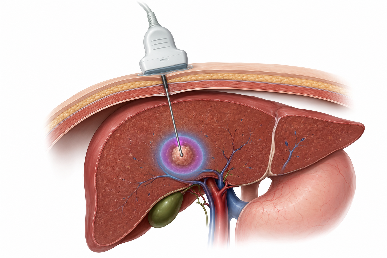
Ablation destroys a tumour in place without removing a large part of the liver. It can be curative for selected small HCCs.
Microwave and radiofrequency ablation are the usual methods. Other techniques may suit selected situations.
Microwave ablation
Uses microwave energy to heat and destroy the tumour. It is commonly used for small HCCs.
Radiofrequency ablation
Uses an electrical current to heat and destroy the tumour. It is commonly used for small HCCs.
Ethanol injection
Injects alcohol directly into the tumour to destroy cancer cells. It is less commonly used where heat-based ablation is available.
Cryoablation
Uses a probe to freeze and destroy the tumour. It is less commonly used for HCC. Ask whether it is suitable for you and available at your centre.
How it is done
A needle or probe is guided into the tumour using scans. It may pass through the skin or be placed during surgery. The aim is to destroy the tumour and a small surrounding margin while protecting nearby organs, bile ducts and the diaphragm, the breathing muscle above the liver.
Choosing the type of ablation
Discuss the type of ablation with your doctor. The safest and most effective choice depends on several factors:
- Tumour size and number. Ablation often works best for a small number of tumours, usually around 3 cm or smaller.
- Safe access. The team needs to reach the tumour with a needle or probe and treat a small margin around it.
- Nearby structures. Tumours close to bile ducts, bowel or major blood vessels need careful planning to reduce injury. Blood flowing through large vessels can also carry heat away, making some heat treatments less effective.
- Liver function and local expertise. Your liver health, the treatments available and the team's experience also help guide the choice.
A follow-up scan using contrast checks whether any active cancer remains in the treated area. Repeat ablation or another treatment may be needed.
TACE, TAE and arterial treatment
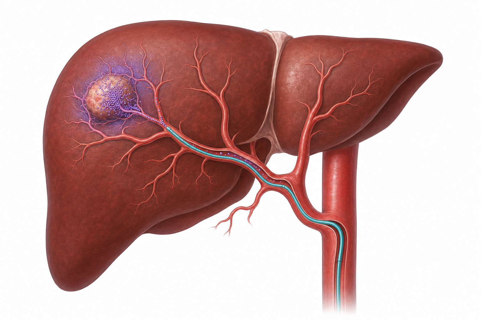
HCC receives much of its blood from the hepatic artery, which carries blood to the liver. Specialists can use a thin tube called a catheter to deliver treatment through this artery. Transarterial embolisation (TAE) blocks the tumour’s blood supply; transarterial chemoembolisation (TACE) also delivers chemotherapy.
What the names mean
TAE (bland embolisation)
Particles block selected tumour-feeding arteries without chemotherapy.
TACE
Chemotherapy is delivered into tumour-feeding arteries and followed by embolisation.
DEB-TACE
Drug-eluting bead transarterial chemoembolisation (DEB-TACE) uses tiny beads that slowly release chemotherapy while blocking the tumour’s blood supply.
Selective / super-selective
A catheter is advanced into smaller branches to spare more healthy liver when anatomy allows.
When it may be used
- Liver-limited HCC that is not suitable for resection or ablation, often in intermediate-stage disease.
- Bridging or downstaging before transplant.
- Selected combinations or sequences with radiation or systemic therapy.
Safety depends on blood flow to the liver, liver function, ascites, the amount of cancer and the healthy liver remaining. TACE is not repeated automatically. The team checks the scans and liver health to decide whether another session or a different treatment is best.
Radioembolisation and focused radiation
Radiation can be delivered from inside the liver artery or from a machine outside the body.
TARE / SIRT / Y-90 radioembolisation
Transarterial radioembolisation (TARE) is also called selective internal radiation therapy (SIRT). Tiny beads containing the radioactive substance yttrium-90 (Y-90) are delivered through a catheter into arteries feeding the tumour.
Before TARE, you usually have a separate planning procedure to map the liver’s blood vessels, followed by a scan using a small amount of radioactive tracer. This includes a lung-shunt assessment to estimate how much of the treatment could reach the lungs, helping the team decide whether TARE is suitable and plan the radiation dose.
- May control HCC confined to the liver, help while waiting for transplant, or shrink cancer to allow another treatment. Treating one small section of liver with a high radiation dose is called radiation segmentectomy.
- May suit some people whose cancer has grown into the portal vein, when treatments that block blood flow are unsuitable.
- Fatigue, abdominal discomfort and temporary liver-test changes are common; ulcers, gallbladder injury and radiation-induced liver disease are uncommon but serious.
External-beam radiotherapy
Stereotactic body radiotherapy (SBRT), also called stereotactic ablative radiotherapy (SABR), gives highly focused radiation over a few sessions. Other techniques shape radiation around the tumour; proton therapy uses a different type of radiation particle. The team may consider these for tumours in difficult locations, cancer in blood vessels, returning cancer or symptoms such as bone pain.
When systemic treatment is needed
Even when surgery cannot remove HCC or it has spread to other organs (metastatic cancer), medicines may help control it and relieve symptoms.
Systemic therapy is usually central when HCC has invaded major vessels, spread outside the liver, progressed despite appropriate local therapy, or is too extensive for safe liver-directed treatment.
Before choosing treatment
- Confirm HCC and map the current disease burden; obtain tissue if the diagnosis is uncertain or testing will change care.
- Assess Child–Pugh and ALBI results, ascites, encephalopathy, kidney function, blood pressure and ability to manage daily activities.
- Check hepatitis B and C; plan antiviral therapy or HBV reactivation prevention where indicated.
- Review bleeding and clotting risk, varices, recent surgery, autoimmune disease, prior organ transplant and all medicines/supplements.
- Discuss treatment goals, expected benefit, visits, side effects, fertility where relevant, access and cost.
Four checks before choosing the first treatment
Is immunotherapy suitable?
The team checks for autoimmune disease, a previous organ transplant and infection. You should know how to recognise side effects caused by the immune system attacking healthy organs.
Are blood-vessel-targeting drugs suitable?
The team checks varices, bleeding and clotting risks, blood pressure, urine protein and wound healing before choosing these medicines.
Liver and organ reserve
Liver scores, ascites, encephalopathy, blood results, kidney health, nutrition and daily activity help show which treatment you can tolerate.
Delivery and monitoring
Discuss visits for drips or tablets at home, other medicines, help taking treatment correctly and how to get advice quickly about side effects.
First-line immunotherapy options
First-line means the first systemic treatment used for this cancer. For many people, this includes immune checkpoint inhibitors, medicines that release the immune system’s brakes so it can attack cancer. Liver function, bleeding risk, autoimmune disease and a previous transplant can change the choice.
Example approach | Why it may be chosen | Key cautions before starting |
|---|---|---|
Atezolizumab + bevacizumab | Atezolizumab is immunotherapy. Bevacizumab blocks vascular endothelial growth factor (VEGF), a signal that helps tumours grow blood vessels. This combination has strong first-line evidence. | Bleeding from varices or elsewhere in the digestive tract, recent surgery, high blood pressure, protein in urine and clotting risks need assessment. A camera test of the food pipe and stomach (upper endoscopy) and treatment of high-risk varices are commonly needed first. |
Durvalumab + tremelimumab | Two immunotherapy medicines. One initial dose of tremelimumab is given with durvalumab, followed by continued durvalumab alone. | The immune system may inflame healthy organs. Autoimmune disease and previous transplant need careful assessment. This regimen does not contain bevacizumab, but immune side effects still need prompt care. |
Nivolumab + ipilimumab | Two immunotherapy medicines given together initially, followed by nivolumab alone. A first-line option for HCC that cannot be removed by surgery or has spread. | Immune side effects can be serious, especially during the early combination. They may affect the liver, bowel, lungs, skin, nerves or hormone-producing glands. |
Durvalumab alone | One immunotherapy medicine that may suit selected people when a combination is unsuitable. | Immune side effects and transplant rejection remain possible. Autoimmune disease still needs assessment, even with one medicine. |
First-line targeted tablets: lenvatinib and sorafenib
Lenvatinib and sorafenib are tyrosine kinase inhibitors (TKIs), targeted tablets that block signals involved in cancer growth and blood-vessel formation. Both are established treatments for HCC that cannot be removed by surgery. They are alternatives, and the choice should be individualised.
Feature | Lenvatinib | Sorafenib |
|---|---|---|
Place in care | Used on its own as a first treatment when immunotherapy is unsuitable or not chosen. | Used on its own as a first treatment when immunotherapy is unsuitable or not chosen. |
How it is taken | Usually once daily, with a starting dose based on body weight. Your team may pause or reduce the dose to manage side effects. | Usually twice daily without food. Follow your prescription exactly; your team may pause or reduce the dose to manage side effects. |
Common problems | High blood pressure, fatigue, diarrhoea, reduced appetite or weight, muscle/joint pain, abdominal discomfort, hand-foot soreness, urine protein, voice change and thyroid slowing. | Diarrhoea, fatigue, hand-foot skin reaction, rash, reduced appetite or weight, hair thinning, abdominal discomfort and high blood pressure. |
Routine monitoring | Blood pressure, urine protein, thyroid, liver and kidney tests, weight and fluid intake. The team reviews other medicines and planned surgery or dental procedures. | Blood pressure, skin and feet, liver tests, blood salts, weight and fluid intake. The team reviews heart and bleeding risks, other medicines and planned procedures. |
Call promptly | Severe diarrhoea or dehydration, bleeding, chest pain, sudden one-sided weakness or trouble speaking, severe headache or vision change, marked breathlessness, reduced urine, jaundice or confusion. | Painful or blistering hand-foot changes, severe rash, bleeding, chest pain, severe diarrhoea/dehydration, worsening jaundice or confusion. |
How the choice is made
- Liver reserve and current symptoms from cirrhosis or HCC.
- Blood-pressure, urine-protein, skin/foot, bowel, nutrition, cardiac and bleeding risks.
- Other medicines, planned procedures, ability to take tablets consistently and access to monitoring.
- Previous treatment, expected side-effect pattern and informed patient preference.
When treatment changes and when further tests help
Later-line treatment means treatment used after an earlier option stops working or causes unacceptable side effects. The choice depends on what you have already received, liver function, blood markers, side effects and your goals.
Options after earlier treatment
- Oral targeted drugs: cabozantinib; regorafenib after prior sorafenib that was tolerated; or an unused lenvatinib/sorafenib strategy in selected sequences.
- Ramucirumab: blocks vascular endothelial growth factor receptor 2 (VEGFR2), which helps new blood vessels form. It may suit selected patients with AFP of at least 400 nanograms per millilitre (ng/mL). The established treatment setting is after sorafenib; the team should explain the evidence for the proposed sequence after any other prior treatment.
- Checkpoint immunotherapy: an unused immunotherapy approach may be considered, but evidence for every sequence after modern combinations is incomplete.
- Local treatment: radiation, ablation or arterial therapy can still help a limited progressing site or a symptom while systemic disease is otherwise controlled.
- Clinical trials: especially valuable because the best sequence after first-line immunotherapy remains an evolving area.
Molecular testing
Molecular testing looks for changes in cancer genes or proteins that might guide treatment. Broad genetic testing is not needed for every typical HCC. It may help when the diagnosis is unusual, usual treatments have been tried, or a clinical trial is being considered.
Rare findings include gene fusions, where parts of two genes join together, involving genes called NTRK or RET. Tests may also find high microsatellite instability (MSI-high) or deficient mismatch repair, signs that cancer cells cannot repair certain genetic errors normally. Your team should explain whether a result opens a suitable treatment option. A negative result does not remove the usual HCC treatments.
Managing side effects
Managing treatment side effects
Cirrhosis, cancer and treatment can cause similar symptoms. Tell your team about changes from how you usually feel; do not wait for them to become severe.
Treatment group | Common problems | Urgent problems |
|---|---|---|
Oral TKIs | Hand-foot soreness, diarrhoea, fatigue, appetite/weight loss, mouth soreness, high blood pressure. | Chest pain, sudden one-sided weakness or trouble speaking, severe bleeding, very high blood pressure, severe diarrhoea or dehydration, or sudden worsening of liver function. |
Bevacizumab / anti-VEGF | High blood pressure, protein in urine, bleeding tendency, slower wound healing. | Vomiting blood, black stools, major bleeding, severe abdominal pain, sudden breathlessness, a new painful swollen leg or a wound that opens or fails to heal. |
Checkpoint immunotherapy | Rash/itch, diarrhoea, fatigue; thyroid and other hormone changes. | New breathlessness/cough, persistent diarrhoea, jaundice, severe weakness, confusion, severe headache/vision change, blistering rash or chest pain. |
Liver-directed treatment | Pain, fever, nausea, fatigue and temporary liver-test changes. | Persistent fever, severe/worsening pain, jaundice, confusion, fainting, breathlessness or rapidly increasing ascites. |
When you call
Give the treatment name, last dose, symptom start, temperature, stool/vomit count, urine change and medicines already taken.
Do not self-treat
Follow the symptom plan your oncology team has given you. Check with them before starting steroids, medicines for diarrhoea or herbal remedies, or changing a treatment dose. Early assessment can prevent serious harm.
Live well
Topics in this section
Protecting the liver during cancer treatment
The remaining liver is part of the treatment plan every day.
- Avoid alcohol. There is no safe 'detox' drink for a damaged liver, and alcohol can worsen cirrhosis, dehydration and drug toxicity.
- Review every medicine. Include prescriptions, painkillers, over-the-counter tablets, vitamins, gym products, Ayurvedic/herbal remedies and supplements. 'Natural' does not mean liver-safe.
- Treat hepatitis. Hepatitis B medicines may be needed to treat infection or prevent it becoming active again during cancer care. The liver and cancer teams should also agree when to treat hepatitis C.
- Use painkillers safely. Ask the liver team which medicine and dose are safe for you. Paracetamol (acetaminophen) and non-steroidal anti-inflammatory drugs (NSAIDs), such as ibuprofen, can cause harm in the wrong situation.
- Prevent infection. Ask about hepatitis A/B, influenza, COVID-19 and pneumococcal vaccination according to your history and treatment. Avoid raw or undercooked shellfish and follow food hygiene.
- Protect kidneys and circulation. Report vomiting, diarrhoea, low urine, dizziness or sudden weight change. Medicines that remove extra fluid (diuretics) and blood-pressure medicines may need adjustment by your team.
When to seek urgent help
Use the 24-hour number provided by your treating centre. If you cannot reach the team and the problem is severe, go to the nearest emergency department or call local emergency services.
Possible bleeding
Vomiting blood, coffee-ground vomit, black/tarry stools, uncontrolled bleeding, fainting or marked dizziness.
Possible encephalopathy
New confusion, reversed sleep pattern, unusual behaviour, marked drowsiness or inability to stay awake.
Possible infection
Fever or chills with feeling unwell, especially after a procedure, on steroids or during systemic therapy.
Possible liver decompensation
New jaundice, rapidly increasing abdominal swelling, breathlessness, very low urine or severe weakness.
Possible treatment emergency
Chest pain; sudden weakness on one side or trouble speaking (possible stroke); severe diarrhoea; new breathlessness; blistering rash; severe headache or vision change; or a severe reaction during a treatment drip.
Possible abdominal complication
Sudden severe abdominal pain, rigid abdomen, repeated vomiting or pain after a procedure that is worsening rather than settling.
Supportive and palliative care
Supportive care treats the person, not just the tumour. It can run alongside surgery, transplant planning, liver-directed therapy or systemic treatment at any stage.
Problem | What the team may consider |
|---|---|
Pain | Cause-specific treatment, liver-safe medicines, nerve blocks, radiation or specialist pain/palliative review. |
Ascites / leg swelling | Salt advice, prescribed diuretics, or draining abdominal fluid through a small tube (paracentesis). The team also checks kidney function, infection and whether another procedure may help. |
Encephalopathy | Find and treat the cause. Medicines such as lactulose or rifaximin can reduce harmful substances from the gut that contribute to confusion. Agree a safety plan with caregivers. New confusion needs urgent assessment. |
Jaundice / itching | Check for liver failure versus bile-duct obstruction; skin care and anti-itch treatment; drainage only when obstruction and benefit justify it. |
Nausea / appetite loss | Anti-sickness medicines, constipation treatment, small frequent meals and dietitian input. |
Breathlessness / fatigue | Check for fluid build-up, anaemia, infection, pain, poor sleep and loss of strength. Gentle activity and rehabilitation may help, alongside treatment of the cause. |
Emotional or spiritual distress | Counselling, mental health care, peer support and family discussions. You can also talk about the care you would want if your health changes (advance care planning). |
Nutrition, muscle and activity
Cirrhosis and HCC can cause loss of muscle even when body weight looks stable because fluid hides weight loss.
Practical nutrition
- Aim for small, frequent meals with enough energy and protein; a late-evening snack may help some people with cirrhosis avoid a long overnight fast.
- Do not restrict protein routinely. Encephalopathy needs medical treatment and individual nutrition advice; severe protein restriction can worsen muscle loss.
- If ascites is present, the team may advise limiting sodium, the main component of salt. Do not restrict fluid unless specifically told to, usually because blood sodium is low.
- Choose safe, familiar foods that you can tolerate. Avoid raw shellfish, unpasteurised products and poorly stored/mouldy grains or nuts.
- Ask before using high-dose vitamins, protein powders or herbal products; some interact with treatment or damage the liver.
Movement
- Short walks, sit-to-stand practice and light resistance work help preserve muscle and independence when safe.
- Build activity gradually. Stop if you feel unwell. Chest pain, new breathlessness, fainting or bleeding needs urgent assessment.
- After surgery, ablation or arterial treatment, follow restrictions on lifting, bathing, driving and wound care.
Follow-up, response and recurrence
Follow-up is planned to detect recurrence, protect liver function, manage late effects and decide whether treatment needs to change.
After curative-intent resection or ablation
Follow-up commonly includes imaging and AFP blood tests every 3–6 months after treatment. Some centres also measure PIVKA-II if it was useful before treatment. Your team will explain the scan type, timing and longer-term schedule based on recurrence risk, kidney and liver health, and any transplant plans. Ongoing follow-up remains important even when scans show no cancer.
During systemic or liver-directed treatment
Scans and blood tests are repeated at intervals suited to your treatment. The team checks tumour size and how it takes up contrast dye, which can help show whether cancer remains active. Effective treatment may reduce contrast uptake before the tumour shrinks. AFP or PIVKA-II trends help only if that marker was useful before treatment.
If cancer returns
- A recurrence may be a new HCC in the at-risk liver or spread from the original tumour; in practice the distinction is not always possible.
- Resection, transplant, ablation, arterial therapy, radiation or systemic treatment may again be considered using the same five decision factors.
- A limited recurrence may still be treated with curative intent. Wider recurrence is treated to control disease and symptoms.
Emotions, family, work and access
There is no correct way to feel. Shock, fear, anger, sadness, relief and uncertainty can sit together or change from day to day.
For patients
Choose one trusted person for appointments; ask the team to explain the plan in manageable steps; tell the team if sleep, panic or low mood is affecting daily life.
For caregivers
Keep a medicine and symptom list, but protect your own rest and health. Know the bleeding/confusion emergency plan.
Work and travel
Ask for realistic recovery and visit schedules. Encephalopathy, sedating medicines and some procedures may temporarily make driving unsafe.
What matters to you
Tell your team what matters most in your daily life, such as time with family, independence or getting back to work. These priorities can help shape your care and support.
Sources and further reading
For more information, explore these resources from the National Cancer Institute (NCI) and other medical organisations. The linked organisations have not reviewed or endorsed this CTI guide. Your care team can explain how the information applies to you.
- NCI: Liver Cancer Treatment
- AASLD: practice guidance on prevention, diagnosis and treatment of hepatocellular carcinoma (2023)
- EASL: clinical practice guidelines on hepatocellular carcinoma (2025)
- ASCO: systemic therapy for advanced hepatocellular carcinoma (2024 guideline update)
- ESMO: hepatocellular carcinoma clinical practice guideline
- Macmillan: liver cancer
- NCI: aflatoxins and liver cancer risk
- RadiologyInfo (ACR/RSNA): radioembolisation and the planning assessment
- NCI: liver cancer diagnosis
- IMbrave050: updated results after complete resection or ablation (2026)
- AASLD: hepatocellular carcinoma guidance and critical update
- FDA: first-line nivolumab with ipilimumab for HCC (2025)
Staging and risk-group references
AJCC 8th edition TNM staging and the BCLC framework; the 2026 BCLC update retains the 2022 stage categories.
Medicine approval and prescribing references
These official records describe licensed uses and prescribing information for the medicines named in this guide. Approval depends on the cancer type, biomarkers, previous treatment, combination and formulation. Some uses discussed in the guide are off-label: outside the medicine’s licence, with evidence from clinical guidelines or studies. FDA or EMA approval does not establish approval or availability in India.
- Atezolizumab — FDA prescribing information · Approval history
- Bevacizumab — FDA prescribing information · Approval history
- Cabozantinib tablets (Cabometyx) — FDA prescribing information · Approval history
- Durvalumab — FDA prescribing information · Approval history
- Ipilimumab — FDA prescribing information · Approval history
- Lenvatinib — FDA prescribing information · Approval history
- Nivolumab — FDA prescribing information · Approval history
- Ramucirumab — FDA prescribing information · Approval history
- Regorafenib — FDA prescribing information · Approval history
- Sorafenib — FDA prescribing information · Approval history
- Tremelimumab — FDA prescribing information · Approval history
Additional medicine approval notices
These records explain the specific medicine uses and eligibility requirements discussed in this guide.
Related care
Meet our doctors



Where to get care