
Share
Start here
Kidney cancer at a glance
- Renal cell carcinoma (RCC) is the most common kidney cancer in adults. About 9 in 10 adult kidney cancers are RCC.
- Clear-cell RCC is the most common subtype, or type of RCC. Other types include papillary and chromophobe RCC. The subtype can affect how the cancer behaves and which treatments help.
- Many small kidney cancers cause no symptoms and are discovered during a scan for another reason.
- Surgery is the main treatment for most localised RCC, meaning cancer confined to the kidney. A partial nephrectomy removes the tumour while saving the rest of the kidney when this can be done safely.
- Some small tumours can be monitored with regular scans. Other options include ablation, which destroys the tumour using heat or freezing, or focused radiation. These may help when surgery carries more risk.
- Advanced RCC has grown into nearby tissues or spread elsewhere. Treatment often uses immunotherapy to help the immune system attack cancer, targeted medicines that block cancer growth, or both. Traditional chemotherapy has little benefit for most RCC.
- Most people can live a full life with one well-functioning kidney, but blood pressure and kidney function need ongoing care.
A statistic describes a group, not an individual. Ask your doctor what features of your cancer and general health are most relevant to your outlook.
Bring your scan images
For your consultation or a second opinion, bring a compact disc (CD) containing your scan images in Digital Imaging and Communications in Medicine (DICOM) format and the software needed to view them, along with the written scan reports. DICOM is the standard format used to store and share medical scans.
Understand
What your kidneys do
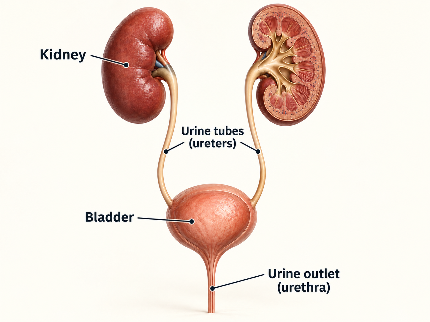
Most people have two kidneys, deep in the upper abdomen on either side of the spine. Each is roughly the size of a fist.
- Tiny filters called nephrons remove waste and extra water from the blood to make urine.
- The kidneys balance fluids, salts and acids, help control blood pressure and signal the body to make red blood cells.
- Urine travels through tubes called ureters to the bladder, where it is stored. The adrenal glands above the kidneys make hormones, the body’s chemical messengers. They are separate organs.
What is kidney cancer?
Cancer begins when abnormal cells keep growing and form a tumour. RCC usually starts in the lining of tiny tubes within the kidney, called tubules, that help form urine.
A kidney mass is an abnormal lump. It may be solid or contain fluid, which is called cystic. Some masses, such as oncocytoma and angiomyolipoma, are benign: they are not cancer.
Scans can suggest the cause. Examining a tissue sample (a biopsy) or tissue removed during surgery may confirm it.
RCC can behave in different ways
- It may remain confined to the kidney for a long time.
- It may grow into nearby fat or lymph nodes, small structures that help fight infection. It may also enter the renal vein, which carries blood away from the kidney, or the vena cava, the large vein that takes this blood towards the heart.
- Cancer cells may travel through the blood or lymphatic system, a network of vessels and nodes. They can form new tumours called metastases, meaning areas of cancer spread. Common sites include the lungs, lymph nodes and bones; the liver or brain can also be affected.
Cancer that started elsewhere and later reached the kidney is a metastasis from the original cancer. It is not classified or treated as primary kidney cancer.
The subtype matters
Histology means the pattern and appearance of cancer cells under a microscope. It is a key part of choosing treatment.
Subtype | Why it matters |
|---|---|
Clear cell | About 70% of RCC. These cells look pale or clear under a microscope. Most standard RCC drug combinations were studied in this subtype. |
Papillary | The cells often form small finger-like shapes. Most papillary RCC develops without an inherited cause; rarely, it is linked to a gene change passed down in a family. |
Chromophobe | A less common subtype identified by how its cells look and stain in the laboratory. It often grows more slowly when confined to the kidney, although behaviour varies. |
Rarer forms | Some rare types are named for the cells involved, such as collecting-duct or renal medullary cancer. Others involve rearranged genes (translocation-associated RCC) or lack a cell protein called fumarate hydratase (FH-deficient RCC). Unclassified RCC does not fit a recognised subtype. |
Features your report may mention
- Sarcomatoid or rhabdoid change: the cells look very abnormal and may grow or spread more quickly. These changes can occur within several RCC subtypes.
- Tumour necrosis: areas of dead tumour tissue. The team considers this alongside the other findings in your tissue report, also called a pathology report.
- Clear-cell component: at least part of the cancer has clear-cell features. This can affect which treatments are suitable after surgery.
For a rare subtype, a pathologist who specialises in kidney and urinary tract cancers should review the tissue. A pathologist is a doctor who diagnoses disease by examining cells and tissue. Specialists can then discuss the findings together and recommend a plan.
Symptoms and warning signs
Many kidney cancers cause no symptoms. Having one of these symptoms does not mean you have cancer, but it should be assessed.
Possible symptoms
- Blood in the urine, even if it happens only once
- Persistent pain in the side or back that is not explained by an injury
- A lump or fullness in the side or abdomen
- Unexplained weight loss, poor appetite, ongoing fever or marked tiredness
- Less commonly, anaemia (too few healthy red blood cells), high blood calcium, leg swelling or symptoms caused by spread to another organ
If you are already receiving cancer treatment, use the 24-hour number given by your treating team. Do not wait for the next routine appointment when symptoms are severe or rapidly worsening.
Risk factors
A risk factor increases the chance of developing cancer, but it cannot explain why one person developed it. Usually there is no single cause. Having kidney cancer is not your fault.
Factors that increase risk
- Smoking
- Excess body weight and high blood pressure
- Severe long-term kidney disease, especially when dialysis is needed to filter waste and extra fluid from the blood
- A parent, brother, sister or child with kidney cancer
- Certain inherited conditions, caused by gene changes passed down in a family, and some long-term workplace chemical exposures
Routine screening is not recommended for people at average risk who have no symptoms. People with a confirmed inherited condition that increases kidney cancer risk need a specialist plan for regular checks.
Genetic counselling helps you understand inherited cancer risk and decide whether testing is useful.
Germline testing uses blood or saliva to look for a gene change present throughout the body that may be passed down in a family. It is different from testing the tumour itself. Counselling also helps explain what the results could mean for relatives.
Tests and diagnosis
Your care team
Kidney cancer care works best when specialists review your diagnosis and treatment options together and one person coordinates the plan.
Team member | Main role |
|---|---|
Uro-oncologist / urologist | A doctor specialising in the urinary tract and its cancers. Assesses and performs kidney surgery and may oversee monitoring of a small tumour. |
Medical oncologist | A doctor who plans cancer medicines, including immunotherapy and targeted therapy. |
Radiation oncologist | A doctor who uses radiation to treat selected kidney tumours or areas of spread. |
Radiologist | A doctor who interprets scans. An interventional radiologist uses scan guidance to take a biopsy or destroy a tumour with ablation. |
Pathologist | Examines tissue to confirm the subtype, grade and whether cancer reaches the cut edge, called the margin. |
Nephrologist | A kidney specialist who helps protect kidney function, especially with long-term kidney disease, protein leaking into urine or difficult-to-control blood pressure. |
Genetic counsellor | Assesses inherited risk and explains testing and family implications. |
Nurses and supportive-care team | Help with symptoms, treatment effects, nutrition, emotions, recovery, daily activities and practical needs. |
Ask who your main contact is for results, side effects and urgent concerns. Save their number in your phone and share it with your caregiver.
A second opinion is reasonable, particularly before complex surgery, when the subtype is rare, or when several treatment options are equally acceptable.
How cancer is diagnosed
You are unlikely to need every possible test. The team chooses tests that answer a specific question.
- Confirm whether there is a kidney mass and show its size, position and relationship to nearby blood vessels and organs.
- Check your general health and kidney function through your medical history, examination, blood pressure, blood tests and urine tests.
- Look for spread with scans of the chest and abdomen. Computed tomography (CT) combines X-rays to create detailed images. Selected questions may need positron emission tomography with CT (PET-CT), which uses a small amount of radioactive tracer to show tissue activity.
- Obtain tissue when the diagnosis or subtype must be confirmed before treatment, or examine the tissue removed at surgery.
- Bring the scans, tissue findings, stage, kidney function and your priorities together in a plan agreed by the specialist team.
Common blood and urine tests
- A full blood count checks red cells, white cells and platelets, which help blood clot. Creatinine is a waste product measured in blood. It helps calculate the estimated glomerular filtration rate (eGFR), an estimate of how well your kidneys filter blood.
- Liver tests and blood calcium help check your general health. Other blood tests may include alkaline phosphatase or lactate dehydrogenase, substances that can change with liver, bone or other tissue problems.
- Urinalysis is a urine test that checks for blood, protein and other changes.
These tests support diagnosis and treatment planning but cannot, by themselves, prove that a kidney mass is cancer.
Scans and staging tests
Test | Typical purpose |
|---|---|
Ultrasound | Uses sound waves to show the kidney. It can detect a lump and help distinguish a solid mass from a fluid-filled cyst. |
Contrast CT | Usually the main staging scan. Contrast is a dye that helps show the kidney, tumour, blood vessels, lymph nodes and other organs more clearly. |
Magnetic resonance imaging (MRI) | Uses a strong magnet and radio waves to create detailed images. It can help when CT contrast is unsuitable or when veins, the liver, spine or the inside of a tumour need a closer look. |
PET-CT | Your team may occasionally recommend PET-CT to answer a specific question about possible spread. Contrast CT or MRI remains the main approach to staging RCC. |
Other tests | Bone scans are usually chosen when symptoms suggest a problem. Brain CT or MRI may be considered when RCC has spread, before starting whole-body treatment or surgery to reduce the amount of cancer. |
Biopsy and molecular testing
A biopsy takes small samples of tissue for examination under a microscope. A needle usually passes through the skin, guided by CT or ultrasound.
A biopsy is commonly considered when
- Imaging is uncertain or a benign condition is possible
- Planned monitoring, ablation or focused radiation is being considered
- The cancer appears advanced and drug treatment will begin without kidney surgery
- A rare subtype or a metastasis needs confirmation
Two different kinds of genetic testing
Tumour profiling
Looks for gene changes within cancer cells. It may help choose treatment for some advanced or rare RCCs. Many tumours confined to the kidney do not need this testing.
Germline testing
Looks for a gene change that may be inherited, using blood or saliva. Results can guide regular checks for you and advice or testing for relatives.
A biopsy can cause pain or bleeding, and sometimes the sample does not give a clear answer. Serious complications are uncommon. Ask how to get help if pain, dizziness, fever or blood in the urine gets worse.
Understanding your pathology report
Stage describes where the cancer is. Grade describes how abnormal the cells look and how aggressively they may behave.
Report item | Plain-language meaning |
|---|---|
Histology | The exact RCC subtype or another diagnosis. |
Tumour size | The widest measurement of the tumour. Size and growth into nearby tissues help determine the tumour category, called T. |
WHO/ISUP grade | The World Health Organization/International Society of Urological Pathology (WHO/ISUP) grade runs from 1 to 4 for clear-cell and papillary RCC. A higher grade means the cells look more abnormal and may behave more aggressively. This system is not routinely used for chromophobe RCC. |
Margins | Whether cancer reaches the cut edge of removed tissue. |
pT and pN | The p means the tissue was examined after surgery. T describes the tumour’s size and local growth; N describes whether nearby lymph nodes contain cancer. |
Vessel / fat invasion | Whether cancer has grown into a kidney vein, the vena cava or nearby fat. |
High-risk features | Very abnormal cell patterns, dead tumour tissue and other findings that may increase the chance of cancer returning. The team considers these alongside stage and subtype. |
A positive margin means cancer cells reach the cut edge of the removed tissue. Your team weighs the need for further treatment against its risks, using the scan and tissue findings and your kidney function. Another operation is not always needed.
Planning your treatment
Understanding your stage
TNM staging for renal cell carcinoma
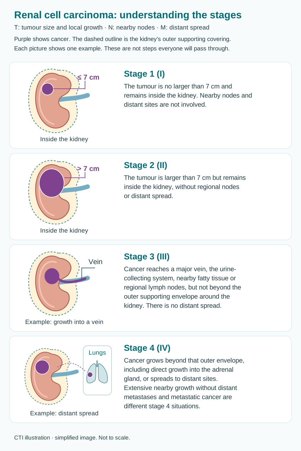
Doctors combine tumour size and local growth (T), nearby lymph node involvement (N) and distant spread (M) to determine the stage. These stages apply to renal cell carcinoma (RCC), which starts in kidney tissue. Cancer of the kidney’s urine-collecting lining uses a different system.
Lymph nodes are small immune-system structures. Regional nodes are those near or draining the cancer’s starting area; distant spread means cancer has reached a site outside that regional area. Stages describe findings, not a sequence that everyone will pass through.
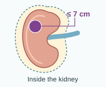
Purple shows cancer. The dashed outline is the kidney’s outer supporting covering, called Gerota’s fascia. Each picture shows one example of a stage; the text explains other ways that stage can occur.
Stage 1 (I)
The tumour is no larger than 7 cm and remains inside the kidney. Nearby nodes and distant sites are not involved.
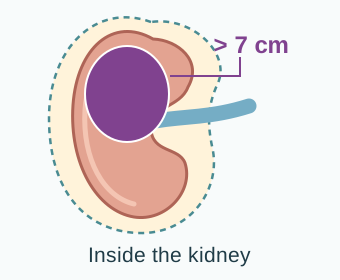
Stage 2 (II)
The tumour is larger than 7 cm but remains inside the kidney, without regional nodes or distant spread.
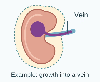
Stage 3 (III)
Cancer reaches a major vein, the urine-collecting system, nearby fatty tissue or regional lymph nodes, but not beyond the outer supporting envelope around the kidney. There is no distant spread.
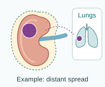
Stage 4 (IV)
Cancer grows beyond that outer envelope, including direct growth into the adrenal gland, or spreads to distant sites. Extensive nearby growth without distant metastases and metastatic cancer are different stage 4 situations.
CTI illustration. Enlarge the staging overview.
Cancer extending along a vein is not automatically distant metastatic disease. The surgeon assesses its extent and whether an operation can remove it safely. Grade and, in metastatic disease, a separate clinical risk score help guide the plan.
Clinical stage comes from examination, biopsies and scans. Pathological stage can add information from surgery; a prefix such as yp means the tissue was assessed after treatment. A revised assessment does not necessarily mean the cancer grew while you were waiting. Grade describes the cells’ behaviour and is different from stage.
How your treatment plan is chosen
Good decisions combine medical evidence with what matters to you.
The team considers
- Stage, subtype, grade, tumour size and position
- Whether the tumour can be removed completely and safely
- Kidney function, one or two working kidneys, blood pressure and other illnesses
- Symptoms, general fitness and previous treatment
- The chance of benefit, possible harms, recovery time, cost and availability
- Your priorities, family responsibilities and comfort with monitoring or uncertainty
Shared decision-making
You and your team choose the plan together. Discuss the reasonable options, including monitoring or delaying treatment when safe. Ask how many people like you are likely to benefit, what the main side effects are and how daily life may change. A second opinion can help if you remain unsure.
Treatment recommendations change. Your oncology team should interpret current evidence for your diagnosis and treatment setting.
Your treatment options at a glance
The first major question is whether treatment needs to focus on the kidney, the whole body, or both.
Supportive and palliative care help manage symptoms and improve comfort and quality of life. They can be part of your care from the beginning, alongside treatment for the cancer.
Treatment options
Topics in this section
Active surveillance
Active surveillance means planned monitoring with regular checks and scans. Treatment can start if the findings or your needs change.
Who may consider it
- People with a very small kidney mass, especially one under about 2 cm
- People whose age, frailty or other illnesses make treatment risk greater than the immediate cancer risk
- People with selected fluid-containing or slow-growing masses, after specialist review
What usually happens
- Initial scans, kidney-function tests and often a discussion about biopsy
- Repeat scans at defined intervals, more frequently at first
- Treatment if the mass grows, scans or symptoms change, or you prefer treatment
Monitoring can avoid or delay treatment side effects. It means attending repeat tests and living with some uncertainty. Most carefully selected small masses grow slowly, but keeping follow-up appointments is essential.
Active surveillance before treatment is different from follow-up surveillance after treatment.
Surgery: partial or radical
The aim is complete cancer removal while preserving as much safe kidney function as possible.
Partial nephrectomy
Removes the tumour and a small rim of tissue, leaving the rest of the kidney. Often preferred for small tumours, especially if you have one kidney, tumours in both kidneys or reduced kidney function.
Radical nephrectomy
Removes the whole kidney and surrounding fatty tissue. It may be safer for a large tumour, one deep in the kidney or one growing into nearby structures when saving part of the kidney would leave cancer behind.
Approach and expertise
Surgery may use one larger cut (open surgery) or several small cuts (laparoscopic, or keyhole, surgery). In robot-assisted surgery, the surgeon controls the instruments. The choice depends on the tumour, your health and the surgeon’s experience. Smaller cuts can shorten recovery in suitable cases.
- The adrenal gland is usually removed only if imaging or the tumour position makes involvement likely.
- Nearby lymph nodes may be sampled or removed if they look suspicious. Removing many normal-looking nodes has not shown a benefit for everyone.
- A tumour in the renal vein or vena cava may require complex surgery at an experienced centre.
Ablation and focused radiation
These options can treat selected tumours without removing the kidney, particularly when surgery carries extra risk.
Thermal ablation
A thin probe passes through the skin to destroy the tumour. Cryoablation freezes it; radiofrequency and microwave ablation heat it. These treatments are mainly used for small tumours, commonly around 3 cm or less. The choice depends on the tumour’s size and location.
Focused radiation
Stereotactic ablative radiotherapy (SABR), also called stereotactic body radiotherapy (SBRT), delivers a few highly focused treatments from a machine outside the body. It may be an option when surgery is unsuitable for a tumour confined to the kidney, or for selected areas of spread.
Important practical points
- A biopsy is usually taken before ablation to confirm the diagnosis.
- Ablation may have a higher chance of needing repeat local treatment than surgery; careful imaging follow-up is essential.
- Focused radiation can affect nearby healthy tissue. Changes after treatment can also make follow-up scans harder to interpret.
- Availability and expertise vary. Ask how many similar procedures the centre performs and how success will be checked.
Arterial embolisation blocks blood vessels feeding the tumour. It may occasionally help control bleeding or other symptoms when surgery is unsuitable.
Lowering the risk of recurrence
The pathology report helps estimate whether surgery alone is enough or whether treatment after surgery should be discussed.
Surveillance remains appropriate for many people
Regular scans and kidney-function checks are the usual approach after many stage 1 and lower-risk tumours. Your team will explain whether further treatment is likely to help.
Adjuvant treatment for selected higher-risk clear-cell RCC
Adjuvant treatment means medicine given after surgery to lower the chance of cancer returning. Pembrolizumab immunotherapy may be offered after complete removal of RCC with clear-cell features when the stage or grade shows a higher recurrence risk. Pembrolizumab alone is usually given for about one year.
Belzutifan plus pembrolizumab is another option for adults with a clear-cell component and an intermediate-high or high recurrence risk after kidney surgery, including selected people whose areas of spread have also been completely removed. Your team weighs the expected benefit against the side effects, including anaemia and low oxygen levels with belzutifan.
Traditional chemotherapy and routine radiation to the area where the kidney was removed are not standard after complete removal of typical RCC. A clinical trial, a research study testing care or treatment, may be an option.
When cancer has spread or returned
Advanced RCC is often treatable even when it cannot be removed completely. The aim is usually long-term control, symptom relief and preserving quality of life.
The plan may combine
- Systemic treatment: medicines that act throughout the body
- Focused radiation, ablation or surgery for troublesome or limited sites
- Supportive and palliative care for symptoms and treatment effects
Is kidney surgery still useful?
Cytoreductive nephrectomy means removing the kidney tumour when cancer has also spread elsewhere. Your team may discuss it if you are well enough and the kidney tumour is causing symptoms, the spread is limited, or medicines have controlled the cancer well.
Many people benefit from starting whole-body treatment first.
For metastatic RCC, doctors may use the International Metastatic Renal Cell Carcinoma Database Consortium (IMDC) risk score. It combines how well you manage daily activities, blood results and the time between diagnosis and starting drug treatment. It helps guide treatment but cannot predict exactly what will happen to you.
Whole-body treatment
Modern RCC treatment commonly combines medicines that activate immune cells with medicines that block cancer growth signals and blood-vessel formation.
Type | What it does |
|---|---|
Immunotherapy | Medicines such as nivolumab, pembrolizumab and ipilimumab block immune checkpoints, proteins that can act as brakes on immune cells. This helps the immune system attack cancer. |
Therapy targeting blood-vessel growth | Vascular endothelial growth factor (VEGF) helps new blood vessels form. Tablets such as axitinib, cabozantinib, lenvatinib, pazopanib and sunitinib block signals that support blood-vessel growth and cancer growth. They belong to a group called tyrosine kinase inhibitors (TKIs). |
Other targeted medicines | Other medicines block proteins that help cancer cells grow or respond to low oxygen. They are used in specific situations, depending on the cancer and previous treatment. |
Chemotherapy | Usually has little benefit for typical RCC. It may help some rare subtypes, such as renal medullary or collecting-duct cancer. |
Common first-treatment combinations for clear-cell metastatic RCC include
- Nivolumab plus ipilimumab, especially for an IMDC intermediate- or poor-risk group
- Pembrolizumab plus axitinib
- Pembrolizumab plus lenvatinib
- Nivolumab plus cabozantinib
Histology changes the plan
Non-clear-cell RCC includes several subtypes. Each may need a different treatment plan.
Non-clear-cell RCC
Papillary, chromophobe and rarer subtypes can respond differently. Treatment may use targeted medicine, immunotherapy, a combination, or chemotherapy for certain rare cancers. If the cancer is linked to an inherited condition, this may also guide the choice. Clinical trials can be particularly useful when standard treatment evidence is limited.
After the first treatment stops working
The next treatment depends on which medicines you have received, how long they worked, your symptoms and how well your organs are working. Options may include a different targeted medicine, immunotherapy if it has not already been used, treatment directed at a few growing areas, or a clinical trial.
Some later treatments depend on previous use of an immune checkpoint inhibitor targeting programmed death receptor 1 (PD-1) or programmed death-ligand 1 (PD-L1). These are proteins that help control immune responses. Your team can check whether your earlier treatment belongs to this group.
Belzutifan alone is an option for adults with advanced RCC after both a PD-1 or PD-L1 inhibitor and a VEGF-targeted TKI. Belzutifan plus lenvatinib is an option for adults with advanced RCC that has a clear-cell component after a PD-1 or PD-L1 inhibitor.
These belzutifan treatments have different eligibility requirements. Both need checks for anaemia and low oxygen levels; the combination also needs blood-pressure and other safety checks.
Your oncologist and cancer pharmacist can explain why a medicine suits your cancer, how to take it, possible interactions with other medicines, and help with access or cost.
Managing side effects
Targeted therapy side effects
TKIs are often tablets taken at home. Side effects are common but can often be managed when reported early.
Other targeted medicines have different risks: belzutifan, for example, can lower red blood cells and oxygen levels. Your team will explain which blood tests and other checks you need.
Common effects
- High blood pressure, tiredness, diarrhoea, nausea or poor appetite
- Sore mouth, taste change, rash, painful hands or feet
- Changes in thyroid function (a gland that helps control the body’s energy use), protein leaking into urine or abnormal liver tests
Your practical safety checklist
- Check blood pressure at home if advised and keep a written log.
- Report diarrhoea early. Follow your team’s advice about fluids and medicines to control it.
- Tell the oncology team before surgery, dental work or a procedure because some drugs delay wound healing.
- Share every prescription, medicine bought without a prescription, vitamin, herb and supplement. Some foods or medicines can change how a cancer drug works.
- Do not reduce, skip or restart a dose without instructions.
A planned treatment pause or dose reduction can help control side effects. The goal is a dose you can take safely and consistently.
Immunotherapy: report early
Checkpoint inhibitors can cause inflammation, an immune reaction that can harm healthy organs. Reactions may begin during treatment or weeks to months after it ends.
Possible immune-related problems
- Skin: rash, itching or blisters
- Bowel: diarrhoea, cramps or blood
- Lungs: new cough or breathlessness
- Liver: abnormal tests, yellow eyes or dark urine
- Hormone glands: severe fatigue, dizziness, headache, thirst, frequent urination or weight change
- Kidneys, heart, muscles, nerves, eyes or brain: uncommon but potentially serious inflammation
- Tell every clinician that you have received immunotherapy, even after treatment has stopped.
- Carry the treatment or immunotherapy alert card provided by your centre.
- Treatment may need to pause. Steroids, medicines that reduce inflammation, or other medicines that calm the immune system may be needed. Early care lowers the risk of permanent harm.
Live well
Supportive and palliative care
Supportive and palliative care help with physical symptoms, emotional strain and practical needs. They can be given alongside cancer treatment at any stage.
It can help with
- Pain, fatigue, appetite, nausea, constipation, sleep and breathlessness
- Anxiety, low mood, family communication, spiritual concerns and caregiver strain
- Rehabilitation, mobility, work, finances and planning ahead
When cancer affects bone or brain
Radiation, surgery or ablation may relieve pain or help protect movement and other functions. Bone-strengthening medicines may help, but your team checks kidney function and often arranges a dental assessment first.
Cancer that has spread to the brain may need steroids, surgery or precisely targeted radiation as well as whole-body treatment.
Ask for a dietitian if weight or appetite is falling, a physiotherapist for safe activity, a psychologist or counsellor for distress, and a pain or palliative specialist when symptoms remain difficult.
A caregiver also needs information, rest and support. Ask the team what tasks are essential and which can be shared.
Follow-up after treatment
Follow-up checks for cancer returning, manages effects that appear or persist after treatment, and protects your kidney and general health. The schedule is based on your own risk and needs.
A follow-up visit may include
- Symptoms, physical examination, blood pressure and medication review
- Kidney-function tests, urine protein and other blood tests when needed
- Chest and abdominal imaging at intervals based on stage, grade, subtype and treatment
- Additional tests guided by symptoms or a hereditary syndrome
If you are receiving treatment for advanced RCC
Scans and blood tests check how well treatment is working and whether it is causing harm. A small change on one scan does not always mean treatment must change. The team also considers your symptoms and how well you are tolerating treatment.
Cancer can sometimes return years later, so long-term review is important. Report new persistent pain, blood in urine, weight loss, cough, breathlessness, weakness, confusion or other new nervous-system symptoms rather than waiting for the next scan.
Anxiety before scans ('scanxiety') is common. Planning how and when results will be shared can reduce uncertainty.
Protecting a remaining kidney
Most people with one healthy kidney live normally. Ongoing care helps protect your kidney, heart and blood vessels.
- Have blood pressure, creatinine/eGFR and urine protein checked as advised.
- Avoid smoking; work towards a healthy weight, regular activity and good control of diabetes and blood pressure.
- Eat a balanced diet and limit salt. If kidney function is reduced, check with a kidney dietitian before following a high-protein diet.
- Drink enough to avoid dehydration, but follow personalised fluid advice if you have kidney or heart disease. More water is not always better.
- Ask before using painkillers such as ibuprofen or diclofenac, herbal remedies, gym supplements or high-dose vitamins. Some can harm kidney function or interact with treatment.
- Tell doctors and radiology teams that you have one kidney or reduced kidney function. Contrast scans can still be possible after an individual risk assessment.
After recovery, most exercise is helpful. If you have one kidney, discuss contact or high-impact sports with your team because an injury could affect that kidney.
Emotional and practical support
There is no correct way to feel. Shock, fear, anger, sadness, relief and uncertainty can appear together or change over time.
For patients
Tell the team if anxiety, low mood, poor sleep or loss of interest lasts or affects daily life. Ask about counselling, peer support, sexual health, recovery and returning to work. If having children matters to you, discuss fertility, the ability to have children, before treatment.
For caregivers
Ask what to monitor, who to call and how to share responsibility. Your own sleep, health, work and emotional support matter too.
Sources and further reading
For more information, explore these resources from the National Cancer Institute (NCI) and other medical organisations. The linked organisations have not reviewed or endorsed this CTI guide. Your care team can explain how the information applies to you.
- NCI: Renal Cell Cancer Treatment (PDQ®)–Patient Version
- Cancer Council Australia: kidney cancer
- Macmillan: kidney cancer
- EAU: renal cell carcinoma diagnostic evaluation
- EAU: renal cell carcinoma treatment guidance
- FDA: belzutifan plus pembrolizumab after surgery for higher-risk clear-cell RCC
- FDA: belzutifan plus lenvatinib after prior immunotherapy
- FDA: belzutifan alone after prior immunotherapy and VEGF-targeted treatment
Staging and risk-group references
AJCC 8th edition.
Medicine approval and prescribing references
These official records describe licensed uses and prescribing information for the medicines named in this guide. Approval depends on the cancer type, biomarkers, previous treatment, combination and formulation. Some uses discussed in the guide are off-label: outside the medicine’s licence, with evidence from clinical guidelines or studies. FDA or EMA approval does not establish approval or availability in India.
- Axitinib — FDA prescribing information · Approval history
- Belzutifan — FDA prescribing information · Approval history
- Cabozantinib tablets (Cabometyx) — FDA prescribing information · Approval history
- Ipilimumab — FDA prescribing information · Approval history
- Lenvatinib — FDA prescribing information · Approval history
- Nivolumab — FDA prescribing information · Approval history
- Pazopanib — FDA prescribing information · Approval history
- Pembrolizumab — FDA prescribing information · Approval history
- Sunitinib — FDA prescribing information · Approval history
Additional medicine approval notices
These records explain the specific medicine uses and eligibility requirements discussed in this guide.
Related care
Meet our doctors



Where to get care