
Share
Start here
Gallbladder and bile duct cancer at a glance
These cancers are uncommon. Where the cancer starts helps determine its symptoms and treatment. Start with the overview below, then read the sections that apply to your diagnosis; you do not need to understand every treatment at once.
- Gallbladder cancer begins in the pear-shaped sac beneath the liver that stores bile.
- Bile duct cancer is called cholangiocarcinoma. It may arise inside the liver, at the liver hilum (where the main bile ducts leave the liver), or farther down the common bile duct.
- Most are adenocarcinomas: cancers that start in gland-forming cells. A specialist examines the tissue to confirm the type, because less common types need different treatment.
- Early cancers may be curable with surgery. Many are found late because small tumours may cause few symptoms.
- A blocked bile duct can cause jaundice and infection. Drainage may be needed before or during cancer treatment.
- For advanced cancer, chemotherapy combined with immunotherapy is a common first treatment. Immunotherapy helps the immune system fight cancer. Tests for changes in the cancer’s genes or proteins, called molecular profiling, may identify a targeted medicine.
The four questions that organise care
1 Exact site
Where did the cancer start: gallbladder, inside the liver, at the hilum or in the lower duct?
2 Extent
Has it spread, and can every visible part be removed safely?
3 Biology
What do the tissue examination and tests of the cancer’s genes or proteins show?
4 Goal
Is the aim to cure the cancer, reduce the chance of it returning, control it for longer or relieve symptoms?
Understand
The gallbladder and biliary tree
The liver makes bile. The gallbladder stores it. A branching system of ducts carries it into the small intestine to help digest fats.
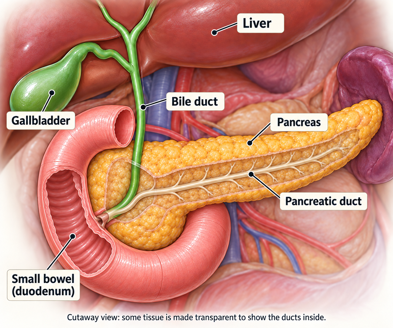
Liver
Makes bile; patient right is shown on the viewer’s left.
Gallbladder
Stores and concentrates bile beneath the liver.
Hepatic ducts
Right and left branches join as bile leaves the liver.
Cystic duct
Links the gallbladder neck to the main duct pathway.
Common bile duct
Descends beside the pancreatic head toward the bowel.
Pancreas & duodenum
The lower duct enters the C-shaped first part of the small bowel.
Four main cancer sites
“Biliary tract cancer” is a general name for cancers of the gallbladder and bile ducts. Their treatment can differ depending on where they start.
1 Gallbladder
Starts in the gallbladder wall beneath the liver.
2 Intrahepatic
Starts in the small bile ducts within the liver.
3 Perihilar
Starts where the right and left hepatic ducts meet at the liver hilum.
4 Distal
Starts in the lower common bile duct near or through the pancreas.
Symptoms and warning signs
Symptoms depend on the site. A tumour inside the liver may cause vague discomfort, while a tumour in a major duct may block bile early.
Possible symptoms
Yellow skin or eyes; dark urine; pale stools; itching; pain in the upper right abdomen; nausea; appetite loss; weight loss; fatigue; fever or chills.
Gallbladder cancer
May be found unexpectedly after surgery for gallstones. Pain, a lump, nausea or weight loss can occur, but early disease may be silent.
Perihilar or distal cancer
Jaundice, itching, dark urine and pale stools are common because a large duct becomes narrowed.
Intrahepatic cancer
May cause a liver mass, upper abdominal pain, weight loss, fatigue or abnormal liver tests without jaundice.
Symptoms are not a diagnosis
Gallstones, inflammation of the liver or pancreas, bile duct narrowing and infections can cause similar problems. Persistent or unexplained symptoms need assessment, but most people with these symptoms do not have cancer.
Useful details to share with your team
Record | Useful detail |
|---|---|
Temperature | The highest reading, when it began and whether there were shaking chills. Do not delay urgent help to collect these details. |
Bile-flow clues | Change in eye/skin colour, urine, stool colour and severity of itching |
Pain and intake | Where the pain is, vomiting, fluids taken and any rapid weight change |
Risk factors
Usually there is no single answer. Risk factors increase probability; they do not prove what caused one person’s cancer, and most people with a risk factor never develop it.
Gallbladder cancer risk factors
- Long-standing gallstones and chronic gallbladder inflammation
- Gallbladder polyps (growths on the lining), especially larger or growing ones, and calcium build-up in the gallbladder wall (porcelain gallbladder)
- An unusual connection between the pancreatic and bile ducts, or bile ducts that are abnormally widened from birth (choledochal cysts)
- Increasing age, obesity and, in some regions, carrying Salmonella bacteria long term
Bile duct cancer risk factors
- Primary sclerosing cholangitis (a condition that scars and narrows bile ducts), choledochal cysts, and repeated bile duct stones or inflammation
- Cirrhosis (long-term liver scarring), chronic hepatitis B or C, and some conditions affecting how the liver processes nutrients
- Infection with liver flukes (parasitic worms) in parts of Asia where they are common
- Inflammatory bowel disease—mainly through its link with primary sclerosing cholangitis
- Smoking, excess alcohol and metabolic risk may contribute, especially for intrahepatic disease
Ask whether relatives need any action; for most families, they do not need special screening.
Jaundice and cholangitis: act early
A blocked bile duct can cause a yellow pigment called bilirubin to build up, causing jaundice. Infection in the bile ducts is called cholangitis and can become life-threatening quickly.
Jaundice
Yellowing of the eyes or skin, dark urine, pale or clay-coloured stools and often severe itching.
Cholangitis
Fever or chills plus jaundice and right-upper abdominal pain. Older or very unwell people may instead become weak, drowsy, confused or faint.
When to seek urgent help
- You have a fever of 38°C (100.4°F) or higher, shaking chills or feel suddenly very unwell.
- Jaundice worsens, urine becomes much darker, or a drainage bag suddenly stops filling.
- Pain rapidly worsens, vomiting prevents fluids, or you become confused, dizzy or faint.
- A stent or external drain moves, leaks, blocks, falls out, or the skin around it becomes red or painful.
Treatment may include
Blood tests and cultures, intravenous fluids, antibiotics and urgent restoration of bile flow with an endoscopic stent or an image-guided drain. Cancer treatment may pause until the infection and liver function improve.
Before leaving hospital, confirm
- What stent or drain was placed, where it sits and when it should be reviewed
- Which symptoms require a same-day call and which number works after hours
- When blood tests, antibiotics or the next oncology appointment are due
Tests and diagnosis
Your care team
This is a rare and technically complex cancer. Decisions are safest when specialists review the scans, pathology, liver function and treatment goals together.
Team member | Main role |
|---|---|
Hepatobiliary surgeon | Assesses whether surgery can remove the cancer and performs gallbladder, liver and bile duct operations |
Gastroenterologist / advanced endoscopist | Endoscopic retrograde cholangiopancreatography (ERCP), endoscopic ultrasound, tissue sampling and internal stents |
Interventional radiologist | Uses scan guidance to take tissue samples, place drains through the skin and perform treatments directed at the liver |
Radiologist | Interprets computed tomography (CT) and magnetic resonance imaging (MRI) scans. MRCP (magnetic resonance cholangiopancreatography) is an MRI examination of the bile and pancreatic ducts. |
Pathologist / molecular laboratory | Confirms cancer type, margins, nodes and biomarkers |
Medical oncologist | Plans chemotherapy, immunotherapy, targeted medicines and clinical trials (research studies of treatments) |
Radiation oncologist | Plans radiotherapy, including highly focused treatment or radiotherapy combined with chemotherapy |
Palliative-care, dietetic and nursing teams | Manage symptoms, nutrition, stents, treatment effects and practical support |
A second opinion is particularly valuable before a major resection, transplant pathway, or a final conclusion that surgery is not possible.
How cancer is diagnosed
No single test answers every question. The sequence may change if jaundice or infection needs treatment first.
Confirm the clinical problem with history, examination, liver tests and an initial ultrasound or scan.
Map the tumour with CT scans taken at different times after contrast dye, and/or MRI with detailed bile duct images (MRCP). Scan the chest and other areas where the cancer might have spread.
Decide whether bile drainage is needed and which ducts should be drained before any procedure.
Obtain tissue when it will change management. The safest method depends on tumour site and whether surgery or transplant is planned.
Review pathology, stage and resectability at a multidisciplinary meeting.
If the cancer cannot be removed by surgery or has spread to distant parts of the body (metastatic cancer), arrange broad molecular testing early enough to help guide later treatment.
Tests often happen in parallel
Blood
Tests of liver and kidney function, blood clotting and blood cell counts. CA 19-9 and CEA are tumour markers that can be measured in blood, but they cannot confirm cancer on their own.
Imaging
Ultrasound, CT, MRI/MRCP; positron emission tomography combined with CT (PET-CT) or laparoscopy in selected situations.
Tissue
Surgery, endoscopy, brushings, needle biopsy or image-guided sampling.
Biology
Examination of tissue under a microscope (histology), tests for particular proteins (immunohistochemistry) and tests for gene changes.
Scans and staging tests
Imaging should define the cancer site, vessels and ducts involved, spread to nodes or organs, and whether enough healthy liver will remain after surgery.
Test | What it can answer |
|---|---|
Ultrasound | A quick first look for gallstones, widened ducts, a gallbladder lump or a liver abnormality |
Multiphasic CT | Tumour size, relation to vessels and organs, lymph nodes, liver spread, chest disease and surgical anatomy |
MRI + MRCP | Detailed liver tissue and a non-invasive map of bile-duct narrowing and branch anatomy |
EUS | Endoscopic ultrasound (EUS) uses a thin flexible tube with an ultrasound probe to view the lower bile duct, pancreas and nearby nodes; it can guide selected biopsies |
ERCP | Uses a flexible tube passed through the mouth into the bowel to reach the bile ducts. It can show or treat a blockage, collect cells or tissue and place a stent (a small tube that keeps the duct open). |
PTC | Percutaneous transhepatic cholangiography (PTC) uses a needle through the skin and liver to show the ducts; a drain may be placed when endoscopic drainage is unsuitable |
PET-CT | May reveal unexpected spread in selected patients; it cannot replace high-quality CT/MRI |
Staging laparoscopy | Keyhole inspection of the abdomen may find small areas of cancer on organ surfaces or the abdominal lining that scans miss before a major operation |
Biopsy and your pathology report
Pathology confirms what the tumour is. It should be reviewed by someone familiar with hepatobiliary cancers, especially when the site of origin is uncertain.
How tissue may be obtained
- From a gallbladder or bile-duct operation
- Brushings or forceps biopsy during ERCP or percutaneous cholangiography
- Endoscopic-ultrasound-guided biopsy of a mass or suspicious lymph node
- Image-guided core biopsy of an intrahepatic mass or metastatic site
What the report should clarify
Item | Why it matters |
|---|---|
Cancer type | Most are adenocarcinomas; neuroendocrine, squamous/adenosquamous and other rare tumours need different discussion |
Grade | How abnormal and fast-growing the cells appear |
Depth and organ involvement | Helps determine the pathological T stage |
Lymph nodes | How many were examined and how many contain cancer |
Margins | R0 means no cancer is found at the cut edges; R1 means cancer cells are seen there under a microscope; R2 means visible cancer remains after surgery |
Biomarkers | May identify an immunotherapy or targeted-treatment option |
Molecular testing: finding possible treatment targets
Cancers starting in the same place can have different gene or protein changes, called biomarkers. These can help doctors choose a treatment. Testing is most important when cancer cannot be removed by surgery, has returned (recurrent cancer) or has spread to distant parts of the body.
Ask for a broad panel
Next-generation sequencing (NGS) can test many genes at once. Testing RNA, a molecule that carries genetic instructions, can help detect gene fusions: changes that join parts of different genes together. Some of these changes can be targeted by medicines.
A blood-based ‘liquid biopsy’ can help when tissue is scarce, but a negative blood test may need tissue confirmation.
Test | Potential relevance |
|---|---|
FGFR2 fusion/rearrangement | Mainly intrahepatic cholangiocarcinoma; FGFR inhibitors after prior therapy |
IDH1 mutation | Mainly intrahepatic disease; ivosidenib after progression |
HER2 IHC/ISH | IHC measures HER2 protein and ISH looks for extra copies of the HER2 gene. These changes are more common in gallbladder and bile duct cancers outside the liver and may guide HER2-targeted treatment. |
MSI/MMR and TMB | Tests of DNA repair (MSI/MMR) and the number of tumour gene changes (TMB) may help predict benefit from immunotherapy |
BRAF V600E | Medicines that block BRAF and MEK signals that help cancer cells grow |
NTRK, RET or NRG1 fusion | Rare changes that may guide a targeted medicine, either for bile duct cancer or under an approval covering several cancer types |
BRCA1/2, PALB2 and other repair genes | May influence platinum use, trials and possible inherited-risk assessment |
Planning your treatment
Understanding your stage
Site-specific TNM staging for biliary cancers
Gallbladder, intrahepatic, perihilar and distal bile duct cancers have separate staging rules. Find the starting site first, then read the matching group. In the descriptions below, stages 1–3 have no spread to distant parts of the body. Stage 4 is not always distant spread.
Lymph nodes are small immune-system structures. Regional nodes are those near or draining the cancer’s starting area; distant spread means cancer has reached a site outside that regional area. Stages describe findings, not a sequence that everyone will pass through.
Gallbladder cancer
Depth of growth and nodes matter more than size alone.
Gallbladder cancer: local tumour growth (T)
These illustrations explain the T part of TNM for gallbladder cancer. T1–T4 are not the same as overall stages I–IV: lymph nodes (N) and distant spread (M) also determine the stage. Bile duct cancers use different rules.
T1a and T1b
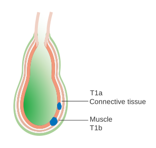
Growth into the supporting tissue beneath the lining (T1a) or the muscle layer (T1b).
T2
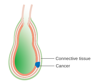
Growth beyond the muscle into surrounding connective tissue. T2a is on the side facing the abdominal cavity; T2b is on the liver side, without growing into the liver.
T3
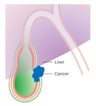
Growth through the outer surface, into the liver or into one nearby organ. This example shows growth towards the liver.
T4
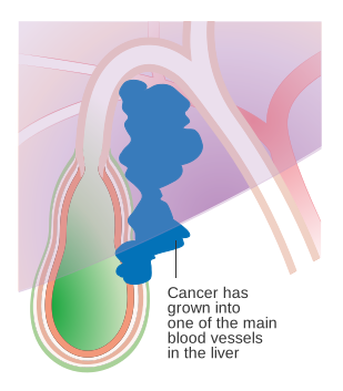
Growth into a main blood vessel supplying the liver, as shown here, or into two or more nearby organs outside the liver.
Stage 1 (I)
Cancer reaches the supporting layer or muscle of the gallbladder wall, with no cancer found in nearby lymph nodes.
Stage 2 (II)
Cancer reaches tissue around the muscle but not through the outer surface or into the liver. No cancer is found in nearby lymph nodes.
Stage 3 (III)
Cancer grows through the surface, into liver or one nearby organ, or involves 1–3 regional nodes, without stage 4 local features.
Stage 4 (IV)
Growth into major blood vessels or several nearby organs can be stage IVA. Cancer in four or more regional lymph nodes, or spread to distant parts of the body, is stage IVB.
Intrahepatic bile duct cancer: inside the liver
Separate liver tumours and blood-vessel involvement affect the stage.
Stage 1 (I)
One tumour, without growth into blood vessels or cancer in nearby lymph nodes.
Stage 2 (II)
Growth into blood vessels within the liver or more than one tumour, without cancer in nearby lymph nodes.
Stage 3 (III)
Growth through the liver covering, into nearby structures outside the liver, or into regional lymph nodes.
Stage 4 (IV)
Cancer has spread to distant parts of the body.
Perihilar bile duct cancer: at the liver’s hilum
The hilum is where the right and left liver ducts join.
Stage 1 (I)
Cancer stays within the duct wall, with no cancer in regional lymph nodes.
Stage 2 (II)
Growth beyond the duct into surrounding fat or liver, without cancer in regional lymph nodes.
Stage 3 (III)
Growth into particular nearby blood vessels or ducts, or cancer in 1–3 regional lymph nodes. Your team can explain which finding determines your stage.
Stage 4 (IV)
Stage IVA means cancer in at least 4 regional lymph nodes. Stage IVB means spread to distant parts of the body.
Distal bile duct cancer: nearer the bowel
Depth is measured under the microscope, not from the tumour’s overall width.
Stage 1 (I)
Growth less than 5 mm deep into the duct wall, with no cancer in regional lymph nodes.
Stage 2 (II)
Deeper growth or cancer in 1–3 regional lymph nodes, without growth into the major arteries that define stage 3.
Stage 3 (III)
Cancer in at least 4 regional lymph nodes, or growth into particular major arteries.
Stage 4 (IV)
Cancer has spread to distant parts of the body.
Stage 0, when used, means non-invasive abnormal cells in the lining. Whether surgery can remove the cancer safely (resectability) is a separate decision involving bile ducts, major vessels, the amount of healthy liver that would remain and your fitness. A small tumour can still sit in a difficult position.
Clinical stage comes from examination, biopsies and scans. Pathological stage can add information from surgery; a prefix such as yp means the tissue was assessed after treatment. A revised assessment does not necessarily mean the cancer grew while you were waiting. Grade describes the cells’ behaviour and is different from stage.
Incidental gallbladder cancer
Sometimes cancer is found only after the gallbladder has been removed for stones or inflammation. The specialist team should promptly review the operation record, tissue slides, cut edges and new scans. If the cancer reached the muscle layer (T1b) or deeper, a second operation may be recommended when there is no spread and complete removal is possible.
Resectability can change
- A tumour considered unsuitable for surgery at one centre may be operable at a specialist liver and bile duct centre that performs these operations regularly.
- Drainage, nutrition and infection control may make liver surgery safer. In selected cases, a procedure called portal-vein embolisation helps the part of the liver that will remain grow before surgery.
- Systemic or local treatment can occasionally shrink disease enough for reconsideration, but this is not guaranteed.
- New distant spread or inadequate future liver function can make surgery unsafe even when the primary tumour is technically removable.
You can also ask whether the team plans to review the possibility of surgery again after treatment.
Your treatment options at a glance
Treatment planning starts with whether all the cancer can be removed safely. Test results, your overall health and your preferences help guide the next steps.
↓
↓
YES — localised and resectable
Surgery for the particular cancer site → review the removed tissue → discuss capecitabine after surgery, or chemotherapy with radiotherapy in selected situations → follow-up checks.
NO — locally advanced or metastatic
Treat jaundice or infection → chemotherapy with immunotherapy, or local treatment in selected situations → review the response → consider treatment matched to test results or another treatment if needed.
Treatment goals
Goal | Meaning |
|---|---|
Cure | Complete removal or, rarely, a transplant-based pathway |
Reduce recurrence | Treatment after surgery to destroy microscopic disease |
Control | Slow growth, shrink disease, prolong life and preserve function |
Relieve symptoms | Restore bile flow, reduce pain/itching and support nutrition and wellbeing |
Treatment options
Topics in this section
- Surgery offers the main chance of cure
- Gallbladder cancer surgery
- Intrahepatic cholangiocarcinoma surgery
- Perihilar and distal bile duct surgery
- Recovery after surgery
- Treatment after surgery
- When the cancer cannot be removed now
- First treatment: chemotherapy with immunotherapy
- When treatment needs to change
- Targeted medicines: FGFR2, IDH1 and BRAF
- Targeted medicines: HER2 and rare gene changes
- Radiotherapy and liver-directed treatment
Surgery offers the main chance of cure
The operation depends on where the cancer started. The aim is to remove it completely, with no cancer seen at the cut edges under a microscope (an R0 resection), while preserving enough healthy liver and digestive function.
Before committing to a major operation
- Complete staging of the chest, abdomen and pelvis, with MRI/MRCP when duct anatomy needs definition
- Review of the main blood vessels supplying the liver (the portal vein and hepatic artery) and the liver tissue that would remain after surgery
- Assessment of liver function, kidney function, nutrition, heart/lung fitness and frailty
- A drainage plan for selected jaundiced patients—not automatic drainage of every blocked duct
- Possible staging laparoscopy to look for small deposits that imaging can miss
- A clear discussion of expected benefit, complications, hospital stay and recovery
Margin and nodes matter
The surgeon usually removes the tumour with a rim of normal tissue and regional lymph nodes. The final pathology determines stage, prognosis and whether additional treatment is advised.
Gallbladder cancer surgery
The operation depends on how deeply the cancer has grown into the gallbladder wall and whether the first gallbladder removal (cholecystectomy) was done for known cancer or suspected gallstones.
Situation | Common approach |
|---|---|
T1a, completely removed | Gallbladder removal alone is usually enough, followed by regular checks |
T1b or deeper, no spread | A wider operation removes nearby liver tissue (a wedge or segments IVb–V) and regional lymph nodes. Bile duct removal is added only when needed to obtain a cancer-free cut edge. |
Incidental cancer after cholecystectomy | Specialist review of the tissue and scans; a second, wider cancer operation may be considered for T1b or deeper disease |
Large or locally advanced but potentially resectable | A larger operation tailored to the cancer; treatment before surgery may be considered at specialist centres or in trials |
Distant spread or unresectable anatomy | Systemic treatment and symptom control rather than radical gallbladder surgery |
What may be removed
- Gallbladder and the liver tissue directly beneath it
- Lymph nodes along the bile duct and hepatic vessels
- The extrahepatic bile duct if the cystic-duct margin or tumour location requires it
- Occasionally an adjacent organ or vascular segment when an R0 resection remains realistic
Intrahepatic cholangiocarcinoma surgery
This operation removes the liver tumour, an adequate margin and regional lymph nodes while leaving a future liver remnant large and healthy enough to function.
Possible operations
Limited liver resection
Removes the tumour and a margin while preserving more liver; suitable anatomy is essential.
Lobectomy or extended resection
Removes a larger liver section when needed to clear major branches or multiple nearby lesions.
Vascular/bile-duct reconstruction
Selected tumours require reconstruction, increasing complexity and risk.
Portal-vein embolisation
Blocks flow to the planned resection side so the future liver remnant grows before surgery.
What influences resectability
- Number and distribution of tumours, vascular invasion and regional nodes
- Underlying cirrhosis, fatty liver or cholangitis and the predicted future liver volume
- Whether disease outside the planned resection can be cleared
- The patient’s overall fitness and ability to recover from major liver surgery
Perihilar and distal bile duct surgery
These tumours are close to the liver hilum or pancreas, so their operations differ substantially.
Perihilar cancer
Usually removes the affected bile ducts, a small part of the liver called the caudate lobe, and a right- or left-sided liver section, along with lymph nodes. The surgeon creates a new route for bile to drain; blood vessels may also need repair or reconstruction.
Distal cancer
Usually requires pancreaticoduodenectomy (Whipple): removal of the pancreatic head, distal bile duct, gallbladder and duodenum, followed by reconstruction.
Preparing the liver for perihilar surgery
- Drain the future liver remnant selectively when jaundice, cholangitis or the operation plan makes this necessary.
- Treat infection and improve nutrition before major resection.
- Use portal-vein embolisation in selected patients when the future liver remnant is too small.
A transplant pathway for selected perihilar cancer
Some people with early, unresectable perihilar cholangiocarcinoma may be considered for a strict protocol involving chemoradiation, staging and liver transplant at a specialist centre. Eligibility is narrow.
The biopsy route and previous operations may affect candidacy, so referral should occur before a needle biopsy through the abdominal lining when transplant is a possibility.
Before a transplant-centre referral
- Do not assume every tissue-sampling route is equivalent; ask the specialist centre first.
- Gather a CD containing the original scan images (DICOM format) with viewing software, the written scan reports, drainage records, pathology and complete procedure notes.
- Clarify whether infection control and biliary drainage are adequate for assessment.
Recovery after surgery
Recovery depends on the operation. Cholecystectomy is very different from a major liver resection or Whipple procedure.
Watch area | What the team monitors |
|---|---|
Liver and bile flow | Bilirubin, liver enzymes, bile leak, jaundice and cholangitis |
Infection and healing | Fever, wound changes, fluid collections, drains and blood counts |
Nutrition and digestion | Appetite, weight, nausea, diarrhoea, fat digestion and vitamin status |
Pancreas after Whipple | Leakage of pancreatic fluid, too few digestive enzymes, and new or changing diabetes |
Clots and mobility | Early walking, breathing exercises and medicines to prevent blood clots when appropriate |
Pathology | Final stage, nodes, margins and the recommendation for adjuvant therapy |
Call the surgical team for
- Fever, chills, increasing jaundice, worsening abdominal pain or repeated vomiting
- Redness, pus, opening of a wound, or a sudden change in drain output
- Breathlessness, chest pain, one-sided leg swelling, fainting or confusion
- Inability to eat or drink, persistent diarrhoea, pale greasy stools or rapid weight loss
Before discharge, know
- Which drains, wounds or medicines need daily attention and who will remove them
- The nutrition, enzyme, diabetes and activity plan for the first weeks at home
- The emergency number and the date for pathology and oncology review
Treatment after surgery
Even when surgery removes all visible cancer and the cut edges are clear, tiny groups of cells may remain elsewhere. Treatment after surgery, called adjuvant treatment, aims to reduce the chance of cancer returning; it does not mean cancer is known to remain.
Capecitabine after surgery
For many resected gallbladder and bile-duct cancers, about six months of oral capecitabine is the preferred adjuvant therapy, based largely on the BILCAP study and international guidelines. The decision considers recovery, stage, nodes, margins, liver function and patient preference.
Situation | Discussion may include |
|---|---|
R0, lower risk | Capecitabine or, in selected low-risk cases, surveillance |
Node-positive or higher stage | Capecitabine; a clinical trial; occasionally another chemotherapy regimen in centre-specific practice |
R1 microscopic margin | Systemic therapy and possible chemoradiation, especially for extrahepatic or gallbladder sites |
R2 visible residual disease | Treat as unresectable disease with systemic and/or local symptom-directed therapy |
Capecitabine practical points
- Tablets are taken in cycles; the dose is adjusted for kidney function and tolerance.
- Report diarrhoea, mouth ulcers, fever, chest pain and painful redness or peeling of hands/feet early.
- Do not change or stop the dose without contacting the oncology team.
When the cancer cannot be removed now
Cancer may be unresectable because of its location or spread, how much healthy liver would remain, or your overall health. Ask whether treatment aims to shrink it enough to reconsider surgery, control it for longer or relieve symptoms.
Address problems that could delay treatment
- Drain a clinically important obstruction and treat cholangitis
- Treat dehydration, kidney problems and imbalances in blood salts (electrolytes)
- Address pain, itching, nausea, weight loss and poor intake
- Obtain adequate tissue and molecular testing
Treatment routes
Systemic therapy
Chemo-immunotherapy is preferred for many fit patients with locally advanced or metastatic disease.
Radiation / chemoradiation
May control a tumour threatening nearby structures, build on the response to earlier treatment in selected cases, or relieve pain and bleeding.
Liver-directed treatment
Ablation, SBRT, embolisation or Y-90 may be considered for selected liver-dominant intrahepatic cancer.
Clinical trial
May be particularly useful for a rare cancer, an unusual test result or treatment aimed at shrinking the cancer enough to reconsider surgery.
First treatment: chemotherapy with immunotherapy
For many adults with untreated, unresectable or metastatic biliary tract cancer, a preferred treatment is gemcitabine plus cisplatin chemotherapy with either durvalumab or pembrolizumab immunotherapy.
Option | Evidence and practical meaning |
|---|---|
Durvalumab + gemcitabine/cisplatin | In the TOPAZ-1 trial, people receiving this combination lived longer on average than those receiving chemotherapy alone. Durvalumab commonly continues after the initial chemotherapy course to help maintain control. |
Pembrolizumab + gemcitabine/cisplatin | In the KEYNOTE-966 trial, adding pembrolizumab also helped people live longer on average than chemotherapy alone. Pembrolizumab continues after the planned cisplatin course when tolerated. |
What a typical schedule looks like
Gemcitabine and cisplatin are commonly given on days 1 and 8 of a 21-day cycle, with the immunotherapy on day 1. Cisplatin is usually limited to an initial course; immunotherapy may continue as maintenance treatment to help keep the cancer under control.
Exact doses, cycle count and maintenance duration follow the chosen protocol and the patient’s tolerance.
Choosing and adapting treatment
- No trial directly comparing durvalumab with pembrolizumab has established that one is better.
- Kidney function, hearing, nerve symptoms such as numbness or tingling (neuropathy), infection, bilirubin levels and how well you manage daily activities may affect treatment timing, dose or choice of chemotherapy.
- Some frail patients benefit more from gentler chemotherapy or symptom-focused care than from a three-drug regimen.
When treatment needs to change
Treatment may change because scans show growth, side effects outweigh benefit, infection or liver dysfunction intervenes, or a biomarker result creates a better option.
Second-line and later treatment
Route | When it may fit |
|---|---|
FOLFOX | Fluorouracil (5-FU), leucovorin (folinic acid) and oxaliplatin. This is the best-established chemotherapy after gemcitabine–cisplatin, supported by the ABC-06 trial. |
FOLFIRI or other fluoropyrimidine/irinotecan therapy | FOLFIRI combines fluorouracil, leucovorin and irinotecan. It or a related combination may be used when oxaliplatin is unsuitable or after other treatments; the evidence is less definitive. |
Liposomal irinotecan + 5-FU/leucovorin | Used in some regions and centres, particularly after gemcitabine-based treatment; availability and guideline position vary |
Biomarker-matched therapy | Often preferred when a validated target such as FGFR2, IDH1, HER2, BRAF V600E, MSI-H/dMMR, NTRK, RET or NRG1 is present |
Clinical trial | Worth discussing when available: established treatments after the first treatment can offer limited benefit, and trials explore other options |
How the next choice is made
- Previous benefit and toxicity, neuropathy, blood counts, kidney/liver function and current fitness
- Cancer pace and location, symptoms, stent reliability and need for rapid shrinkage
- Molecular results, medicine access, cost and the patient’s priorities
Targeted medicines: FGFR2, IDH1 and BRAF
These changes are found mainly in bile duct cancers inside the liver. Treatment needs a reliable laboratory result and careful monitoring for side effects.
Biomarker | Treatment examples | Important monitoring |
|---|---|---|
FGFR2 fusion | Pemigatinib after previous systemic therapy. Futibatinib is an option for adults with previously treated, unresectable, locally advanced or metastatic intrahepatic cholangiocarcinoma with an FGFR2 fusion or rearrangement. Lirafugratinib is another option for adults with previously treated, unresectable locally advanced or metastatic cholangiocarcinoma with an FGFR2 fusion or rearrangement. | Phosphate level, eyes/retina, mouth, nails, skin, liver tests and drug interactions |
IDH1 mutation | Ivosidenib after progression | Heart tracing (ECG), including the QT interval (part of the heart’s electrical cycle), blood salts, liver tests, medicine interactions and symptoms of an abnormal heartbeat |
BRAF V600E | Dabrafenib + trametinib | Fever, rash, heart function, eyes, blood pressure and secondary skin changes |
FGFR treatment practical points
- FGFR2 fusions are different from isolated FGFR changes of uncertain significance; the exact report matters.
- Phosphate, a mineral in the blood, can rise during treatment. This may need dietary advice, medicines that reduce phosphate absorption or a dose adjustment.
- Blurred vision, flashes or new floaters need prompt eye review.
- Resistance can emerge; a clinical trial or repeat molecular test may sometimes clarify a new option.
Targeted medicines: HER2 and rare gene changes
Some biliary cancers make unusually large amounts of HER2 protein or have a rare gene fusion. These findings may guide treatment. You do not need every medicine listed below: your oncologist will explain which, if any, matches your test results and previous treatment.
Result | Possible approach |
|---|---|
HER2-positive | Zanidatamab is an option for previously treated unresectable/metastatic HER2 IHC 3+ biliary tract cancer. Trastuzumab-based combinations or trastuzumab deruxtecan may be considered in selected settings. |
MSI-H / dMMR or TMB-high | Checkpoint immunotherapy may be effective; previous first-line immunotherapy and treatment-specific eligibility influence the choice. |
NTRK fusion | Larotrectinib or entrectinib under approvals based on the gene change rather than the organ where the cancer began |
RET fusion | Selpercatinib under a gene-based approval covering several cancer types, after previous treatment or when no satisfactory option exists |
NRG1 fusion | Zenocutuzumab is an option for adults with advanced unresectable or metastatic cholangiocarcinoma that has an NRG1 fusion and has progressed during or after previous systemic treatment. |
BRCA/PALB2 or other repair gene | May help guide platinum chemotherapy, genetic counselling or a clinical trial. PARP inhibitors (medicines affecting DNA repair) are not an established treatment for all bile duct cancers with these changes. |
HER2 safety differs by drug
HER2 antibodies can cause infusion reactions and reduced heart function. Trastuzumab deruxtecan can cause serious lung inflammation (interstitial lung disease); new cough or breathlessness needs urgent assessment. Zanidatamab commonly causes diarrhoea and infusion reactions and can harm an unborn baby.
Radiotherapy and liver-directed treatment
Local treatment can complement systemic therapy when disease is confined, liver-dominant, threatening a structure or causing symptoms.
Approach | Possible role |
|---|---|
External-beam radiotherapy | Local control after selected R1 surgery, control of unresectable disease, or relief of pain/bleeding |
Chemoradiation | Radiotherapy with a fluoropyrimidine chemotherapy medicine that makes cancer cells more sensitive to radiation, in selected gallbladder or bile duct cancers outside the liver |
SBRT | Stereotactic body radiotherapy (SBRT) gives highly focused radiation to a limited tumour or area of spread, when nearby bowel and ducts can be protected |
Thermal ablation | Uses heat to destroy selected small tumours inside the liver, away from vulnerable bile ducts or blood vessels |
TACE / bland embolisation | Blocks the tumour’s blood supply, with chemotherapy (TACE) or without it (bland embolisation). Used in selected cancers mainly within the liver; evidence and practice vary. |
Y-90 radioembolisation | Delivers tiny radioactive beads through arteries supplying the liver, for selected cancers mainly within the liver |
Planning protects normal tissue
- Previous operations, stents and infections affect radiation and embolisation risk.
- The liver, stomach, duodenum, bowel, kidneys and spinal cord have dose limits.
- Combining local and systemic therapy requires a deliberate sequence and recovery plan.
Managing side effects
During chemo-immunotherapy
Side effects vary. Reporting symptoms early helps the team offer medicines, adjust doses and treat inflammation caused by immunotherapy before it becomes severe.
Treatment | Common or important effects |
|---|---|
Gemcitabine | Low blood counts, fever/infection risk, fatigue, nausea, rash, flu-like symptoms and liver-test changes |
Cisplatin | Nausea, kidney injury, low magnesium, hearing change, ringing in the ears (tinnitus) and nerve damage causing numbness or tingling |
Durvalumab / pembrolizumab | Immune inflammation of thyroid, bowel, liver, lungs, skin, kidneys, pituitary or other organs; can occur during or after treatment |
Before each cycle
- Blood count, kidney/liver function and electrolytes; thyroid tests at intervals
- Review of fever, jaundice, stent/drain function, new breathlessness, diarrhoea, rash or unusual fatigue
- Hydration and anti-sickness plan; hearing assessment when clinically indicated
- Dose and timing adjusted to recovery—not simply to the calendar
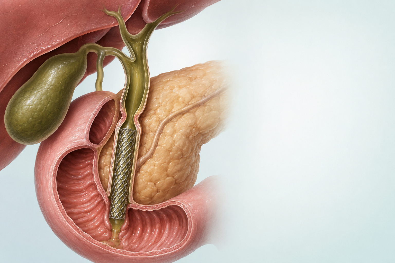
Biliary drainage and stents
Drainage can relieve jaundice, treat or prevent infection, improve liver function and make surgery or systemic treatment safer—but it also carries risks.
Method | Typical use |
|---|---|
ERCP stent | A plastic or metal tube keeps the duct open. It is placed using a flexible tube passed through the mouth into the bowel, often for a lower bile duct blockage. |
Percutaneous drain/stent | Placed through the liver by interventional radiology, often for high or complex obstruction |
EUS-guided drainage | Specialist alternative when standard ERCP is not possible |
Surgical bypass | Creates another route for bile in selected operative or palliative situations |
Live well
Supportive care and nutrition
Supportive and palliative care helps with symptoms, emotional wellbeing and practical needs. It can be provided alongside cancer treatment at any stage.
Problem | Ways the team can help |
|---|---|
Itching / jaundice | Drainage when appropriate, skin care, prescribed medicines and treatment of obstruction |
Pain | Pain-relieving medicines, nerve blocks, radiotherapy, drainage and specialist palliative care |
Nausea / poor appetite | Anti-sickness medicines, small frequent meals, review of obstruction/infection and dietetic support |
Weight and muscle loss | Protein-rich food, strength/activity plan, supplements when useful and treatment of reversible causes |
After Whipple | Pancreatic enzyme replacement for pale, greasy or floating stools; diabetes monitoring |
Fatigue and mood | Treat anaemia, infection, sleep and pain; graded activity; counselling and family support |
Fluid in the abdomen (ascites) / swelling | Salt and fluid advice when relevant, medicines, drainage and liver-function review |
Eating with biliary cancer
- Do not adopt a severe low-fat or ‘anti-cancer’ diet without advice; unnecessary restriction can worsen weight loss.
- Aim for frequent energy- and protein-rich meals, enough fluid and safe food handling during low blood counts.
- Tell the team about herbal or Ayurvedic products because some can injure the liver or interact with treatment.
Follow-up after treatment
Follow-up checks for signs that cancer has returned, manages lasting treatment effects, monitors stents and supports recovery. Your team will agree a schedule with you. Report new symptoms between visits rather than waiting for the next appointment.
After curative-intent treatment
A common approach is clinical review, imaging and tumour markers such as CA 19-9 and CEA every 3–6 months for the first 2 years, then every 6–12 months up to 5 years, adjusted for site, stage, treatment and symptoms.
CA 19-9 is most useful when it was high before treatment. Jaundice or infection can also raise it, so your team interprets the result alongside symptoms and scans.
Sources and further reading
For more information, explore these resources from the National Cancer Institute (NCI) and other medical organisations. The linked organisations have not reviewed or endorsed this CTI guide. Your care team can explain how the information applies to you.
- NCI: Gallbladder Cancer Treatment (PDQ®)–Patient Version
- NCI: Bile Duct Cancer Treatment
- Cancer Council Australia: gallbladder and bile duct cancer
Staging and risk-group references
AJCC 8th edition.
Medicine approval and prescribing references
These official records describe licensed uses and prescribing information for the medicines named in this guide. Approval depends on the cancer type, biomarkers, previous treatment, combination and formulation. Some uses discussed in the guide are off-label: outside the medicine’s licence, with evidence from clinical guidelines or studies. FDA or EMA approval does not establish approval or availability in India.
- Capecitabine — FDA prescribing information · Approval history. Adjuvant bile-duct/gallbladder treatment is outside the FDA-labelled cancer indications; see the clinical guidance.
- Cisplatin — FDA prescribing information · Approval history
- Dabrafenib — FDA prescribing information · Approval history
- Durvalumab — FDA prescribing information · Approval history
- Entrectinib — FDA prescribing information · Approval history
- Fluorouracil — FDA prescribing information · Approval history
- Futibatinib — FDA prescribing information · Approval history
- Gemcitabine — FDA prescribing information · Approval history
- Infigratinib — FDA withdrawal record. The FDA cholangiocarcinoma indication was withdrawn in May 2024; listed here as a withdrawal reference.
- Irinotecan — FDA prescribing information · Approval history
- Ivosidenib — FDA prescribing information · Approval history
- Larotrectinib — FDA prescribing information · Approval history
- Leucovorin (folinic acid) — FDA prescribing information · Approval history
- Liposomal irinotecan — FDA prescribing information · Approval history. The linked FDA approval is for pancreatic cancer, not bile-duct cancer.
- Lirafugratinib — FDA prescribing information · Approval history
- Oxaliplatin — FDA prescribing information · Approval history
- Pembrolizumab — FDA prescribing information · Approval history
- Pemigatinib — FDA prescribing information · Approval history
- Selpercatinib — FDA prescribing information · Approval history
- Trametinib — FDA prescribing information · Approval history
- Trastuzumab — FDA prescribing information · Approval history. The single-antibody label does not establish a bile-duct cancer approval.
- Trastuzumab deruxtecan — FDA prescribing information · Approval history. A tumour-agnostic indication applies to eligible HER2 IHC 3+ disease.
- Zanidatamab — FDA prescribing information · Approval history
- Zenocutuzumab — FDA prescribing information · Approval history
Additional medicine approval notices
These records explain the specific medicine uses and eligibility requirements discussed in this guide.
Related care
Meet our doctors



Where to get care