
Share
Start here
Colon cancer at a glance
This guide covers adenocarcinoma, the most common type of colon cancer, which starts in the gland-forming cells of the bowel lining. Neuroendocrine tumours, gastrointestinal stromal tumours, lymphomas, sarcomas and anal cancers follow different pathways.
Colon cancer starts in the large bowel above the rectum. It is often curable when confined to the bowel and nearby lymph nodes, and some cancers that have spread can still be treated with the aim of a cure.
What stage means
Stage describes depth through the bowel wall, lymph nodes and spread to distant organs.
Main curative treatment
Surgery removes the affected colon segment and its draining lymph nodes.
Why biomarkers matter
Mismatch repair (MMR) and microsatellite instability (MSI) tests check how cancer cells repair DNA errors. These and other gene or protein tests can guide treatment after surgery and when cancer has spread.
Colon is not the same as rectum
Colon cancer | Rectal cancer |
|---|---|
Usually treated first with surgery when localised. | Pelvic magnetic resonance imaging (MRI), radiotherapy and treatment before surgery are often central. |
Radiotherapy has little routine role. | Radiotherapy may reduce pelvic recurrence or help avoid surgery in selected responders. |
The part removed during colon surgery (colectomy) depends on where the cancer is. | Surgery follows the tissue layer around the rectum and aims to protect the muscles that control bowel movements and the nearby pelvic nerves where possible. |
Bring your scan images
For your consultation or a second opinion, bring a CD containing your scan images (DICOM format) and the software needed to view them, along with the written scan reports.
Understand
The colon: route, segments and function
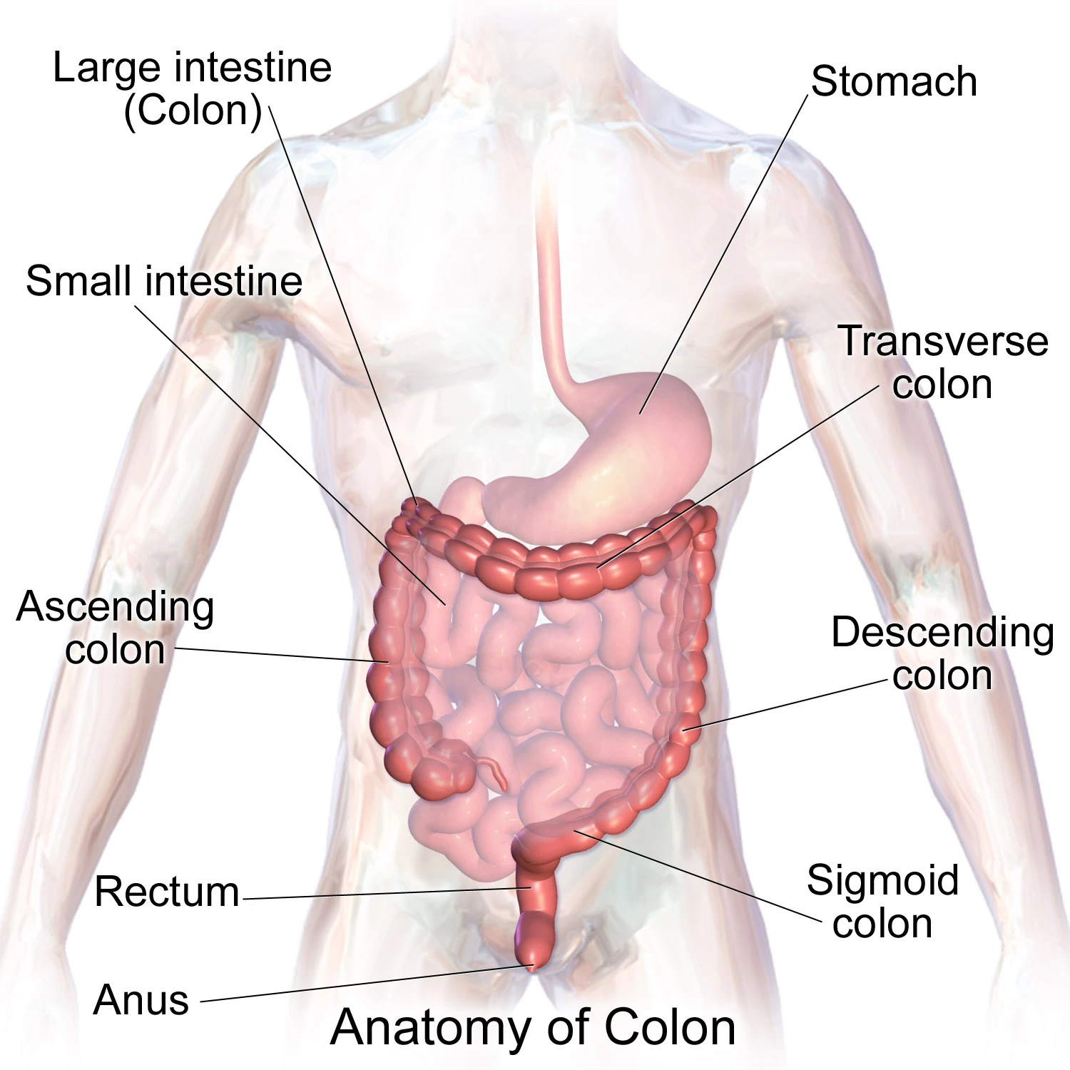
The colon absorbs water and salts, turns bowel contents into formed stool and moves stool towards the rectum. The side of the tumour can influence symptoms and metastatic treatment choices.
Segment | Practical point |
|---|---|
Caecum and ascending colon | Right-sided; bleeding may be hidden and present as iron-deficiency anaemia. |
Transverse and descending colon | Symptoms vary; narrowing may cause changing bowel habit or cramps. |
Sigmoid colon | Left-sided; the narrower passage can make blockage and visible bleeding more likely. |
Rectum | A separate pelvic organ with a different local-treatment pathway; not covered here. |
From normal lining to invasive cancer
Most colon cancers develop through accumulated cell changes over years. Removing pre-cancerous polyps during colonoscopy can prevent some cancers.
Normal lining
Millions of lining cells renew in a controlled way.
A polyp forms
An adenoma or serrated lesion grows from the inner lining. Most polyps are not cancer.
High-grade dysplasia
Cells look very abnormal but have not grown through the thin muscle layer within the bowel lining (muscularis mucosae).
Invasive adenocarcinoma
Cancer crosses into deeper bowel-wall layers and can enter lymphatic or blood vessels.
Regional or distant spread
Cells may reach nearby lymph nodes, the liver, lungs, peritoneum or other sites.
A cancer found inside a removed polyp
Some malignant polyps need no further surgery if excision is complete and pathological risk is low. Deep submucosal invasion, poor differentiation, lymphovascular invasion, tumour budding, a close/positive margin or piecemeal removal may favour formal colectomy with lymph-node removal.
Symptoms can differ on the right and left
Colon cancer may cause no symptoms early. Persistent or unexplained changes deserve assessment even when piles, diet or infection seem possible explanations.
More typical of right-sided cancer | More typical of left-sided cancer |
|---|---|
Iron-deficiency anaemia, fatigue, breathlessness or paleness | Visible red or dark blood mixed with stool |
Vague abdominal discomfort, weight loss or a mass | New constipation, diarrhoea, alternating habit or narrower stool |
Bleeding that is not visible in the stool, without a major change in stool shape | Cramping, bloating and progressive obstruction |
Symptoms anywhere in the colon
- Unexplained weight loss or reduced appetite.
- Persistent abdominal pain or bloating.
- A feeling that bowel habit has changed from your normal.
- A positive stool blood test, even if you feel well.
Risk, prevention and screening
Most people with colon cancer have no single cause. Risk factors change probability; they do not assign blame and do not prove why one person developed cancer.
Higher-risk history
Previous colorectal cancer or advanced polyps; longstanding ulcerative colitis or Crohn's colitis; a strong family history or inherited syndrome.
Age and biology
Risk rises with age, but younger adults can develop colon cancer. Persistent symptoms should not be dismissed because of age.
Modifiable associations
Smoking, excess alcohol, obesity, low activity, high processed-meat intake and low fibre are associated with higher risk.
Protective habits
Regular physical activity, healthy weight, avoiding tobacco and a fibre-rich diet support general and bowel health.
Screening is for people without symptoms
Programmes use stool testing and/or colonoscopy according to age and risk. A positive stool test is not a cancer diagnosis; it identifies who needs colonoscopy. People with symptoms or high inherited risk need diagnostic or surveillance pathways, not routine screening alone.
Lynch syndrome, polyposis and germline testing
An inherited gene change that increases cancer risk can affect surgery, follow-up, treatment and advice for relatives. Tumour testing and germline testing answer different questions.
Condition | Clues and implications |
|---|---|
Lynch syndrome | Pathogenic variants in MLH1, MSH2, MSH6, PMS2 or EPCAM. Often few polyps; increased colorectal and several other cancer risks. Tumour MMR testing is the usual first clue. |
Familial adenomatous polyposis (FAP) | APC-related; many adenomatous polyps, often from youth. Colectomy and lifelong upper/lower gastrointestinal surveillance may be needed. |
MUTYH-associated polyposis | Usually requires pathogenic variants in both MUTYH copies; polyp burden varies. Siblings may be at risk. |
Other syndromes | POLE/POLD1, NTHL1, hamartomatous-polyposis and other rare syndromes require specialist interpretation. |
When to ask for genetic counselling
- Cancer at a young age, multiple primary cancers or many polyps.
- A close relative with colorectal, endometrial or related cancers, especially young.
- Loss of an MMR protein or MSI-high tumour without a clearly non-inherited explanation.
- A known familial pathogenic variant or a pattern that concerns your clinician.
Tests and diagnosis
How cancer is diagnosed
Diagnosis has three separate jobs: confirm cancer in tissue, define how far it has spread, and understand features that may change treatment.
Clinical assessment
Your symptoms, bowel and medical history, family history, medicines, examination and ability to carry out everyday activities.
Blood tests
Blood counts, iron levels, kidney and liver function, and a starting carcinoembryonic antigen (CEA) level. CEA is a blood marker used for later comparison; it can be normal even when cancer is present.
Colonoscopy
Inspect the whole colon where possible, biopsy the lesion, remove other polyps and mark the site when helpful.
Pathology
Confirm adenocarcinoma and report differentiation; later, the resection specimen defines depth, nodes and margins.
Staging scans
Computed tomography (CT) chest/abdomen/pelvis is usual. Liver MRI, positron emission tomography combined with CT (PET-CT) or other tests answer selected questions.
Multidisciplinary team (MDT) review
Surgeons, radiologists, pathologists and oncologists integrate results and treatment goals.
Colonoscopy, biopsy and the first report
Colonoscopy shows where the lesion is, whether it narrows the bowel and whether other polyps or cancers are present. The biopsy identifies the tumour type but cannot fully stage the bowel wall or lymph nodes.
Report item | What it tells you |
|---|---|
Site and distance landmarks | Which colon segment is involved and what operation may be needed. |
Appearance and obstruction | Length, circumferential involvement, narrowing and whether the scope could pass. |
Biopsy histology | Usually adenocarcinoma; uncommon tumour types need different management. |
Differentiation | How gland-forming the cells look. Grade from a small biopsy can change in the full specimen. |
Synchronous lesions | A second cancer or important polyps elsewhere in the colon. |
If the scope cannot pass
CT colonography or colonoscopy after surgery may be used to assess the remaining colon. The plan depends on urgency and whether obstruction is present.
Scans and staging tests
Imaging looks for spread, resectability and complications. No single scan answers every question, and tiny deposits may remain below imaging resolution.
Test | Main question | Important limitation |
|---|---|---|
Contrast CT chest/abdomen/pelvis | Liver, lung, nodes, peritoneum, local complications | Small lesions can be indeterminate or missed. |
Liver MRI | Are liver lesions metastases, and are they resectable/ablatable? | Usually selected when CT is uncertain or curative local treatment is considered. |
PET-CT | Is there otherwise hidden active disease in a selected curative-intent situation? | Inflammation can light up; small or mucinous disease can be less avid. Not routine for everyone. |
CEA | A baseline tumour marker for later comparison | Normal does not exclude cancer; smoking and benign conditions can raise it. |
Before contrast or treatment
- Tell the team about previous contrast reactions, kidney disease, diabetes medicines and pregnancy possibility.
- Baseline blood counts and kidney/liver function guide scan safety and drug dosing.
- Frailty, nutrition, heart/lung function and daily activity can be as important as age.
Understanding your pathology report after surgery
The surgical specimen supplies the most reliable local stage. Ask for the pT, pN, margins, number of nodes examined and high-risk features as separate items.
Feature | Plain-language meaning |
|---|---|
pT | How deeply the tumour has grown through the colon wall or into adjacent structures. |
pN and nodes examined | How many regional nodes contain cancer. Assessment of at least 12 nodes is an important quality benchmark when feasible. |
Margins | Whether cancer reaches a cut or radial surface. R0 means no microscopic cancer at the margin. |
Lymphovascular/perineural invasion | Cancer in vessels or around nerves; raises recurrence risk. |
Tumour deposits/budding | Additional patterns associated with recurrence risk and sometimes nodal staging. |
Grade and subtype | Poor differentiation, signet-ring and some mucinous patterns can influence risk interpretation. |
Planning your treatment
Understanding your stage
TNM staging for colon cancer
Think of the bowel wall as several layers: an inner lining, supporting tissue, muscle and outer tissue. Stage depends on the depth of growth, nearby lymph nodes and distant spread—not simply on the width of a tumour.
Lymph nodes are small immune-system structures. Regional nodes are those near or draining the cancer’s starting area; distant spread means cancer has reached a site outside that regional area. Stages describe findings, not a sequence that everyone will pass through.
T — Tumour depth
How far the cancer grows into or through the bowel wall. The width of the tumour alone does not determine T.
N — Nearby lymph nodes
Whether cancer is found in regional lymph nodes. Certain tumour deposits in nearby tissue also affect the N category.
M — Distant spread
Whether cancer has reached a distant part of the body, such as the liver, lungs or the lining of the abdominal cavity.
CTI illustration.
Stage 0
Abnormal or cancerous cells remain within the inner lining and have not entered the supporting layer beneath it. This is also called in situ disease.
Stage 1 (I)
Cancer has entered the supporting layer or muscle of the colon wall, but has not grown through the muscle. Nearby nodes and distant sites are not involved.
Stage 2 (II)
Cancer has grown through the muscle into outer tissue, the outer covering or an adjacent organ. No regional lymph-node spread is found. Some stage 2 cancers have features that increase recurrence risk.
Stage 3 (III)
Cancer involves regional lymph nodes, or particular deposits in nearby bowel-supporting tissue. It has not spread to distant sites. The depth of growth and number of involved nodes determine the subgroup.
Stage 4 (IV)
Cancer has reached a distant site, such as the liver, lung or abdominal lining. Direct growth into an organ next to the bowel is different from a separate distant deposit.
Surgery and examination of the removed bowel and nodes often refine the stage first estimated from scans. Selected people with a small amount of distant spread may still be offered treatment aiming to remove or destroy all visible disease. This needs a specialist assessment.
Clinical stage comes from examination, biopsies and scans. Pathological stage can add information from surgery; a prefix such as yp means the tissue was assessed after treatment. A revised assessment does not necessarily mean the cancer grew while you were waiting. Grade describes the cells’ behaviour and is different from stage.
How your treatment plan is chosen
The best plan combines anatomy, pathology, tumour biology, fitness and personal priorities. A single scan or single speciality should not decide a complex pathway alone.
Team member | Main contribution |
|---|---|
Colorectal surgeon | Operability, type of colectomy, anastomosis, stoma and surgical risk. |
Radiologist | Stage, liver/lung/peritoneal disease, complications and response. |
Pathologist | Histology, pTNM, margins, high-risk features and MMR. |
Medical oncologist | Adjuvant benefit, systemic therapy, biomarkers, toxicity and trials. |
Liver/thoracic/interventional team | Local treatment of limited metastases. |
Stoma, nutrition, rehabilitation and supportive teams | Preparation, recovery, symptoms, function, costs and quality of life. |
Questions to ask your care team
- Is the goal cure, lower recurrence risk, conversion to resection, disease control or symptom relief?
- Is treatment urgent, and is there obstruction, perforation or severe anaemia?
- Which uncertainties could change the plan, and what test resolves them?
- What matters most to you: the chance of cure, avoiding nerve damage (neuropathy), work, fertility, avoiding a stoma or treatment close to home?
Treatment options
Topics in this section
- Colectomy: what may be removed
- Preparing, recovering and living with a stoma
- Stage II: do I need chemotherapy?
- Stage III: treatment after surgery
- First-line treatment for metastatic colon cancer
- How biomarkers guide treatment after cancer has spread
- Newer treatment options
- Liver- or lung-limited metastatic disease
- Later-line treatment, trials and supportive care
Colectomy: what may be removed
The surgeon removes the affected part of the colon together with its nearby lymph nodes, following the blood vessels and lymph drainage. The aim is to remove the cancer with a clear edge of healthy tissue around it and, when safe, join the remaining bowel.
What cancer surgery aims to achieve
- Remove the tumour-bearing colon segment with adequate margins.
- Remove the associated mesocolon and draining lymph nodes intact.
- Examine enough nodes for reliable staging.
- Restore bowel continuity when safe, or create a temporary/permanent stoma when needed.
Operation | Typical location |
|---|---|
Right hemicolectomy | Caecum, ascending colon and sometimes proximal transverse colon. |
Extended right/left hemicolectomy | Tumours near flexures or transverse colon, depending on vascular drainage. |
Left hemicolectomy | Descending colon and selected distal transverse lesions. |
Sigmoid colectomy | Sigmoid colon; commonly joined to the upper rectum. |
Subtotal/total colectomy | Selected synchronous cancers, polyposis, extensive colitis or complex anatomy. |
Open, laparoscopic and robotic approaches
All three can be oncologically sound when performed by an appropriately trained team. Minimally invasive surgery often reduces wound pain and shortens recovery; open surgery may be safest for obstruction, bulky invasion, adhesions or multivisceral resection. Robotic surgery can help in selected anatomy but is not automatically superior.
Before agreeing to surgery
- Which part of the bowel and which lymph nodes will be removed.
- Chance of conversion to open surgery or removal of another organ.
- Risks of a leak where the bowel is joined, bleeding, infection, blood clots or a temporary slowing of bowel movement (ileus).
- Possibility and likely duration of a stoma.
- Enhanced recovery, pain plan and expected hospital stay.
Preparing, recovering and living with a stoma
Preparing your health and activity before surgery, then following a recovery plan, can reduce complications. A stoma is an opening made in the abdomen so stool can pass into a bag. It may be temporary or permanent, but many colon operations do not require one.
Before surgery
Correct anaemia, optimise diabetes and medicines, stop smoking, improve protein intake, walk/strengthen and plan home support.
Stoma marking
A stoma nurse marks a site that works with sitting, clothing, skin folds and hand function before surgery when possible.
Early recovery
Pain control, breathing exercises, early mobilisation, eating as tolerated and clot prevention are typical enhanced-recovery elements.
At home
Increase activity gradually, protect the wound, hydrate, follow diet advice and know whom to call for fever, vomiting or reduced output.
Stoma warning signs
- No output with cramps, swelling or vomiting.
- Very high watery output, dizziness, reduced urine or marked thirst.
- A dark/black stoma, severe pain, heavy bleeding or rapid swelling.
- Persistent leakage, broken skin or inability to keep the pouch on.
Stage II: do I need chemotherapy?
After the cancer has been completely removed, chemotherapy may treat cancer cells too small to see. In stage II, the extra benefit is often small. Your team will help you weigh the chance of the cancer returning against treatment side effects.
High-risk stage II features
- A tumour growing through the bowel’s outer covering or into a nearby organ (T4), a hole in the bowel (perforation) or bowel blockage.
- Fewer than 12 nodes examined when no clear explanation.
- Lymphovascular or perineural invasion, poor differentiation in an appropriate context, tumour budding or close/positive margin.
- Cancers with proficient mismatch repair (pMMR) or microsatellite stability (MSS) need a different treatment discussion from dMMR/MSI-H cancers.
Confirm pathology quality
pT stage, at least 12 nodes where feasible, margins, obstruction/perforation, invasion and tumour budding.
Check MMR/MSI
dMMR/MSI-H usually has a better stage II prognosis and changes fluoropyrimidine-alone benefit.
Estimate absolute benefit
Ask for benefit in percentage points, not only relative-risk language.
Match treatment intensity
Options include monitoring, a fluoropyrimidine medicine alone, or chemotherapy combinations called CAPOX or FOLFOX (explained below), depending on recurrence risk and overall health.
Include your priorities
Consider nerve damage, work, other health conditions, fertility, travel and which side effects you feel able to manage.
Where ctDNA fits
Circulating tumour DNA (ctDNA) is DNA released by cancer cells that can be found in the blood. Finding it after surgery is linked to a higher risk of the cancer returning. It is increasingly used in trials and selected centres, but a negative result does not prove zero risk and should not automatically override established pathological features. Ask whether the proposed use is guideline-supported or research-based.
Stage III: treatment after surgery
Treatment after surgery aims to lower the risk of cancer returning. The choice depends on your MMR result, recurrence risk and overall health.
Chemotherapy options
Oxaliplatin combined with a fluoropyrimidine medicine, such as fluorouracil or capecitabine, lowers the risk of cancer returning after stage III surgery. CAPOX is oral capecitabine plus oxaliplatin; FOLFOX uses infusional fluorouracil, leucovorin and oxaliplatin.
Issue | CAPOX | FOLFOX |
|---|---|---|
Schedule | Oxaliplatin every 3 weeks plus capecitabine tablets for 14 days | Infusion every 2 weeks, usually through a central line/pump |
Practical fit | Fewer infusion visits; tablets must be taken as prescribed, with dosing checked against kidney function | More visits and line care; useful when tablets are unsuitable |
Shared side effects | Neuropathy, low counts, fatigue, nausea, diarrhoea | Neuropathy, low counts, fatigue, nausea, diarrhoea |
Distinct concerns | Hand-foot syndrome and capecitabine-related diarrhoea | Mouth soreness, pump/line complications |
How long does chemotherapy alone last?
For chemotherapy without atezolizumab, three months of CAPOX may be suitable for lower-risk stage III cancer (often T1–3, N1). This reduces nerve damage from oxaliplatin, with a small difference in cancer-control outcomes. Higher-risk cancer (T4 and/or N2) more often needs six months, particularly with FOLFOX. Your team will explain how your stage, treatment combination and side effects affect the choice.
Adding immunotherapy for stage III dMMR cancer
For stage III colon cancer with deficient mismatch repair (dMMR), atezolizumab with fluoropyrimidine and oxaliplatin chemotherapy is now an FDA-approved standard treatment option after the cancer has been completely removed by surgery. The approval was granted on 8 October 2026.
The ATOMIC trial tested atezolizumab with mFOLFOX6 for six months, followed by atezolizumab alone for a further six months. mFOLFOX6 is a schedule of fluorouracil, leucovorin and oxaliplatin. Adding atezolizumab reduced the risk of cancer returning or death compared with chemotherapy alone.
Your oncologist will confirm the dMMR result and whether this treatment suits you. Atezolizumab can cause the immune system to inflame healthy organs, so monitoring is important. Treatment may need to stop earlier if the cancer returns or side effects become unacceptable.
First-line treatment for metastatic colon cancer
The first decision is whether all the cancer can be removed by surgery (whether it is resectable). If not, systemic treatment is chosen from tumour biology, side, symptoms, fitness, organ function and the goal of therapy.
Building block | Examples and selection |
|---|---|
Chemotherapy backbone | FOLFOX or CAPOX, described above, or FOLFIRI (fluorouracil, leucovorin and irinotecan). Selected people fit enough for a more intensive combination may receive FOLFOXIRI, which also includes oxaliplatin. |
Blocking blood-vessel growth | Bevacizumab blocks vascular endothelial growth factor (VEGF), a signal that helps tumours grow blood vessels. It or another medicine targeting this pathway may accompany chemotherapy when suitable. |
Blocking growth signals | Cetuximab and panitumumab block the epidermal growth factor receptor (EGFR). Their clearest first-line benefit is in left-sided cancers without the relevant RAS/BRAF gene changes (called wild-type). |
MSI-H/dMMR | Checkpoint immunotherapy is preferred for many unresectable/metastatic cancers with this biomarker. |
BRAF V600E | First-line options include encorafenib, cetuximab and FOLFOX/fluorouracil-based chemotherapy. |
Right versus left matters
For RAS/BRAF wild-type disease, anti-EGFR therapy gives its clearest first-line benefit in left-sided primary tumours. Right-sided disease more often uses a chemotherapy plus anti-VEGF approach unless a biomarker-directed strategy applies.
How biomarkers guide treatment after cancer has spread
Tests for gene and protein changes in the cancer should be available early enough to guide treatment. These changes, called biomarkers, can help identify which treatments may work and in what order. “Wild-type” means that the tested gene does not have the particular changes being looked for.
Biomarker | Why it matters |
|---|---|
MMR/MSI | MSI-H/dMMR predicts benefit from checkpoint immunotherapy and may suggest Lynch syndrome. |
KRAS/NRAS | A pathogenic RAS mutation predicts lack of benefit from cetuximab/panitumumab; KRAS G12C has specific inhibitor combinations after prior therapy. |
BRAF V600E | Poorer-risk biology and a target for encorafenib plus cetuximab, now with first-line chemotherapy options. |
HER2 amplification/overexpression | Selected RAS wild-type cancers may receive trastuzumab-based combinations or an antibody-drug conjugate. |
NTRK/RET fusions and other rare changes | May open tumour-agnostic targeted treatment; confirm with a reliable assay. |
POLE/POLD1 changes or very high tumour mutational burden (TMB) | A very high number of gene changes in the cancer may support specialist consideration of immunotherapy. |
HER2 options
Options include tucatinib plus trastuzumab for previously treated RAS wild-type HER2-positive unresectable/metastatic colorectal cancer. Trastuzumab deruxtecan is an antibody-drug conjugate with FDA accelerated approval for adults with HER2 IHC 3+ solid tumours that cannot be removed by surgery or have spread, after previous systemic treatment when no satisfactory alternative remains. This approval covers several cancer types, including colorectal cancer. Lung inflammation (interstitial lung disease) is an important risk that needs urgent assessment if suspected.
Newer treatment options
Treatment evidence is not a personal recommendation; biomarker confirmation, prior treatment and safety criteria still apply.
Treatment option | Patient-relevant meaning |
|---|---|
Encorafenib + cetuximab + fluorouracil-based chemotherapy for BRAF V600E metastatic colorectal cancer | An option for adults, including first-line treatment. Encorafenib with cetuximab without chemotherapy remains an option after prior systemic therapy. |
Nivolumab + ipilimumab for MSI-H/dMMR colorectal cancer that cannot be removed or has spread | Approved from age 12; age-specific eligibility and safety criteria apply. |
Sotorasib + panitumumab for KRAS G12C metastatic colorectal cancer | After prior fluoropyrimidine, oxaliplatin and irinotecan. |
Fruquintinib for previously treated metastatic colorectal cancer | An oral later-line anti-VEGF-receptor option; exact prior-treatment wording differs. |
Tucatinib + trastuzumab for HER2-positive, RAS wild-type metastatic colorectal cancer | Colorectal indication after standard chemotherapy. |
Trastuzumab deruxtecan for HER2 IHC 3+ solid tumours | After prior treatment when no satisfactory option remains; urgent action is needed for possible lung toxicity. |
Liver- or lung-limited metastatic disease
Selected metastatic colon cancers can be treated with curative intent. The pathway needs coordinated oncology, liver/thoracic surgery, radiology and interventional expertise.
Resectable now
All visible bowel and metastatic disease can be removed or ablated with acceptable organ reserve.
Potentially convertible
Systemic therapy may shrink or stabilise disease enough for later local treatment.
Unresectable but controllable
Treatment aims to prolong life, preserve function and prevent symptoms.
Symptom-priority care
Obstruction, bleeding, pain, infection or declining function may need urgent local and supportive treatment.
Common sites
Site | Selected local approaches |
|---|---|
Liver | Resection, thermal ablation, stereotactic radiotherapy or embolisation in a specialist MDT. |
Lung | Resection, ablation or stereotactic radiotherapy for selected limited deposits. |
Peritoneum | Surgery to remove visible cancer deposits may help carefully selected people. Heated chemotherapy delivered into the abdomen (hyperthermic intraperitoneal chemotherapy, or HIPEC) is not routinely added. |
Primary colon tumour | Surgery/stent if obstructed or bleeding; an asymptomatic primary is not always removed first when metastases are unresectable. |
Map all disease
High-quality CT; liver MRI and selected PET-CT when they change a curative local plan.
Define technical and biological resectability
Can all visible disease be cleared while preserving adequate liver or lung function?
Choose sequence
Colon-first, liver-first, simultaneous surgery or systemic therapy first according to symptoms, burden and biology.
Use local tools deliberately
Resection is established; ablation or stereotactic radiotherapy can complement surgery in selected lesions.
Reassess early
Conversion therapy needs scheduled MDT review before too many cycles or avoidable liver injury/neuropathy.
Plan surveillance
Recurrence can still be treatable; follow-up should retain surgical review when new limited disease appears.
Questions to ask the specialist team
- Is every lesion technically removable or ablatable now?
- How much working liver would remain after surgery? Would a procedure to help that part grow before surgery (portal-vein embolisation) be needed?
- Will chemotherapy improve resectability, or could it delay definitive surgery?
- What is the plan if scans show mixed response?
Later-line treatment, trials and supportive care
The next treatment depends on what has helped before, side effects, biomarker results, remaining nerve symptoms, organ function, current symptoms and time since previous treatment.
Approach | Examples |
|---|---|
Switch chemotherapy class | FOLFOX/CAPOX to FOLFIRI or the reverse; reuse selected agents after a meaningful interval. |
Change biological partner | Anti-VEGF pathway agents or anti-EGFR therapy when molecular and sidedness criteria fit. |
Biomarker-directed | BRAF V600E, KRAS G12C, HER2, MSI-H/dMMR, NTRK and other rare targets. |
Later-line medicines | Trifluridine-tipiracil plus bevacizumab, fruquintinib or regorafenib in appropriate previously treated disease. |
Local/palliative procedures | Stent, surgery, radiotherapy, ablation, drainage or nerve/pain procedures for specific problems. |
Clinical trial | Earlier access to an investigational strategy; ask about phase, randomisation, extra visits and costs. |
Supportive care is active treatment
- Pain, nausea, bowel function, appetite, sleep and mood.
- Anaemia, ascites, neuropathy, stoma and skin care.
- Advance-care planning and caregiver support alongside anticancer treatment.
- Honest discussion when treatment burden exceeds likely benefit.
Managing side effects
Chemotherapy side effects: act early
Most side effects can be reduced when reported early. Your treatment centre's 24-hour number and temperature threshold take priority over general advice.
Problem | What to watch for | What to do |
|---|---|---|
Infection/neutropenia | Fever, chills, confusion, sudden deterioration | Contact the oncology team immediately for 38°C (100.4°F) or higher, shaking chills or feeling suddenly unwell; follow any lower threshold your team gives you. Do not mask fever and wait. |
Diarrhoea/dehydration | Frequent watery stool, dizziness, reduced urine | Call early; fluids and anti-diarrhoeals must follow the treatment plan. |
Chest/coronary symptoms | Chest pain, pressure, breathlessness during fluoropyrimidine | Stop activity and seek urgent assessment. |
Neuropathy | Cold-triggered tingling, persistent numbness, functional difficulty | Avoid cold exposure initially; report cumulative symptoms before the next cycle. |
Hand-foot syndrome | Red, painful, peeling palms/soles | Moisturise, reduce friction/heat and report before skin breaks. |
Common supportive measures
- Anti-sickness medicines taken as prescribed, not only after severe nausea.
- Mouth care, hydration, gentle activity and nutrition support.
- Dose delay or reduction when recovery is incomplete; this is safety, not failure.
- Discuss fertility preservation before treatment when relevant.
Immunotherapy side effects
If you receive immunotherapy, contact your oncology team promptly about new or worsening diarrhoea, cough, breathlessness, yellowing of the skin or eyes, or severe unusual tiredness. Immune side effects can begin during or after treatment. Severe breathlessness, chest pain, confusion or collapse need urgent medical assessment.
Live well
Nutrition, bowel habit and physical activity
After colectomy, bowel function often changes before settling. The goal is adequate nutrition and hydration, then gradual return towards a varied high-quality diet rather than a permanently restrictive 'cancer diet'.
Issue | Practical approach |
|---|---|
Frequent/loose stool | Smaller meals, adequate fluids, soluble fibre introduced gradually and medication if prescribed. |
Constipation | Hydration, regular movement, gradual fibre and clinician review before laxatives if obstruction is possible. |
Gas/bloating | Eat slowly, identify individual triggers and avoid unnecessary long-term exclusions. |
Weight or muscle loss | Early dietitian review; protein at each meal/snack and resistance exercise as recovery allows. |
High-output ileostomy | Urgent stoma-team plan for oral rehydration, salt, medicines and kidney monitoring. |
Activity is part of recovery
- Short frequent walks early; build time before intensity.
- Strength training supports muscle, function and treatment tolerance when cleared.
- After localised colon cancer, regular structured exercise is now specifically reinforced in current guidance.
- Stop and seek advice for chest pain, fainting, new breathlessness, calf swelling or severe abdominal pain.
Function, feelings, fertility and work
Recovery includes more than scans. Fatigue, neuropathy, bowel confidence, fear of recurrence, relationships, work and financial pressure deserve routine discussion.
Emotional health
Anxiety often peaks around scans or new symptoms. Psycho-oncology, peer support and practical coping plans can help.
Sexual health
Body image, fatigue, neuropathy and stoma concerns can affect intimacy. Ask directly; support is available.
Fertility
Discuss sperm, egg or embryo preservation before chemotherapy when relevant; do not delay an urgent cancer plan without MDT agreement.
Work and driving
Plan graded return, neuropathy-safe duties, stoma access and legal/insurance advice appropriate to your setting.
Caregivers also need a plan
- A named contact in your care team.
- Shared medication and appointment record.
- Respite, sleep and help from family/community.
- Support to ask the team what to expect while respecting the choices of the person you care for.
Follow-up after treatment
Follow-up aims to find treatable recurrence, detect a new bowel lesion, manage late effects and restore general health. The exact schedule depends on stage and treatment.
Element | Typical principle after curative-intent treatment |
|---|---|
Clinical review and CEA | Regular visits for the first 5 years, closer early; CEA is useful only when interpreted with the whole picture. |
CT imaging | Periodic chest/abdomen/pelvis imaging for stage II/III and selected resected stage IV according to recurrence risk. |
Colonoscopy | Usually about 1 year after surgery, or earlier if the full colon was not examined; later intervals depend on findings. |
Health recovery | Bowel/stoma function, neuropathy, nutrition, activity, mood, cardiovascular health and age-appropriate screening. |
Symptoms to report between appointments
- Persistent new abdominal pain, swelling or altered bowel habit.
- Blood in stool, unexplained weight loss, breathlessness or worsening fatigue.
- Jaundice, persistent cough, bone pain, new neurological symptoms or declining function.
- Any obstruction or treatment-emergency signs listed earlier.
Sources and further reading
For more information, explore these resources from the National Cancer Institute (NCI) and other medical organisations. The linked organisations have not reviewed or endorsed this CTI guide. Your care team can explain how the information applies to you.
- NCI: Colon Cancer Treatment (PDQ®)–Patient Version
- ATOMIC: atezolizumab with FOLFOX after surgery for stage III dMMR colon cancer (NEJM, 2026)
- Cancer Council Australia: bowel cancer
- Macmillan: bowel cancer
Staging and risk-group references
AJCC 8th edition.
Medicine approval and prescribing references
These official records describe licensed uses and prescribing information for the medicines named in this guide. Approval depends on the cancer type, biomarkers, previous treatment, combination and formulation. Some uses discussed in the guide are off-label: outside the medicine’s licence, with evidence from clinical guidelines or studies. FDA or EMA approval does not establish approval or availability in India.
- Atezolizumab — FDA prescribing information · Approval history. FDA approval for atezolizumab with fluoropyrimidine and oxaliplatin after surgery for stage III dMMR colon cancer was granted on 8 October 2026; see the ATOMIC approval notice below.
- Bevacizumab — FDA prescribing information · Approval history
- Capecitabine — FDA prescribing information · Approval history
- Cetuximab — FDA prescribing information · Approval history
- Encorafenib — FDA prescribing information · Approval history
- Fluorouracil — FDA prescribing information · Approval history
- Fruquintinib — FDA prescribing information · Approval history
- Ipilimumab — FDA prescribing information · Approval history
- Irinotecan — FDA prescribing information · Approval history
- Leucovorin (folinic acid) — FDA prescribing information · Approval history
- Nivolumab — FDA prescribing information · Approval history
- Oxaliplatin — FDA prescribing information · Approval history
- Panitumumab — FDA prescribing information · Approval history
- Regorafenib — FDA prescribing information · Approval history
- Sotorasib — FDA prescribing information · Approval history
- Trastuzumab — FDA prescribing information · Approval history. The colorectal combination approval is recorded in the tucatinib reference.
- Trastuzumab deruxtecan — FDA prescribing information · Approval history. The tumour-agnostic FDA indication requires HER2 IHC 3+, prior systemic treatment and no satisfactory alternative.
- Trifluridine with tipiracil — FDA prescribing information · Approval history
- Tucatinib — FDA prescribing information · Approval history
Additional medicine approval notices
These records explain the specific medicine uses and eligibility requirements discussed in this guide.
- EMA: Nivolumab with ipilimumab for MSI-high/dMMR colorectal cancer
- FDA prescribing information: oxaliplatin with fluorouracil and leucovorin after stage III colon surgery
- FDA: capecitabine, including combination chemotherapy after stage III colon surgery
- FDA: encorafenib with cetuximab and fluorouracil-based chemotherapy for BRAF V600E metastatic colorectal cancer
- FDA: nivolumab with ipilimumab for unresectable or metastatic MSI-H/dMMR colorectal cancer
- FDA: sotorasib with panitumumab for previously treated KRAS G12C metastatic colorectal cancer
- FDA: fruquintinib for previously treated metastatic colorectal cancer
- FDA accelerated approval: tucatinib with trastuzumab for previously treated HER2-positive, RAS wild-type colorectal cancer
- FDA accelerated approval: trastuzumab deruxtecan for previously treated, unresectable or metastatic HER2 IHC 3+ solid tumours
- FDA: atezolizumab with chemotherapy after surgery for stage III dMMR colon cancer — 8 October 2026 (ATOMIC)
Medicine safety recommendations
Related care
Meet our doctors



Where to get care