
Share
Start here
About this guide
A cervical cancer diagnosis can feel overwhelming. You do not need to understand every treatment at once. Start by understanding your biopsy (tissue sample) result, what the scans show and which specialist is coordinating your care.
Your team will explain whether treatment aims to cure the cancer, control it or relieve symptoms. If future pregnancy matters to you, raise this before treatment starts: some fertility options are time-sensitive.
Bring your medicine and allergy lists, previous cervical reports and someone you trust if helpful. Write down your questions and ask which result or appointment comes next. The sections below explain the choices in more detail.
Cervical cancer at a glance
Cervical cancer begins in the cervix, the lower, narrow part of the womb (uterus) that opens into the vagina. Most cancers develop after a long-lasting infection with a high-risk type of human papillomavirus (HPV), a common virus.
Often preventable
HPV vaccination and screening can prevent many cancers by preventing infection or finding abnormal cells before they become cancer.
Often curable when localised
Early cancers may be cured with surgery. Larger cancers or those that have grown nearby are commonly treated with chemotherapy (cancer medicines) and radiotherapy (radiation treatment) together, called chemoradiation, plus internal radiotherapy (brachytherapy).
Stage helps guide treatment
Stage describes how far cancer has grown or spread. Tumour size, growth into nearby tissues, lymph nodes (small structures in the immune system) and distant spread help determine the stage.
Tests can guide medicine choices
Tests for particular genes or proteins in the cancer, called biomarkers, can help choose medicines if cancer returns or spreads.
The four questions that organise care
What exactly is it?
Confirm the type of cervical cancer from the biopsy, a tissue sample examined under a microscope. The main types and why they matter are explained below.
Where is it?
Check the size of the tumour in the cervix and whether it has grown into the vagina, the supporting tissue beside the cervix (parametrium), the bladder or the rectum. Also check for spread to lymph nodes in the pelvis or higher in the abdomen, near the aorta (para-aortic nodes), and to distant organs.
Can it be cured locally?
When cancer has not spread to distant parts of the body, treatment can often aim for cure. Depending on the stage, extent of the cancer and your health, this may involve surgery or radiotherapy, often combined with chemotherapy.
What matters to you?
Tell your team about plans for pregnancy, menopause symptoms, sexual health, work, travel, caregiving and access to treatment.
Bring your scan images
For your consultation or a second opinion, bring a compact disc (CD) containing your scan images in Digital Imaging and Communications in Medicine (DICOM) format and the software needed to view them, along with the written scan reports.
Understand
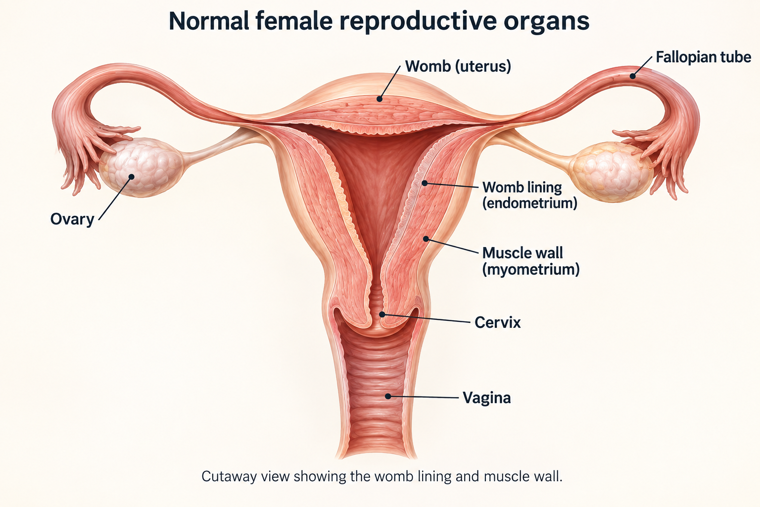
Where the cervix sits
The cervix is the neck of the womb, between the bladder and rectum (the last part of the bowel). This explains why cancer or treatment in the pelvis can affect passing urine, bowel movements and sexual health.
Structure | Why it matters |
|---|---|
Cervix | The lower part of the womb; most cancers start where two types of cells meet, called the transformation zone. |
Parametrium | Supportive tissue beside the cervix; involvement makes a cancer stage IIB. |
Vagina | Which part of the vagina is affected helps determine the stage and the area to treat with radiotherapy. |
Bladder and rectum | Cancer growing into the inner lining of the bladder or rectum is stage IVA. Treatment planning also aims to protect these organs. |
Ureters | Tubes that carry urine from the kidneys to the bladder. A tumour blocking a ureter can make a kidney swell (hydronephrosis) or stop working; this can make the cancer stage IIIB. |
How cervical cancer develops
Most cancers begin at the transformation zone, where mucus-producing glandular cells inside the cervix meet flat squamous cells on its outer surface.
High-risk HPV infection
HPV is common and usually clears naturally. Infection alone does not mean cancer.
Persistent infection
A high-risk HPV type remains for years and changes the way cervical cells grow.
Precancer
Precancer means abnormal cells that have not grown into deeper tissue. Reports may call these high-grade squamous intraepithelial lesions (HSIL), cervical intraepithelial neoplasia (CIN) grades 2–3, or adenocarcinoma in situ (abnormal glandular cells). These changes can be found and treated before cancer develops.
Invasive cancer
Cancer cells have grown through the thin boundary beneath the surface cells into deeper cervical tissue. They may spread to nearby tissues, lymph nodes or other parts of the body.
Prevention still matters after diagnosis
HPV vaccination
Vaccination prevents infection with important cancer-causing HPV types. It does not treat an existing cancer, but family members may benefit according to age and national guidance.
Screening
HPV testing and cervical screening identify high-risk infection or cell changes before symptoms. Screening schedules differ by country and individual risk.
Stopping smoking
Stopping smoking improves general health and may reduce complications during surgery, radiotherapy and treatment with cancer medicines.
Coordinating infection and cancer care
Human immunodeficiency virus (HIV) weakens the immune system. People living with HIV need coordinated cancer and HIV care. Do not stop HIV medicines without specialist advice.
Symptoms and risk factors
Early cervical cancer can cause no symptoms. Persistent bleeding or discharge should not be dismissed, especially after sex or after menopause.
Possible symptom | What to do |
|---|---|
Bleeding after sex | Arrange a pelvic examination; repeated bleeding needs assessment. |
Bleeding between periods or after menopause | Seek prompt medical review. Heavy or ongoing bleeding may need urgent care. |
Watery, blood-stained or foul-smelling discharge | Report a new persistent change, especially with bleeding or pain. |
Pelvic or lower back pain | Assess if persistent, worsening, associated with bleeding, leg swelling or urinary symptoms. |
Pain during sex | Discuss openly; examination can identify cancer and many non-cancer causes. |
Leg swelling, pain near the kidneys or reduced urine | These can be signs of blocked lymph-fluid drainage or a blocked ureter and need urgent assessment. |
Factors that increase risk
Persistent high-risk HPV
The central cause of almost all cervical cancers.
Weakened immunity
HIV, transplant medicines and other causes of reduced immunity can make it harder for the body to clear HPV.
Smoking
Tobacco exposure makes persistent HPV-related cell damage more likely.
Missed screening or follow-up
Precancer can progress if screening, follow-up examination or treatment is delayed.
Track the pattern
Note timing, amount, clots, discharge, pain, fever and triggers such as sex.
Do not self-treat repeatedly
Persistent discharge or bleeding needs examination, not repeated antibiotics or antifungal treatment without a diagnosis.
Tests and diagnosis
How cancer is diagnosed
Symptoms or an abnormal screening result lead to further tests. Whenever safely possible, a biopsy must confirm invasive cancer before cancer treatment is chosen.
Pelvic examination
The clinician examines the cervix, vagina and pelvis, estimates tumour size and mobility, and checks for bleeding or infection.
Colposcopy and biopsy
Colposcopy uses a magnifying instrument to examine the cervix. Small tissue samples are taken from suspicious areas to check for cancer.
Cone biopsy when needed
A cone biopsy removes a cone-shaped piece of cervical tissue, using a surgical blade (cold-knife conisation) or a heated wire loop (loop electrosurgical excision procedure, or LEEP). It can diagnose tiny cancers and treat selected stage IA cancers.
Pathology review
The pathology report describes the cancer type, how deeply and widely it has grown, and whether the edges of removed tissue (margins) are clear.
It also checks for cancer cells in small blood vessels or lymph channels, called lymphovascular space invasion (LVSI). Specialist review can help when findings are unusual or could change treatment.
Imaging and stage
Magnetic resonance imaging (MRI) gives detailed pictures of the pelvis. Computed tomography (CT) and positron emission tomography combined with CT (PET-CT) can help assess lymph nodes and other organs. Your team chooses the scans needed to establish the stage.
Tests often done before treatment
Blood tests
A complete blood count (CBC), kidney and liver function tests, and electrolytes (salts in the blood). Pregnancy and HIV tests may also be offered when appropriate.
Anaesthetic assessment
An assessment of whether medicines used to keep you comfortable or asleep during a procedure are safe for you. This is needed before cone biopsy, major surgery or brachytherapy.
Cystoscopy or proctoscopy
These use a thin viewing instrument to look inside the bladder or rectum when cancer may have grown into its lining. They are not needed for everyone.
Distress and support assessment
Emotional, social, financial and practical needs should be identified early.
Understanding your pathology report
The report should answer what the tumour is and, for a cone or operation specimen, how far it has invaded and whether it was completely removed.
Report item | Why it matters |
|---|---|
Histology | Histology means the type of cancer seen under the microscope. Squamous cell carcinoma starts in flat surface cells; adenocarcinoma starts in glandular cells; adenosquamous carcinoma has features of both. Their treatment often follows similar stage-based pathways. |
Neuroendocrine carcinoma | A rare cancer with features of hormone-producing and nerve-like cells. It can grow quickly and usually needs specialist care combining platinum-based chemotherapy and etoposide with other treatments. |
Grade | Describes how abnormal the cells look; stage and other risk features usually guide treatment more strongly. |
Depth and horizontal extent | The depth of growth determines microscopic stage IA1 or IA2. The International Federation of Gynecology and Obstetrics (FIGO) staging system no longer uses horizontal width to assign a stage, although tumour measurements still help plan care. |
LVSI | Cancer cells in lymphatic or blood spaces raise the risk of lymph-node spread and influence node assessment and postoperative treatment. |
Margins | A positive margin means cancer reaches a cut edge and may require further surgery or chemoradiation. |
Stromal invasion | This means growth into the supporting tissue of the cervix. The depth, together with other findings, helps decide whether treatment is needed after surgery. |
Nodes and parametrium | Cancer in lymph nodes or the parametrium raises the risk of cancer returning after radical surgery, which removes the womb, cervix and surrounding tissue. |
Useful questions
- Is the diagnosis invasive cancer or precancer only?
- Are the cone margins clear, and is LVSI present?
- Does the pathology match the appearance on MRI and examination?
Scans and staging tests
Scans show the size of the cervical tumour, whether nearby tissues are affected, and whether cancer has reached lymph nodes or distant organs. No single scan answers every question.
Test | Best use | Important limits |
|---|---|---|
Pelvic MRI | Measures the tumour and shows nearby tissues to help plan treatment | Very small lesions and microscopic spread may not be visible |
PET-CT | Checks pelvic and para-aortic nodes and distant organs; helps plan the radiotherapy area | Uses a radioactive sugar tracer called fluorodeoxyglucose (FDG). Inflammation can also take up the tracer; very small cancer deposits may be missed |
CT with contrast dye | Checks the chest, abdomen and pelvis when PET-CT is unavailable or during follow-up | Less detailed than MRI for local cervical anatomy |
Kidney imaging | Checks for a swollen kidney or blocked urine flow and assesses kidney function | May involve ultrasound or CT; a blockage may need a procedure to drain urine |
Cystoscopy / proctoscopy | Looks directly at the inner lining of the bladder or rectum if cancer involvement is suspected | Not routinely required for every patient |
Before a scan
Kidney function
Contrast dye helps some structures show more clearly on a scan. Tell your team about kidney disease, dehydration, diabetes and previous reactions to contrast dye.
Pregnancy
Tell your team if you could be pregnant before scans that use radiation or contrast dye.
Blood glucose
High blood sugar can affect PET-CT results. Follow the preparation instructions from your scanning centre.
Implants
Tell MRI staff about pacemakers, metal implants, clips or a severe fear of enclosed spaces.
Ask for comparison
Have the radiologist compare all prior pelvic and chest imaging, not just the latest report.
Understanding the radiotherapy area
Ask whether a suspicious lymph node needs a biopsy, monitoring, an extra radiation dose or inclusion in a wider treatment area.
Planning your treatment
Understanding your stage
FIGO 2018 staging for cervical cancer
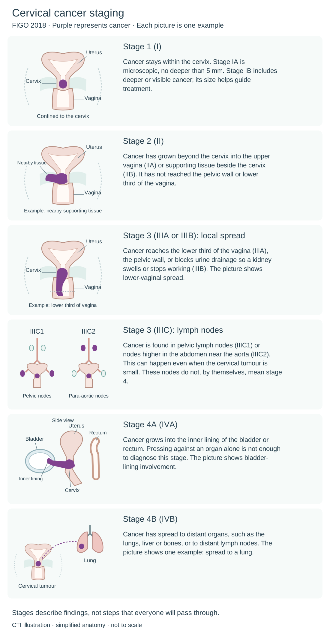
FIGO staging describes where cervical cancer has grown or spread. Examination, scans and tissue findings help establish the stage. Each picture shows one example; each stage can occur in more than one way.
Lymph nodes are small immune-system structures. Regional nodes are those near or draining the cancer’s starting area; distant spread means cancer has reached a site outside that regional area. Stages describe findings, not a sequence that everyone will pass through.
Purple represents cancer. These are examples, not a sequence everyone will pass through.
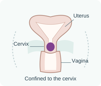
Stage 1 (I)
Cancer stays within the cervix. Stage IA is microscopic, no deeper than 5 mm. Stage IB includes deeper or visible cancer; its size helps guide treatment.
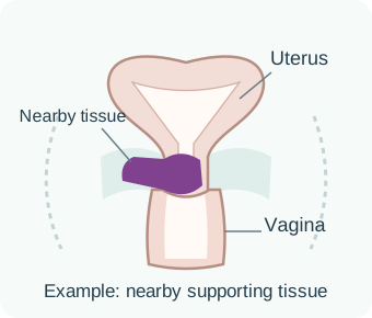
Stage 2 (II)
Cancer has grown beyond the cervix into the upper vagina (IIA) or supporting tissue beside the cervix (IIB). It has not reached the pelvic wall or lower third of the vagina.
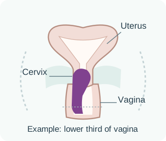
Stage 3 (IIIA or IIIB): local spread
Cancer reaches the lower third of the vagina (IIIA), the pelvic wall, or blocks urine drainage so a kidney swells or stops working (IIIB). The picture shows lower-vaginal spread.
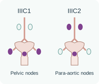
Stage 3 (IIIC): lymph nodes
Cancer is found in pelvic lymph nodes (IIIC1) or nodes higher in the abdomen near the aorta (IIIC2). This can happen even when the cervical tumour is small. These nodes do not, by themselves, mean stage 4.
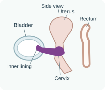
Stage 4A (IVA)
Cancer grows into the inner lining of the bladder or rectum. Pressing against an organ alone is not enough to diagnose this stage. The picture shows bladder-lining involvement.
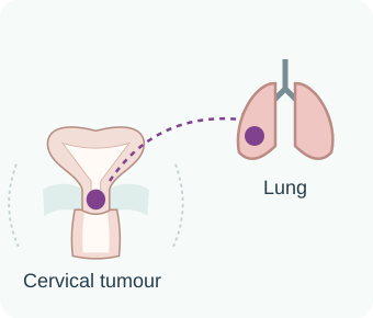
Stage 4B (IVB)
Cancer has spread to distant organs, such as the lungs, liver or bones, or to distant lymph nodes. The picture shows one example: spread to a lung.
Stage 3 and some stage 4A cancers can still be treated with the aim of cure. The stage alone cannot tell you your personal outlook. After IIIC, the letter r means lymph-node findings came from imaging; p means they were confirmed by tissue testing.
Clinical stage comes from examination, biopsies and scans. Tissue examined after surgery can add information. Letters such as yp mean the tissue was assessed after treatment given before surgery.
A revised assessment does not necessarily mean the cancer grew while you were waiting. Grade describes how abnormal the cells look and is different from stage.
Stage is not the whole story
Tumour size
Within the same stage, size can change whether surgery or radiation is favoured.
Lymph nodes
Pelvic nodes are IIIC1; para-aortic nodes are IIIC2. Number, size and location influence radiation fields and outlook.
Histology
Rare neuroendocrine or non-HPV-related adenocarcinomas may behave differently.
Your general health
Kidney function, hearing, nerve symptoms such as numbness or tingling (neuropathy), safety of anaesthesia, HIV control and how well you manage daily activities all influence treatment.
Stage I in more detail
Stage IA is visible only under a microscope. IA1 means invasion no deeper than 3 mm; IA2 means more than 3 mm and no deeper than 5 mm. For stage IB, tumour size separates IB1 (up to 2 cm), IB2 (more than 2 cm and up to 4 cm) and IB3 (more than 4 cm).
The early-stage treatment section explains how these findings guide the choice of treatment.
Why the choice can be difficult
Reducing combined side effects
If radiotherapy is very likely to be needed after surgery, starting with chemoradiation may avoid the added side effects of having both major surgery and pelvic radiotherapy.
Checking lymph nodes first
Checking the first lymph nodes that drain the cervix (sentinel nodes), or other pelvic nodes, may reveal cancer that changes the operation or treatment plan.
Less extensive surgery for some cancers
Some small, low-risk cancers may be treated with less extensive surgery. Your specialist will check whether this is safe for you.
Discuss fertility early
If having a pregnancy in the future matters to you, discuss this before treatment starts. For selected small cancers confined to the cervix, fertility-preserving surgery may be possible. Your team will assess whether this is safe and how it affects your treatment choices.
Common staging questions
Node-positive is not automatically stage IV
Pelvic and para-aortic nodes are stage IIIC in FIGO 2018. Curative chemoradiation may still be possible.
Hydronephrosis needs action
A blocked ureter can damage the kidney and affect whether cisplatin can be given safely. A small tube inside the ureter (stent) or a drainage tube through the back into the kidney (nephrostomy) may be needed promptly.
Some stage IVA cancers may still be curable
Cancer in the inner lining of the bladder or rectum is serious, but it is local spread rather than spread to distant organs.
Stage IVB can vary
A single distant lymph node or lung deposit differs from cancer in many parts of the body. A multidisciplinary team (MDT), specialists who plan care together, may combine medicines that treat cancer throughout the body with treatment to particular sites.
Fertility, pregnancy and menopause
Treatment can permanently affect the ability to become pregnant and the hormones made by the ovaries. Discuss your wishes before surgery, pelvic radiotherapy or chemotherapy begins.
Fertility-sparing surgery
For selected small stage I cancers, a cone biopsy or radical trachelectomy may preserve the womb. A trachelectomy removes the cervix; a radical trachelectomy also removes nearby tissue and part of the upper vagina.
Egg or embryo freezing
Eggs may be collected and frozen, or fertilised to create embryos for freezing. Whether there is time before treatment depends on how urgently cancer care is needed and specialist advice.
Ovarian transposition
Moving ovaries above the planned radiation field may preserve hormone function, but does not protect the uterus from pelvic radiation.
Pregnancy at diagnosis
Care depends on the cancer stage, how far the pregnancy has progressed and your priorities. Cancer and pregnancy specialists should plan care together.
Questions before treatment
- Do I want the option of pregnancy, even if I am not ready for pregnancy now?
- Is fertility-preserving surgery safe for my cancer type and stage?
- Will my ovaries be removed, moved, shielded, or exposed to radiation?
- Would it help to see a fertility, high-risk pregnancy or menopause specialist now?
- What contraception is appropriate during and after treatment?
Menopause planning
Discuss hot flushes, bone and heart health, vaginal dryness and whether menopausal hormone therapy is appropriate.
Future pregnancy
Pregnancy after trachelectomy needs specialist monitoring and often delivery by caesarean section.
How your treatment plan is chosen
Specialists in gynaecological surgery, radiotherapy, cancer medicines, scans, pathology and supportive care work together to recommend treatment. Your wishes and overall health are part of the decision.
Decision | What to discuss |
|---|---|
Treatment goal | Whether the aim is cure, cancer control, symptom relief or a combination |
Primary local treatment | Whether surgery or chemoradiation is preferred, and why |
Lymph nodes | How suspicious pelvic and para-aortic nodes will be confirmed or treated |
Brachytherapy access | Where treatment will be given and whether the centre can use needles or small tubes in the tissue if needed |
Additional medicine treatment | Whether chemotherapy before chemoradiation or pembrolizumab with chemoradiation is appropriate |
Fertility and ovarian function | Your wishes, whether preservation is safe, and whether there is time before treatment |
Other health conditions | Kidney function, hearing, nerve symptoms, HIV care, nutrition and anaesthetic safety |
Practical arrangements | Start date, treatment duration, travel, accommodation, possible transfusions and costs |
Questions about the recommended plan
- What benefit is expected from this treatment, and what is the main alternative?
- Could this plan lead to both radical surgery and pelvic radiation?
- What result would stop, delay or change treatment?
- Who should I call after hours if bleeding, fever or urinary obstruction develops?
Treatment options
Topics in this section
- Choosing treatment for early-stage cancer
- Fertility-sparing surgery
- Hysterectomy and lymph-node surgery
- Do I need treatment after surgery?
- Chemoradiation with the aim of cure
- External-beam radiation and cisplatin
- Brachytherapy is a core treatment
- When extra treatment may help
- When cancer returns in the pelvis
- Biomarkers that can change treatment
- First medicines when cancer cannot be cured locally
- When first-line treatment stops working
- Precision treatment and clinical trials
The first sections explain treatment that aims to cure cancer. Later sections cover options if cancer returns or spreads. Your team can help you focus on the parts that apply to you.
Choosing treatment for early-stage cancer
The aim is cure with the fewest possible long-term harms. Tumour size and depth, LVSI, lymph-node risk, pregnancy plans and your fitness for surgery all matter. A hysterectomy removes the womb and cervix; more extensive operations also remove surrounding tissue.
Situation | Usual treatment options |
|---|---|
IA1, clear cone margins, no LVSI | Follow-up after a cone biopsy that has removed the cancer, if preserving fertility is important; otherwise, a simple hysterectomy may be offered |
IA1 with LVSI / IA2 | Surgery that preserves the womb, or hysterectomy with assessment of sentinel or other pelvic nodes, depending on risk |
Small low-risk IA2-IB1 | Less extensive surgery with lymph-node assessment may be possible after specialist review |
IB1 suitable for fertility preservation | Cone biopsy or radical trachelectomy with lymph-node assessment, depending on tumour size and other findings |
IB1-IB2, fertility not required | Hysterectomy with the extent of surgery tailored to the cancer, plus lymph-node assessment; radiotherapy-based treatment if surgery is unsuitable |
IB3 or large IIA2 | Usually chemoradiation and brachytherapy with the aim of cure; alternatives require specialist assessment |
Treatment is not chosen from stage alone
Margin status
A positive cone margin may require repeat cone, trachelectomy, hysterectomy or radiation depending on the whole picture.
LVSI and nodes
LVSI increases concern about lymph-node spread. Cancer found in lymph nodes usually changes the plan to chemoradiation.
Histology
Usual squamous, adenocarcinoma and adenosquamous tumours share pathways; neuroendocrine and other rare types need different plans.
Treatment that may be needed after surgery
If treatment after surgery is likely to be needed, starting with chemoradiation may reduce the side effects of combining major surgery and radiotherapy.
Fertility-sparing surgery
Preserving the uterus can be safe for carefully selected small stage I cancers, but it requires precise pathology, negative nodes and specialist follow-up.
Procedure | What is removed | Key point |
|---|---|---|
Cone biopsy | Cone of cervix including transformation zone | May be the only treatment needed for IA1 or selected very low-risk cancers if the tissue edges are clear |
Simple trachelectomy | Cervix only | For selected low-risk cancers; lymph nodes are also assessed when needed |
Radical trachelectomy | Cervix, parametrial tissue and upper vagina; uterus retained | For selected IA2–IB1 cancers, commonly under 2 cm, after checking that lymph nodes are clear |
Ovarian preservation | Ovaries left in place | Keeps the ovaries’ hormone production, but cannot preserve the ability to carry a pregnancy if the womb is removed or treated with radiotherapy |
What must be checked
- Tumour size and location, histology, depth, LVSI and clear margins.
- Pelvic nodes - and para-aortic nodes if clinically indicated - before completing fertility-sparing surgery.
- A plan to convert to hysterectomy or chemoradiation if nodes, margins or other features are unfavourable.
- Future pregnancy risks, including infertility, miscarriage, preterm birth and likely caesarean delivery after radical trachelectomy.
After cone
Possible risks include narrowing of the cervix, bleeding and premature birth in a future pregnancy.
After trachelectomy
A permanent cervical stitch may be placed; future pregnancy needs specialist monitoring.
Hysterectomy and lymph-node surgery
The amount of tissue removed depends on the cancer. More extensive surgery removes more tissue beside the cervix and in the upper vagina, which can increase bladder, bowel and sexual side effects.
Operation | Typical role | Main long-term considerations |
|---|---|---|
Simple / type A hysterectomy | Very early or selected low-risk IA2-IB1 disease | Pregnancy no longer possible; ovaries may be preserved |
Modified radical / type B | Selected early cancers where more surrounding tissue needs to be removed | Bladder and sexual effects generally less than type C |
Radical / type C1 hysterectomy | Many operable IB and selected IIA tumours | Difficulty emptying the bladder, swelling from poor lymph drainage (lymphoedema), and sexual changes; protecting nearby nerves may help |
Sentinel-node mapping | Identifies the first draining pelvic nodes | Detailed microscope testing can find tiny cancer deposits. If mapping does not find the sentinel nodes, other pelvic nodes on that side may need to be removed |
Removal of pelvic lymph nodes | When sentinel-node assessment is unsuitable or more lymph-node information is needed | A collection of lymph fluid (lymphocele), infection, blood-vessel or nerve injury, and leg lymphoedema |
Before discharge
Bladder plan
Ask whether you need a catheter, a tube that drains urine, how bladder emptying will be checked, and whom to call if urine stops.
Clot prevention
Early walking, compression stockings or devices, and blood-thinning medicines may be advised.
Pathology date
Book the visit to review the tissue results and whether further treatment is needed.
Recovery limits
Clarify lifting, driving, work, bathing, vaginal intercourse and wound care.
Do I need treatment after surgery?
Tests on tissue removed during surgery may show risks that were not clear beforehand. Treatment after surgery, called adjuvant treatment, can reduce the chance of cancer returning but also adds side effects.
Pathology finding | Usual implication |
|---|---|
Negative nodes, margins and parametrium; low-risk features | Regular follow-up without immediate further cancer treatment |
Intermediate-risk combination | Radiotherapy to the pelvis may be recommended based on tumour size, LVSI and depth of growth into cervical tissue |
Positive pelvic node | Chemoradiation after surgery; the treatment area depends on which lymph nodes are affected |
Positive para-aortic node | Chemoradiation covering a wider area may be considered |
Positive surgical margin | Chemoradiation; brachytherapy or an additional dose to the affected area may be considered |
Parametrial involvement | Postoperative chemoradiation |
The trade-off
Potential benefit
A lower chance of cancer returning in the pelvis and better cancer control for appropriately selected patients.
Added burden
More bowel, bladder, sexual, ovarian and lymphatic effects when pelvic radiation follows radical surgery.
Timing
Healing, kidney function and blood counts are checked before postoperative treatment begins.
Second opinion
Reasonable when risk classification is borderline or the radiation field and brachytherapy need are unclear.
- Ask which exact pathology feature triggers treatment.
- Ask whether chemotherapy adds benefit to radiation in your risk group.
- Ask whether vaginal brachytherapy is needed and why.
Chemoradiation with the aim of cure
For most stage IIB, III and IVA cancers, and many larger IB3 or IIA2 tumours, treatment combines external-beam radiotherapy (EBRT), cisplatin chemotherapy given during radiotherapy, and brachytherapy. EBRT directs radiation from a machine outside the body.
Check the extent of cancer and protect the kidneys
MRI and PET-CT or CT help map the cancer. Your team checks kidney function and treats blocked urine flow with a stent or nephrostomy when needed.
Plan the full radiation course
Agree the area to treat, the brachytherapy method, the radiation dose and the total treatment time before starting.
External-beam radiation
Usually given five days a week for about five to six weeks. Scans help position treatment accurately and limit radiation to nearby healthy organs.
Chemotherapy given during radiotherapy
Cisplatin is commonly given weekly to help radiation work better. Carboplatin may be considered if cisplatin is unsafe.
Brachytherapy
Internal radiotherapy delivers a high dose close to the cervical tumour. Whenever feasible, it is an essential part of treatment and should not be replaced by extra external-beam radiation.
Response assessment
Examination and scans, commonly around three to six months after treatment, assess the response and provide a baseline for later comparisons.
Pelvic nodes
Usually receive an extra radiation dose if cancer is present.
Para-aortic nodes
May need to be included in a wider radiotherapy area.
External-beam radiation and cisplatin
Radiotherapy treats the cervix, supporting tissue and nearby lymph-node areas. Cisplatin given during radiotherapy helps the cancer respond to radiation.
Part of care | What to expect | What is monitored |
|---|---|---|
Planning session | A planning CT and positioning supports, plus bladder and bowel instructions. MRI or PET-CT images may be combined with the planning scan | Target coverage, bowel, bladder, rectum, kidneys and bone marrow |
Daily EBRT | Short weekday sessions; treatment itself is painless | Position and internal organ changes using image guidance |
Weekly cisplatin | Fluids through a vein (intravenous, or IV), anti-sickness medicines and chemotherapy during radiotherapy | Kidney function, blood salts such as magnesium and potassium, blood counts, hearing and nerve symptoms |
Extra dose to affected lymph nodes | Extra dose to involved nodes within or after the pelvic plan | Radiation dose to the bowel and other nearby healthy organs |
Treatment review | Regular radiation and oncology visits | Diarrhoea, urinary symptoms, bleeding, pain, weight and hydration |
Common early effects
Bowel
Loose stools, urgency, cramps and nausea.
Bladder
Frequency, urgency, burning and occasional infection.
Blood counts
Low red blood cells (anaemia), white blood cells or platelets (which help blood clot) may need treatment or a change in the schedule.
Fatigue
Often builds through treatment; anaemia, sleep, nutrition and distress may contribute.
If cisplatin is paused
Radiation usually continues unless the radiation team identifies a separate reason to pause.
If kidney function falls
Treat dehydration or obstruction, review medicines and decide whether carboplatin or radiation alone is safer.
Brachytherapy is a core treatment
Brachytherapy places the radiation source close to the cervix tumour. It creates a high, shaped dose that external-beam radiation alone usually cannot deliver safely.
Technique | How it is used |
|---|---|
Inside the womb and vagina (intracavitary) | Special applicators, such as a tandem with a ring or ovoids, are placed through the vagina into the womb and against the cervix to guide the radiation source |
Within the tissue (interstitial) | Needles or small tubes are placed in the tumour or nearby tissue when extra coverage is needed because of its size, shape, location or remaining tumour after initial treatment |
A combination (hybrid) | Combines intracavitary applicators with interstitial needles |
Image-guided | MRI and/or CT define residual tumour and organs at risk for each application |
High dose-rate | Several short treatment sessions; anaesthesia or deep sedation is often used to place the applicators |
Before, during and after
Preparation
Review anaesthetic safety, blood counts, blood-thinning medicines, infection, fasting and bladder or bowel preparation.
Applicator placement
The radiotherapy team places the applicator. Ultrasound guidance can reduce the risk of making a hole in the womb.
Imaging and plan
CT or MRI checks the applicator position, the treatment area and the dose to the bladder, rectum and other bowel.
Treatment and removal
The radioactive source travels through closed applicator channels; the patient is not radioactive after removal.
When extra treatment may help
Some patients with cancer that has grown nearby may benefit from additional medicines. Discuss these options before radiotherapy starts; they are not suitable for everyone.
Strategy | Evidence-based setting | Practical cautions |
|---|---|---|
Pembrolizumab with chemoradiation | Pembrolizumab, an immunotherapy that helps the immune system attack cancer, may be added for FIGO 2014 stage III–IVA disease. It continues for a planned period after chemoradiation. Eligibility uses the 2014 stage definitions, which differ from the 2018 staging explained above | Possible immune-related inflammation. The team checks autoimmune conditions, previous organ transplant, steroid use, and thyroid, liver and kidney function. Benefit was less clear in the lower-stage, node-positive group in the initial KEYNOTE-A18 trial analysis |
Chemotherapy before chemoradiation | Six weekly cycles of carboplatin and paclitaxel before chemoradiation may be considered for suitable patients with locally advanced cancer. In the INTERLACE trial, this improved five-year survival and the time without cancer worsening | Chemoradiation must start promptly afterwards. Blood counts, nerve symptoms, general health, brachytherapy appointments and travel arrangements all need checking |
Questions to ask your care team
- Which staging system and exact stage make me eligible?
- How much benefit is expected for someone like me?
- Will this delay brachytherapy or extend total treatment time?
- Which side effects would mean treatment should pause or stop?
- Is this strategy appropriate for my cancer and treatment setting, and what is the full course cost?
Radiation quality first
High-quality EBRT and image-guided brachytherapy remain essential.
Clinical trial
A trial may answer uncertainty and improve access.
When cancer returns in the pelvis
If cancer returns in the pelvis, the options depend on its location, earlier surgery or radiotherapy, whether it can be removed, and whether it has spread elsewhere.
Situation | Potential approach |
|---|---|
Pelvic recurrence after surgery, no previous radiation | Curative chemoradiation plus brachytherapy when feasible; surgery in selected cases |
Central recurrence after prior radiation | Pelvic exenteration, a major operation explained below, may offer cure in carefully selected patients without distant spread or cancer in the pelvic sidewall that cannot be removed |
Small accessible recurrence after radiation | Surgery, highly focused radiotherapy or repeat radiotherapy may be considered by an experienced specialist team for selected patients |
Cancer in the pelvis that cannot be removed | Medicines to treat cancer throughout the body (systemic therapy), individually planned radiotherapy, procedures for bleeding or urine problems, pain relief and clinical trials |
Recurrence plus distant metastases | Cancer medicines are the main treatment; treatment to individual areas may relieve symptoms or control a small number of sites |
Pelvic exenteration
This major operation removes cancer that has returned and may involve removing the bladder, rectum and reproductive organs. New openings in the abdomen (stomas) may be needed to collect urine or stool in a bag; vaginal reconstruction may also be needed.
It can offer cure only for carefully selected patients.
Before surgery
Confirm no distant disease, likelihood of clear margins, fitness, nutrition and patient goals.
Life after
Discuss managing a stoma, sexual health, mobility, body image, work and caregiver support before surgery.
Can the cancer be completely removed?
The surgeon should state whether clear margins appear achievable and which organs may need removal.
Whole-person fitness
Nutrition, heart and lung health, preparation for a stoma, emotional support and help from caregivers all affect recovery.
Biomarkers that can change treatment
If cancer remains after treatment, returns or spreads, testing its tissue may help choose medicines. Arrange testing early enough for results to guide treatment. A biomarker result does not automatically mean a particular medicine will help.
Biomarker | How it may guide treatment |
|---|---|
Protein score for immunotherapy | Programmed death-ligand 1 (PD-L1) is a protein measured using a combined positive score (CPS). A CPS of 1 or higher can support pembrolizumab with initial platinum-based and taxane chemotherapy, with or without bevacizumab. After chemotherapy, pembrolizumab alone may be an option if it has not already been used |
Mismatch repair (MMR) / microsatellite instability (MSI) | These tests look for problems with the cells’ repair of genetic material. Deficient MMR (dMMR) or high MSI (MSI-high) may support immunotherapy under indications that cover several cancer types; previous treatment and other eligibility criteria matter |
Tumour mutational burden (TMB) | The number of genetic changes in cancer cells. A high result may support pembrolizumab for selected patients after previous treatment, under an indication that covers several cancer types |
Human epidermal growth factor receptor 2 (HER2) | A protein that can be measured by tissue staining, called immunohistochemistry (IHC). A strongly positive result (IHC 3+) may support an antibody–drug conjugate (ADC), a medicine that links an antibody to a cancer-killing drug, after prior treatment. Suitability and access need review |
NTRK / RET gene fusions | NTRK and RET are gene names. Rare changes that join one of these genes to another gene (fusions) may allow targeted treatment. The exact fusion and treatment criteria must be confirmed |
Next-generation sequencing (NGS) | A test that checks many genes at once. It may find uncommon treatment targets or clinical trials, but many results do not have a proven treatment |
A result is not a prescription
Check what the result means
Your team checks the test method, sample quality and the level needed for a positive result.
Match the treatment to your situation
A medicine used after previous treatment may not be suitable as the first treatment.
Consider previous treatment
Previous immunotherapy or ADC treatment changes the likely benefit and trial options.
Check access
Discuss the treatment schedule, cost and monitoring with your team.
First medicines when cancer cannot be cured locally
Systemic therapy treats cancer throughout the body. It is used when cancer has spread, remains after treatment or returns and cannot be cured with treatment to one area. The choice depends on PD-L1 results, previous cisplatin, general health and whether bevacizumab can be used safely.
Clinical situation | Common first-line approach |
|---|---|
PD-L1 CPS 1 or higher | Pembrolizumab plus paclitaxel with cisplatin or carboplatin; bevacizumab may be added when safe |
PD-L1 CPS below 1 or unknown | Platinum-based chemotherapy with a taxane, often with bevacizumab if safe. If PD-L1 has not been tested, arrange the test promptly |
Previous cisplatin with curative radiation | Carboplatin is often chosen; the earlier treatment and time since it was given matter |
Bevacizumab unsuitable | Platinum-based chemotherapy and a taxane with pembrolizumab for PD-L1-positive cancer, or chemotherapy alone for PD-L1-negative cancer |
Frailty or reduced organ function | A gentler treatment plan tailored to your health, treatment to relieve symptoms in particular areas, supportive care or a clinical trial |
Safety checks before bevacizumab
Bleeding and wound healing
Avoid or delay around major surgery; uncontrolled bleeding is a major concern.
Fistula or perforation
Bevacizumab can increase the risk of a fistula (an abnormal connection between organs) or a hole in the bowel wall. Risk is higher after pelvic radiotherapy, with cancer involving the bladder or bowel, a blockage or an existing fistula.
Blood pressure and kidneys
Check blood pressure and test urine for protein.
Clotting
Discuss any previous blood clots and current blood-thinning medicines.
Before cycle 1
Record nerve symptoms, blood pressure, urine protein, bleeding or fistula risk, kidney function and ability to manage daily activities.
At each reassessment
Review symptoms and scans, then decide whether to continue, stop one medicine, use ongoing maintenance treatment or change the plan.
When first-line treatment stops working
The next treatment depends on medicines already given, biomarker results, nerve symptoms, bleeding risk and general health. Ask whether a clinical trial is suitable when treatment changes.
Option | When it fits | Main safety considerations |
|---|---|---|
Tisotumab vedotin | An established option after chemotherapy for cervical cancer that has returned or spread | Eye injury, neuropathy and bleeding; preventive eye care and examinations are mandatory |
Immunotherapy | Pembrolizumab may be an option after chemotherapy for PD-L1-positive, MSI-high/dMMR or TMB-high cancer if it has not already been given and the relevant treatment criteria are met. Cemiplimab is an option when cancer has returned or spread and worsens during or after platinum chemotherapy, regardless of PD-L1. Earlier immunotherapy affects the choice. | Immune inflammation of thyroid, lung, liver, bowel, kidney, skin and other organs |
Other chemotherapy | Clinician-selected agents when preferred options are unavailable or unsuitable | Responses may be limited. Blood counts, kidney function, nerve symptoms and symptoms from cancer help guide the choice |
HER2-directed ADC | An option for selected previously treated cancers with HER2 IHC 3+ results, under an indication covering several cancer types | Lung inflammation (interstitial lung disease), nausea and reduced blood-cell production |
Fusion-targeted therapy | Rare confirmed NTRK or RET fusion | Medicine-specific risks to the liver, nerves, heart or circulation, and interactions with other medicines |
Clinical trial | May be considered at any treatment stage, especially after immunotherapy or an ADC | Eligibility, travel, extra testing and uncertain benefit |
Review before changing treatment
- Is there a painful, bleeding or threatening area that needs treatment now?
- Would another biopsy or biomarker test help choose the next treatment?
- How do the likely benefits compare with the side effects, visits and costs?
Precision treatment and clinical trials
Your specialist will explain whether a molecular test result has a proven treatment, points to a clinical trial or has no clear treatment implication yet.
Result category | What it means | Next step |
|---|---|---|
Established treatment marker | Linked to an established treatment for this type and stage of cancer | Check suitability, previous treatment and safety |
Marker shared across cancer types | Linked to a treatment across cancer types | Check the test threshold, evidence in cervical cancer and treatment availability |
Trial-only marker | Promising but not established standard care | Look for a trial matched to the test result |
Uncertain variant | The meaning of the genetic change or its effect on treatment is not known | Do not choose treatment based on this result outside a clinical trial |
No result that guides treatment | This is common and does not mean the test was done incorrectly | Use established treatments that do not need a matching biomarker, or consider a clinical trial |
Managing side effects
Staying safe during chemoradiation
Report side effects early. Treating them helps you stay hydrated and nourished and may prevent avoidable breaks in treatment.
Problem | Early action | When to seek urgent help |
|---|---|---|
Diarrhoea | A temporary low-fibre diet if advised, fluids to drink and prescribed medicines for diarrhoea | Dizziness, very little urine, blood in stools, fever or inability to drink |
Nausea / vomiting | Take anti-sickness medicines as prescribed; try small, frequent meals and drinks | Repeated vomiting or missed fluids/medicines |
Urinary burning | Urine test; hydration; pain relief; antibiotics only if indicated | Fever, pain near the kidneys or inability to pass urine |
Bleeding | Track pads and clots; report any increase | Bleeding that soaks pads, faintness, breathlessness or low blood pressure |
Anaemia | Check CBC; treat the cause; transfusion when clinically indicated | Chest pain, fainting, severe breathlessness |
Low white cells | Check temperature and follow your team’s advice about blood tests and treatment | Fever 38°C (100.4°F) or higher, shaking chills |
Skin / vulval soreness | Gentle care, prescribed creams, loose clothing | Blistering, infection or uncontrolled pain |
Protect the treatment schedule
Bring a medicine list
Include supplements and traditional medicines; some interact with treatment.
Measure what matters
Weight, intake, urine, stools, temperature, pain and bleeding help the team act early.
Do not self-stop
Call before missing chemotherapy, radiation or brachytherapy unless there is an immediate emergency.
Plan transport
Fatigue and anaesthesia make reliable travel and a support person important.
Weekly question
What is the one side effect most likely to interrupt treatment this week?
Weekly action
Before leaving the centre, check prescriptions, blood-test dates, any transfusion plans, transport and brachytherapy instructions.
Procedures that can relieve urgent problems
Local procedures can control bleeding, protect kidney function, relieve obstruction and reduce pain while cancer treatment is planned or continued.
Problem | Possible intervention | Purpose |
|---|---|---|
Heavy vaginal bleeding | Packing to control vaginal bleeding, medicines that help stop bleeding, transfusion, urgent radiotherapy, a procedure to block bleeding blood vessels (embolisation), or surgery | Stabilise circulation and control the bleeding source |
Blocked ureter / hydronephrosis | A ureteric stent or a nephrostomy tube to drain urine | Drain the kidney, relieve infection or pain and protect kidney function |
Painful bone metastasis | Pain medicines, radiotherapy for symptom relief, procedures to support the bone, or bone-strengthening treatment when appropriate | Reduce pain and fracture or spinal-cord risk |
Bowel obstruction | Fluids, a tube to relieve pressure, medicines, a stent, surgery to redirect the bowel or another operation | Relieve vomiting and pain; restore passage when feasible |
Fistula | Treatment of infection, wound and skin care, rerouting urine or stool, or surgery in selected patients | Control leakage, infection, odour and skin damage |
Fluid around the lung | Drainage, a longer-term drainage tube, or a procedure to seal the space where fluid collects (pleurodesis) | Relieve breathlessness |
When to seek urgent help
Bleeding
Heavy flow, large clots, fainting or chest symptoms.
Infection
Fever, shaking chills, confusion or very low urine.
Spinal cord
New leg weakness, numbness, loss of bladder or bowel control.
Blood clot
Sudden breathlessness, chest pain or one swollen painful leg.
Live well
After treatment with the aim of cure
The tumour may continue to shrink after radiation. Assessment is timed to avoid mistaking treatment inflammation for persistent cancer.
Treatment summary
Keep a summary of radiotherapy areas, doses and dates, cancer medicines, transfusions, procedures and any complications.
Early clinical review
Check recovery, bleeding, bladder and bowel symptoms, pain, nutrition, menopause and emotional wellbeing.
Response imaging
For stage II-IVA or after chemoradiation, PET-CT or contrast CT is commonly performed around 3-6 months; pelvic MRI may add local detail.
Review residual findings
Your team considers examination, scans and time since treatment together. A biopsy may be needed to confirm remaining cancer before major further treatment.
Plan ongoing care
Agree follow-up appointments, checks for lasting side effects, vaccinations, general health care and whom to contact about new symptoms.
What a scan can show
Complete response
No convincing signs of active cancer; begin regular follow-up.
Uncertain scan activity
Inflammation or healing can look like cancer. Team review and a later scan may be safer than immediate major treatment.
Persistent local disease
Confirm where the cancer is and whether it can be removed; obtain a biopsy if it would change further treatment.
New distant disease
When possible, biopsy a new site and check biomarkers before choosing cancer medicines.
Nutrition and activity during treatment
The goal is not a perfect diet. It is to maintain hydration, protein, energy and function well enough to complete treatment and recover.
Need | Practical approach |
|---|---|
Hydration | Sip regularly; use oral rehydration when diarrhoea or vomiting is significant; follow fluid restrictions if you have heart or kidney disease |
Protein | Include dal, beans, eggs, dairy, fish, chicken, soy, nut pastes or supplements according to preference and tolerance |
Energy | Use small frequent meals; enrich food with oil, ghee, milk powder or nut paste when weight is falling and clinically appropriate |
Diarrhoea | If diarrhoea worsens, your team may advise temporarily reducing rough fibre, very fatty or spicy foods, or lactose (milk sugar). Gradually broaden your diet as symptoms settle |
Anaemia | Do not assume low red blood cells mean iron deficiency. Bleeding, inflammation, treatment effects on blood-cell production and nutrition can all contribute. Check the cause before taking iron |
Food safety | Use safe water, freshly cooked food, refrigeration and hand hygiene, especially if infection-fighting white blood cells are low (neutropenia) |
Move safely
Daily activity
Short walks and light activity help fatigue, mood, bowel function and clot prevention.
Strength
Gentle strengthening exercises may help when you feel able. Ask for physiotherapy advice after surgery.
Pelvic floor
A physiotherapist can help with exercises for the muscles that support the bladder and bowel, including symptoms affecting sexual health.
Stop and call
New chest pain, severe breathlessness, faintness, fever or rapidly worsening weakness.
Life after pelvic treatment
Recovery includes bladder, bowel, hormone, sexual and lymph-drainage problems. Many of these effects can be helped; you can discuss them without embarrassment.
Late effect | What may help |
|---|---|
Vaginal dryness or narrowing | Moisturisers, lubricants, vaginal dilators after healing, pelvic-floor therapy and local hormonal treatment when appropriate |
Pain or fear with sex | Check for treatable causes, return to intimacy gradually, and consider counselling or sexual-health support |
Early menopause | Discuss symptoms, bone and heart health, exercise, calcium and vitamin D needs, and whether menopausal hormone therapy is suitable |
Bladder urgency or leakage | Urine test, bladder training, pelvic-floor therapy, medicines or urology referral |
Bowel urgency or bleeding | Diet review, medicines and gastroenterology assessment; do not assume all bleeding is radiation damage |
Leg or genital lymphoedema | Early lymphoedema therapy, compression, skin care, exercise and infection prevention |
Neuropathy | Foot care, fall prevention, physiotherapy and symptom medicines |
Emotional and relationship health
Body image
Surgery, scars, stomas, bleeding and sexual changes can alter confidence.
Trauma and fear
Anxiety before scans or pelvic examinations is common; psychological support can help.
Partners
Clear consent, slow pacing and non-penetrative intimacy can support recovery.
Work and finances
Ask early about social-work support, insurance questions and adjustments at work.
Follow-up after treatment
Follow-up aims to detect treatable problems, identify symptoms of recurrence, manage late effects and support general health. Routine scanning is not needed forever for everyone.
Time after treatment | Usual appointment schedule |
|---|---|
Years 1-2 | Clinical review about every 3-6 months, individualised by risk and treatment |
Years 3-5 | Clinical review about every 6-12 months |
After year 5 | Yearly or an agreed survivorship schedule |
After chemoradiation | Response imaging commonly at 3-6 months; later imaging for symptoms or specific high-risk findings |
After fertility-sparing surgery | More frequent cervical examinations, cell-sample or HPV tests, and pelvic MRI according to your specialist’s plan |
Symptoms to report between appointments
- New bleeding, persistent discharge, pelvic or back pain, leg swelling, cough, breathlessness, weight loss or reduced appetite.
- Blood in urine or stool, change in bladder or bowel function, recurrent infections or kidney-area pain.
- New numbness, weakness or other nerve symptoms, bone pain, severe tiredness or a rapid decline in daily activities.
Sources and further reading
For more information, explore these resources from the National Cancer Institute (NCI) and other medical organisations. The linked organisations have not reviewed or endorsed this CTI guide. Your care team can explain how the information applies to you.
- NCI: Cervical Cancer Treatment
- Cancer Council Australia: cervical cancer
- Macmillan: cervical cancer
- NCI: Cervical cancer diagnosis
- NCI: Cervical cancer treatment by stage and fertility considerations
- NCI: Biomarker testing for cancer treatment
Staging and risk-group references
FIGO 2018 cervical staging.
Medicine approval and prescribing references
These official records describe licensed uses and prescribing information for the medicines named in this guide. Approval depends on the cancer type, biomarkers, previous treatment, combination and formulation. Some uses discussed in the guide are off-label: outside the medicine’s licence, with evidence from clinical guidelines or studies. FDA or EMA approval does not establish approval or availability in India.
- Bevacizumab — FDA prescribing information · Approval history
- Carboplatin — FDA prescribing information · Approval history
- Cemiplimab — EMA approval record. EU approval for recurrent or metastatic cervical cancer after platinum treatment.
- Cisplatin — FDA prescribing information · Approval history. Use as a radiosensitiser in cervical chemoradiotherapy is supported by guidelines; the individual FDA label does not establish that indication.
- Paclitaxel — FDA prescribing information · Approval history
- Pembrolizumab — FDA prescribing information · Approval history
- Tisotumab vedotin — FDA prescribing information · Approval history
Additional medicine approval notices
These records explain the specific medicine uses and eligibility requirements discussed in this guide.
Related care
Meet our doctors



Where to get care