

Share
Start here
Bladder cancer at a glance
- The bladder is a hollow organ with a muscular wall, low in the abdomen, that stores urine made by the kidneys. Start with this overview, then read the sections that match your diagnosis and treatment.
- Most bladder cancers start in the inner lining and are called urothelial carcinomas. A specialist examines the tissue to confirm the type, because less common types may need different treatment.
- Visible blood in the urine is the most common warning sign. It can come and go and may be painless. It always deserves medical assessment.
- A camera test called cystoscopy looks inside the bladder. An operation called transurethral resection of bladder tumour (TURBT) removes visible tumour through the urine passage (urethra) and provides tissue to examine.
- Non-muscle-invasive bladder cancer (NMIBC) has not entered the bladder muscle. It often returns, so repeated cystoscopy is important.
- Muscle-invasive bladder cancer (MIBC) has grown into the muscle layer. Treatment aiming for cure usually combines medicines that work throughout the body with bladder-removal surgery (radical cystectomy). For selected people, removing the visible tumour and combining chemotherapy with radiotherapy may preserve the bladder.
- Advanced urothelial cancer can often be controlled with medicines and care to relieve symptoms. Options include chemotherapy, immunotherapy (which helps the immune system fight cancer), targeted medicines and antibody-drug conjugates (ADCs), which carry a cancer-killing drug to cells with a particular target.
A statistic describes a group, not an individual. Ask which features of your tumour, general health and treatment response matter most for your outlook.
Bring your scan images
For your consultation or a second opinion, bring a CD containing your scan images (DICOM format) and the software needed to view them, along with the written scan reports.
Understand
Topics in this section
Your urinary system and bladder
The kidneys make urine, the ureters carry it to the bladder, and the urethra carries it out of the body.
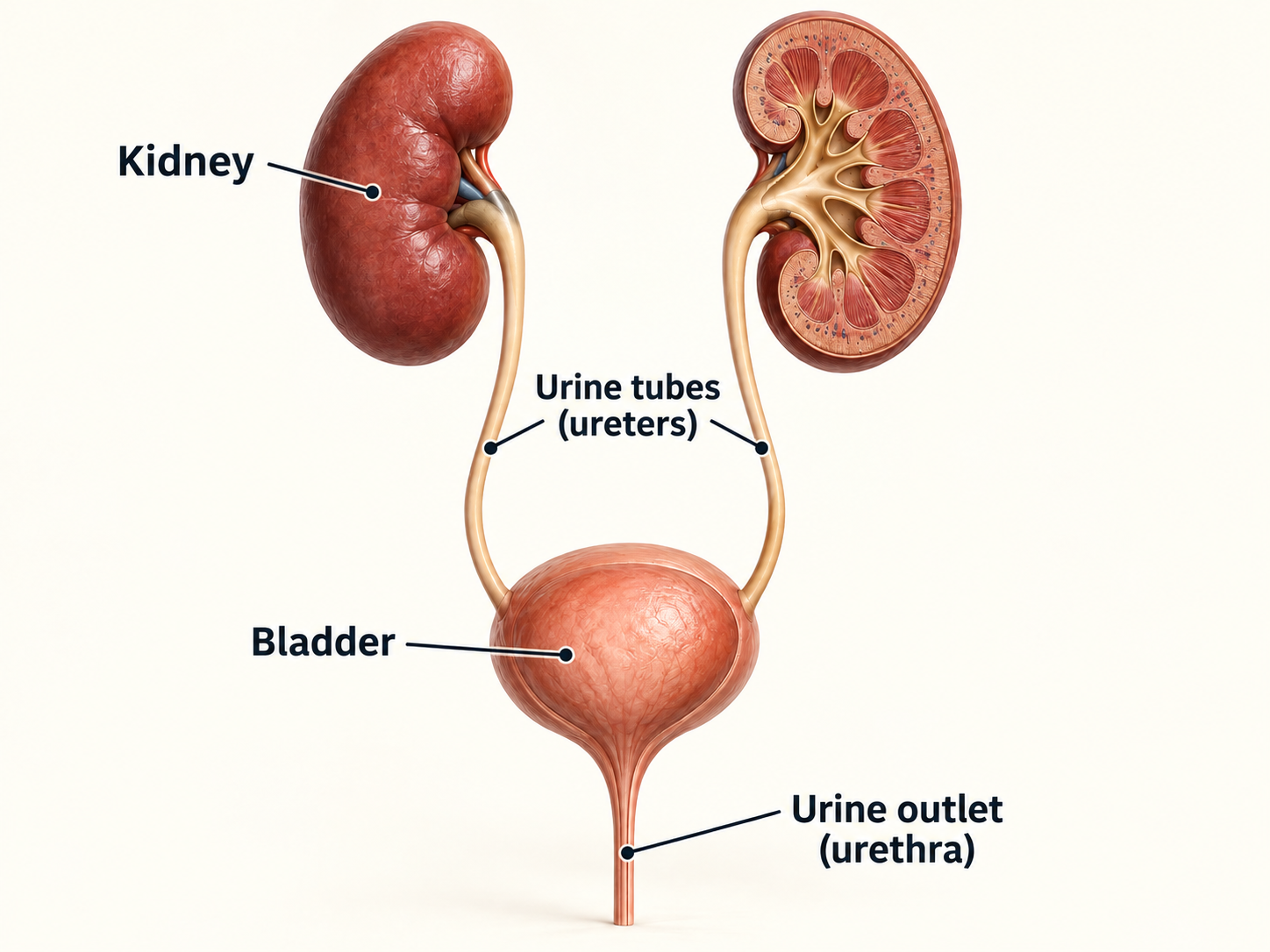
How the bladder works
- A stretchy inner lining keeps urine from leaking into tissue.
- The muscle wall relaxes as the bladder fills and contracts when you pass urine.
Why muscle depth matters
- A tumour confined to the lining behaves differently from one growing into muscle.
- Tissue from a good-quality TURBT is needed to judge this accurately.
Urothelial cancer elsewhere in the urinary tract
The same type of lining runs from the urine-collecting part of each kidney (the renal pelvis), through the ureters and bladder, into part of the urethra. Where the cancer starts affects the tests, surgery and follow-up needed.
Upper urinary tract
Cancer in the renal pelvis or ureter is called upper-tract urothelial carcinoma. It is assessed with scans and sometimes a thin camera passed into the ureter (ureteroscopy); treatment may involve the kidney and ureter.
Urethra and urachus
Cancer can also start in the urethra (the urine passage) or the urachus (a remnant of a channel present before birth, between the bladder and belly button). These uncommon cancers need specialist review because their treatment differs from usual bladder cancer.
Why the distinction matters
- A bladder tumour can coexist with or later recur elsewhere in the urothelial lining.
- Computed tomography (CT) urography and selected endoscopic tests check areas that an ordinary camera examination of the bladder (cystoscopy) cannot see.
- Pain in the side or back, a blockage or bleeding may require checks of the kidneys and ureters as well as the bladder.
- Follow-up may include upper-tract imaging or urethral assessment according to risk and treatment.
What bladder cancer is - and is not
Cancer begins when bladder cells acquire changes that let them grow and survive when they should not. The abnormal cells may form a finger-like (papillary) growth projecting into the space where urine collects, a flat high-grade area called carcinoma in situ (CIS), or an invasive mass.
The main cancer types
Type | What it means |
|---|---|
Urothelial carcinoma | The commonest type. It may be low-grade or high-grade and may show special variant patterns. |
Squamous carcinoma | Uncommon; may be linked with long-standing irritation or a parasitic infection called schistosomiasis. |
Adenocarcinoma | Uncommon cancer with gland-forming features. Primary bladder origin must be distinguished from spread from another organ. |
Small-cell / neuroendocrine | Rare, faster-growing type that usually needs chemotherapy travelling through the bloodstream as part of treatment. |
A pathology review can change the plan
Where possible, a pathologist specialising in urinary cancers should review high-grade T1 cancer, CIS, unusual or rare cell types, or cancer cells in small blood or lymph vessels (lymphovascular invasion). A tumour can contain more than one cell type, and a small biopsy may not show them all.
Bladder cancer is not contagious. It cannot be passed to family members through touch, food, sex or sharing a bathroom.
Symptoms and warning signs
Never ignore blood in the urine - even if it happens once, is painless, clears by itself or occurs while taking a blood thinner.
Possible symptoms
- Urine that is pink, red, cola-coloured or contains clots
- Blood seen only on a urine test
- Needing to pass urine more often or urgently
- Burning or pain when passing urine
- Difficulty passing urine or a weak stream
- Pelvic, side or back pain, unexplained weight loss, fatigue or bone pain in more advanced disease
When to seek urgent help
- Heavy bleeding or clots prevent urine from passing
- You cannot pass urine, or urine output falls sharply after surgery or with a surgically created route for urine to leave the body (a urinary diversion)
- You have fever, shivering, confusion, severe weakness, breathing difficulty or uncontrolled pain
- One leg suddenly swells, you develop chest pain, or you cough blood
If symptoms persist after treatment for a presumed urinary infection, or return soon afterwards, ask whether cystoscopy and upper urinary tract imaging are needed.
Risk factors
Usually there is no single cause. A risk factor changes probability; it does not prove why one person developed cancer. This is not your fault.
Factors with clear links
- Tobacco smoking - the most important preventable risk
- Workplace exposure to aromatic amines used historically in dye, rubber, leather, paint and related industries
- Older age and male sex
Other recognised links
- Prior pelvic radiotherapy
- Previous cyclophosphamide
- Long-standing irritation, stones or catheters
- Schistosomiasis, a parasitic infection acquired from contaminated freshwater in regions where it occurs
- Arsenic exposure in contaminated water
Questions your team may ask
- Do you currently smoke, or did you smoke in the past?
- What work have you done, and were dyes, solvents, fuels or industrial chemicals involved?
- Have you had pelvic radiotherapy or cyclophosphamide?
- Have you lived in or travelled to an area where urinary schistosomiasis occurs?
- Is there a pattern of bowel, womb, upper urinary tract or related cancers in you or your family that could suggest Lynch syndrome, an inherited condition that raises the risk of several cancers?
The key divide: NMIBC or MIBC
The bladder wall has an inner lining, a supporting layer beneath it and a deeper, thick muscle layer. This muscle, called the detrusor, squeezes to empty the bladder. The key question is whether cancer has grown into this muscle.
NMIBC means non-muscle-invasive bladder cancer. The cancer is in the lining or the supporting tissue beneath it, but has not reached the thick muscle layer. This includes Ta, Tis (also called carcinoma in situ, or CIS) and T1 tumours. T1 has grown into the supporting tissue, so “non-muscle-invasive” does not always mean “non-invasive”. Some NMIBC is high risk and can return or grow deeper, so treatment and regular bladder checks are important.
MIBC means muscle-invasive bladder cancer. The cancer has grown into the thick muscle layer (T2) or beyond it (T3–T4). This does not automatically mean it has spread to distant parts of the body. Scans and other tests help establish the full extent.
Why this changes treatment: NMIBC is often treated by removing the tumour through the urine passage, with medicines placed into the bladder when appropriate. Higher-risk cases may need more intensive treatment. MIBC usually needs a combination of treatments, which may include bladder-removal surgery, chemotherapy or radiotherapy. Your team uses the tissue results, scans and overall health to recommend a plan; the Treatment options section explains these choices.
Layer / term | Plain-language meaning |
|---|---|
Ta | A tumour growing from the lining into the bladder, often with finger-like projections. It has not grown into the supporting tissue or thick muscle layer: NMIBC. |
Tis / CIS | Carcinoma in situ (CIS): flat, high-grade cancer cells confined to the lining. It is NMIBC but can be high risk. |
T1 | Cancer has grown into the supporting layer beneath the lining, but not the thick muscle layer: NMIBC. |
T2 | Cancer has grown into the thick muscle layer: MIBC. |
T3-T4 | Cancer has grown beyond the thick muscle into surrounding fat (T3) or nearby structures (T4). |
Tests and diagnosis
Topics in this section
Your care team
Several specialists may be involved in your care. They discuss results together at a multidisciplinary team meeting, sometimes called a tumour board. Ask who coordinates your care and how the team will explain its recommendations.
Team member | Role |
|---|---|
Urologist / uro-oncologist | Performs cystoscopy, TURBT, cystectomy and urinary diversion; leads much of the diagnostic pathway. |
Medical oncologist | Plans chemotherapy, immunotherapy, antibody-drug conjugates and targeted treatment. |
Radiation oncologist | Plans bladder-preserving or symptom-directed radiotherapy. |
Pathologist | Confirms type, grade, depth, CIS, variant histology and other risk features. |
Radiologist / nuclear medicine | Interprets CT, magnetic resonance imaging (MRI), PET-CT (positron emission tomography combined with CT) when appropriate, and other scans to assess the cancer’s extent or response to treatment. |
Specialist nurse / stoma therapist | Teaches you how to care for a catheter, manage bladder treatments and, if needed, look after a stoma (an opening on the abdomen) or another urinary diversion. |
Supportive care team | Manages symptoms, nutrition, rehabilitation, sexual health, emotional support and practical needs. |
Before a major decision
- Confirm the pathology and whether bladder muscle was present in the TURBT specimen.
- Review scans in the clinical context, not as isolated reports.
- Discuss kidney function, hearing, nerve symptoms such as numbness or tingling (neuropathy), heart health, mobility, strength and your own priorities.
- Consider a second opinion for high-risk NMIBC, rare histology, bladder preservation or a major urinary diversion decision.
How cancer is diagnosed
Each test answers a different question. No urine test or scan can replace tissue diagnosis when a suspicious bladder lesion is found.
History, examination and urine testing
Checks bleeding pattern, infection, kidney function, blood count, medicines, tobacco and occupational exposure.
Imaging of the urinary tract
CT urography is commonly used to assess kidneys and ureters and look for obstruction, stones, masses or spread. MRI or ultrasound may be used in selected situations.
Flexible cystoscopy
A thin camera passes through the urethra to inspect the bladder lining. Local anaesthetic gel is commonly used.
TURBT and pathology
Visible tumour is removed through the urethra under anaesthesia. Tissue determines cancer type, grade and depth.
Staging and planning
Chest/abdomen/pelvis imaging, examination under anaesthesia and selected biomarkers guide the treatment plan.
Blue-light cystoscopy or narrow-band imaging may reveal lesions that are difficult to see with white light. Availability varies by centre.
Cystoscopy and TURBT
Cystoscopy finds the lesion. TURBT removes visible tumour and provides the tissue needed to plan treatment.
What a good TURBT aims to do
- Map the number, size and location of tumours and note any suspicious flat areas
- Remove visible papillary tumour safely and sample its base
- Include detrusor muscle when clinically appropriate so invasion can be assessed
- Avoid bladder perforation and control bleeding
- Record whether the resection appears complete
Soon afterwards
A catheter (a thin tube that drains urine) and gentle flushing of the bladder may be needed until the urine clears. Mild burning and some blood can occur.
Call urgently for
Inability to pass urine, heavy clots, increasing pain, fever, shivering, faintness or worsening bleeding.
After TURBT: recovery and getting the result
TURBT is both an operation and the diagnostic step that defines what happens next. Ask when the pathology will be reviewed and who will contact you.
The first few days
- Mild burning, urgency and pink urine are common. Follow the centre's advice on fluids, activity, driving and pain relief.
- A catheter may remain temporarily if bleeding, swelling or a deep resection makes drainage safer.
- Avoid heavy lifting and strenuous exercise until the urology team says the bladder has healed.
- Take antibiotics only if prescribed; burning alone does not prove infection.
The result should clarify
- The tumour type and grade
- Whether detrusor muscle was present and involved
- Whether the resection appeared complete
- Whether carcinoma in situ (CIS) was present
- Whether repeat TURBT is recommended
Understanding your pathology report
Do not rely on the word cancer alone. Several report details directly change treatment.
Report item | Why it matters |
|---|---|
Histological type | Usually urothelial carcinoma; rare or variant types may need a different plan. |
Grade | Low-grade tumours are more likely to return in the lining than to grow deeper or spread. High-grade tumours have a greater risk of growing deeper or spreading. |
Depth / T category | Separates Ta, T1 and muscle-invasive disease. |
Detrusor muscle | Knowing whether the sample contains bladder muscle, and whether cancer has grown into it, helps establish the stage accurately. |
CIS | Flat high-grade disease can be present beside or away from a visible tumour. |
Lymphovascular invasion | Cancer cells in small vessels can indicate higher risk. |
Variant morphology | Micropapillary, plasmacytoid, sarcomatoid, nested, neuroendocrine and other patterns may influence urgency and treatment. |
Ask for clarification if
- The report says T1 high-grade but no muscle is present
- The tumour could not be completely resected
- There is disagreement between the operative impression and pathology
- A rare type, variant pattern or lymphovascular invasion is reported
Scans, biomarkers and inherited risk
What common scans add
Test | Main question |
|---|---|
CT urography | Is there a bladder mass, obstruction, upper urinary tract lesion, enlarged node or other cause of bleeding? |
CT chest/abdomen/pelvis | Has muscle-invasive or high-risk disease spread beyond the bladder? |
MRI pelvis | Can local tumour depth and nearby organ involvement be defined more clearly? |
Bone scan / PET-CT | Is there suspected spread not fully answered by routine imaging? These are selected, not automatic, tests. |
Molecular testing and markers
- Changes in a gene called FGFR3 may identify a targeted medicine for advanced urothelial cancer.
- The amount of HER2 protein on cancer cells may help identify an antibody-drug conjugate treatment after previous treatments in selected advanced cancers.
- Tests may also assess PD-L1 (a protein involved in immune responses), DNA repair through mismatch repair or microsatellite instability testing, and the number of gene changes in a tumour (tumour mutational burden). These can guide treatment in selected situations, alongside the stage and your overall health.
- After bladder-removal surgery, a blood test can sometimes find tiny fragments of cancer DNA, called circulating tumour DNA (ctDNA), before cancer is visible on a scan. This finding is called molecular residual disease (MRD). Atezolizumab is an option after surgery for adults with MIBC who have ctDNA-MRD detected using an authorised companion test, a test approved to help select patients for that treatment.
Planning your treatment
Understanding your stage
TNM staging for bladder cancer
Bladder stage is mainly about how deeply cancer has entered the bladder wall, which lymph nodes are involved and whether it has reached other parts of the body. Grade describes how abnormal the cells look; a high-grade tumour can still be an early-stage cancer.
Lymph nodes are small immune-system structures. Regional nodes are those near or draining the cancer’s starting area; distant spread means cancer has reached a site outside that regional area. Stages describe findings, not a sequence that everyone will pass through.
Stage 0
Cancer is on the inner surface, without growing into the tissue beneath it. Ta forms papillary growths; Tis, also called carcinoma in situ, is flat and high-grade. Neither has invaded the bladder muscle.
Stage 1 (I)
Cancer has entered the supporting tissue just beneath the lining, but not the main muscle layer. It remains non-muscle-invasive, although high-grade T1 disease can carry substantial risk.
Stage 2 (II)
Cancer has reached the main bladder muscle. No regional lymph-node or distant spread is identified.
Stage 3 (III)
Cancer has grown into tissue around the bladder, certain nearby reproductive organs, or regional lymph nodes. There is no distant spread.
Stage 4 (IV)
Stage IVA means invasion of the pelvic or abdominal wall, or spread to distant lymph nodes. Stage IVB means spread to distant organs, such as bone, liver or lungs. Stage IVA is therefore not always distant organ spread.
The TURBT specimen must show which layers were sampled. If muscle is missing, or results suggest high-risk disease, a repeat procedure may be needed before the team can confidently recommend treatment. Non-muscle-invasive risk groups and muscle-invasive stage are different ways of answering different questions.
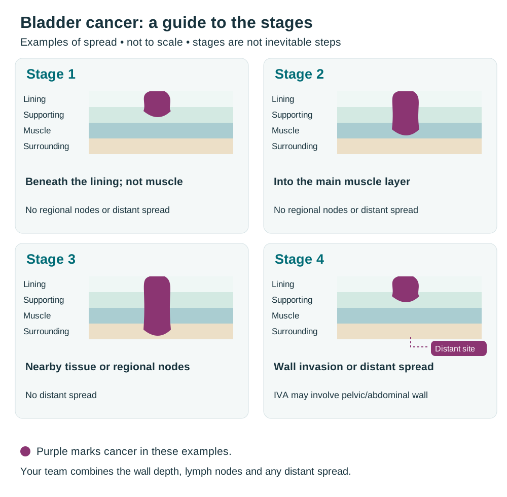
CTI illustration. Enlarge the staging overview.
Clinical stage comes from examination, biopsies and scans. Pathological stage can add information from surgery; a prefix such as yp means the tissue was assessed after treatment. A revised assessment does not necessarily mean the cancer grew while you were waiting. Grade describes the cells’ behaviour and is different from stage.
NMIBC risk is not based on stage alone
Low, intermediate, high and very high risk reflect a combination of stage, grade, CIS, tumour size, number, recurrence pattern, age and selected pathology features. Different international systems do not classify every tumour identically.
Main recurrence question
How likely is the tumour to return somewhere in the bladder?
Main progression question
How likely is the tumour to grow into muscle or spread?
How your treatment plan is chosen
Good decisions combine medical evidence with what matters to you. The first safe plan is not always the only reasonable plan.
Questions about the cancer
- Is disease NMIBC, MIBC, locally advanced or metastatic?
- What are the grade, CIS status, variant features, lymphovascular invasion and NMIBC risk group?
- Was TURBT complete, and was adequate muscle sampled?
- Is the bladder functioning well enough to preserve?
Your health and priorities
- Kidney function and ability to receive cisplatin
- Hearing, neuropathy, diabetes, heart/lung health, frailty and mobility
- Prior pelvic radiotherapy, bowel disease or autoimmune illness
- Values concerning cancer control, bladder preservation, stoma, sexual function, fertility, travel, work and cost
If a decision is not urgent, ask how much time you safely have to obtain pathology review, optimise health, stop smoking, discuss fertility and meet a stoma therapist or radiation oncologist.
Treatment options
Topics in this section
- Low-risk NMIBC
- Intermediate-risk NMIBC
- High- and very-high-risk NMIBC
- BCG: what to expect
- When high-risk NMIBC does not respond to BCG
- Muscle-invasive bladder cancer: the curative pathways
- Medicines before and after bladder-removal surgery
- Radical cystectomy and lymph-node surgery
- Preparing for and recovering from cystectomy
- Choosing a urinary diversion
- Bladder-preserving trimodality treatment
- Locally advanced or metastatic urothelial cancer
- Treatment if cancer grows or returns
Low-risk NMIBC
The usual goal is to remove tumour, reduce recurrence and avoid more treatment than the cancer requires.
Complete TURBT
Visible tumour is removed and pathology confirms a low-grade Ta pattern without high-risk features.
Chemotherapy placed into the bladder soon after TURBT
A single dose soon after TURBT may reduce recurrence when there is no perforation or major bleeding. Gemcitabine or mitomycin are common examples.
Follow-up matched to your risk
Cystoscopy is usually performed at about three months. Later intervals depend on findings and local guidance.
Treat small recurrences sensibly
For carefully selected small, low-grade tumours that return, options may include another TURBT, treatment with heat or a laser in clinic, or close monitoring before deciding whether treatment is needed.
What low risk does not mean
- It does not mean no follow-up is needed.
- A recurrence still needs assessment; grade or risk can change over time.
- Persistent bleeding, pain or urinary symptoms should not be assumed to be harmless.
Smoking cessation is part of treatment, not a judgement. Ask for structured support rather than relying on willpower alone.
Intermediate-risk NMIBC
Intermediate risk is a broad group. The number, size, grade, timing and frequency of tumours help decide how intensive treatment should be.
Common approaches after complete TURBT
Chemotherapy placed into the bladder
- A course placed into the bladder over weeks
- Examples include gemcitabine or mitomycin
- Maintenance may continue for selected patients, generally up to one year
BCG (Bacillus Calmette–Guérin) immunotherapy
- May be chosen when risk is closer to high risk
- Usually an initial course of six weekly treatments, called induction
- Maintenance may be used according to risk, tolerance and supply
The plan may be adjusted by
- Multiple tumours, size at least 3 cm, early or frequent recurrence and prior intravesical treatment
- Any high-grade component, T1 disease or CIS - these can move the person into a high-risk pathway
- Bladder symptoms, catheter tolerance, kidney function and availability of medicines
- Quality of the first TURBT and whether repeat resection is required
Low-grade recurrence and high-grade progression are not the same event. Ask which outcome your recommended treatment is intended to prevent.
High- and very-high-risk NMIBC
The priority is to prevent muscle invasion and metastatic spread while discussing honestly when early bladder removal offers the safest cancer control.
Standard high-risk pathway
- Confirm high-quality TURBT and consider repeat resection, especially for T1 high-grade disease.
- Discuss your diagnosis and treatment options with a multidisciplinary team and ask whether an expert pathology review would help.
- Offer an initial BCG course, followed by further courses to help maintain its effect when appropriate and tolerated.
- Arrange close follow-up with bladder camera checks, tests for cancer cells in urine and scans of the kidneys and ureters.
When early radical cystectomy deserves serious discussion
- Very-high-risk T1 disease, extensive or persistent high-grade disease, or associated CIS
- Variant histology, lymphovascular invasion or tumour involving the prostatic urethra
- A tumour that cannot be fully resected or is repeatedly understaged
- Progression or disease unlikely to respond safely to more intravesical treatment
A decision to preserve the bladder should include a clear monitoring plan and an agreement about which findings would make bladder removal advisable.
BCG: what to expect
BCG is a live weakened bacterium placed into the bladder to stimulate an immune response against high-risk NMIBC.
Typical treatment rhythm
- Induction is commonly once weekly for six weeks.
- Maintenance adds shorter courses over months or years. The exact schedule depends on risk, response, side effects and BCG supply.
- BCG is delayed after TURBT until the bladder has healed and is not given if inserting the catheter has injured the urine passage or with significant visible bleeding.
Common temporary effects
- Urgency and frequency
- Burning when passing urine
- Mild blood in urine
- Tiredness, aches or low fever
Contact the team urgently
- A temperature of 38°C (100.4°F) or higher, shaking chills or feeling suddenly unwell: contact your treatment team straight away. Follow any lower threshold your team gives you; do not wait 48 hours to seek advice.
- High fever with shivering or severe weakness
- Breathing difficulty, jaundice, confusion
- Inability to pass urine or heavy bleeding
Practical safety
- Tell the team about infection symptoms, immune suppression, tuberculosis history and medicines that affect immunity.
- Follow the centre's instructions for fluid restriction before treatment and safe toilet hygiene afterwards.
- Do not dismiss persistent symptoms as normal BCG irritation; infection and recurrence may need to be excluded.
When high-risk NMIBC does not respond to BCG
“BCG-unresponsive” describes certain high-grade cancers that persist or return despite adequate BCG treatment. The timing and amount of treatment matter. Your team will check whether this definition applies, because giving more BCG alone is unlikely to help this group.
Preferred cancer-control option
Radical cystectomy is the standard preferred treatment for a fit person with BCG-unresponsive high-risk NMIBC. When done before progression to MIBC, it offers the strongest chance of cure. Delaying surgery can allow an otherwise curable cancer to progress.
If cystectomy is not possible or is declined
- Clinical trial participation where available
- Intravesical nadofaragene firadenovec gene therapy for CIS with or without papillary tumour
- Nogapendekin alfa inbakicept with BCG for CIS-containing disease
- Intravesical gemcitabine-delivery system for CIS-containing disease
- Intravenous pembrolizumab for selected CIS-containing disease
- Sequential intravesical gemcitabine and docetaxel, a commonly used bladder-sparing approach with largely non-randomised evidence
Muscle-invasive bladder cancer: the curative pathways
When MIBC is confined to the bladder and nearby area, treatment usually addresses the bladder tumour and any tiny groups of cancer cells elsewhere that tests cannot detect. Medicines that travel through the bloodstream are called systemic therapy.
Path A: surgery-based
- Systemic treatment before surgery when suitable
- Bladder removal with removal of lymph nodes in the pelvis
- Adjuvant treatment or molecular monitoring in selected high-risk cases
Path B: bladder-preserving
- Maximal TURBT
- Chemotherapy given during radiotherapy
- Lifelong bladder camera checks
- Bladder removal if invasive cancer returns after bladder-preserving treatment
Before choosing
- Confirm clinical stage with imaging and pathology review.
- Assess kidney function, hearing, neuropathy, diabetes, autoimmune disease, frailty and surgical risk.
- Review whether the tumour is solitary, completely resectable, associated with extensive CIS or causing kidney swelling because urine cannot drain normally (hydronephrosis).
- Meet both a urologic surgeon and radiation oncologist if bladder preservation is being considered.
- Discuss urinary diversion, sexual function, fertility and rehabilitation before treatment starts.
The best route depends on tumour factors, health, local expertise and your informed preference. Both pathways need careful planning and follow-up.
Medicines before and after bladder-removal surgery
Medicines given before surgery (neoadjuvant treatment) can treat tiny groups of cancer cells early. Treatment after surgery is called adjuvant treatment; treatment given before and after surgery is called perioperative treatment.
Treatment choices before and after surgery
Approach | Who may discuss it |
|---|---|
Enfortumab vedotin (EV) + pembrolizumab before and after cystectomy | An option for adults with MIBC who are candidates for cystectomy, including eligible patients who can receive cisplatin. |
Gemcitabine + cisplatin + durvalumab before surgery, then durvalumab | A perioperative option for cisplatin- and immunotherapy-eligible resectable MIBC. |
Neoadjuvant cisplatin-based chemotherapy | A long-established treatment that remains appropriate when combinations involving immunotherapy or antibody-drug conjugates are unsuitable or unavailable. |
After surgery
- Pathological stage and lymph nodes estimate recurrence risk.
- Adjuvant nivolumab is an established option for selected high-risk urothelial carcinoma after radical surgery.
- Adjuvant atezolizumab is an option for ctDNA-MRD-positive MIBC after cystectomy using an authorised companion test.
- Additional chemotherapy may be considered when no effective neoadjuvant cisplatin was given and pathology shows high-risk disease.
Radical cystectomy and lymph-node surgery
Radical cystectomy removes the bladder and nearby lymph nodes. During the same operation, the surgeon creates a new way for urine to leave the body, called a urinary diversion. The main choices are explained below.
What may be removed
In male anatomy
The bladder, nearby lymph nodes and usually the prostate and seminal vesicles. Urethral and nerve-sparing choices are individualised.
In female anatomy
The bladder, nearby lymph nodes and sometimes the uterus, ovaries, fallopian tubes, part of the vagina and urethra. Organ-sparing may be possible in selected cases.
The exact operation depends on tumour location, urethral risk, prior treatment, anatomy, fertility goals and surgical judgement. Ask what is planned in your own operation rather than assuming a standard list.
Preparing well
- Meet the stoma/continence nurse before surgery and mark a suitable stoma site even if another diversion is planned.
- Discuss nutrition, anaemia, diabetes, smoking, exercise/prehabilitation and medicines that affect bleeding.
- Ask about open, laparoscopic or robotic surgery. The surgeon's experience and safe lymph-node dissection matter more than the size of the skin incision alone.
- Plan pain control, early mobilisation, breathing exercises and blood-clot prevention.
Main risks
Bleeding, infection, bowel leak or blockage, urine leak, blood clots, heart/lung problems, wound complications, kidney injury and readmission can occur. Recovery commonly takes weeks to months.
Preparing for and recovering from cystectomy
Cystectomy is a major operation. Preparation, experienced teams and a clear diversion-care plan can reduce avoidable problems.
Ask how often the centre performs this operation, how the team supports recovery and manages complications, and what long-term help is available for your urinary diversion.
Before admission
- Build walking and strength as able; correct anaemia, dehydration, poor nutrition and uncontrolled diabetes; stop tobacco.
- Review blood thinners, diabetes medicines, supplements and allergies with the team.
- Meet the stoma/continence nurse, practise with sample appliances and mark a stoma site while sitting, standing and bending.
- Plan help at home, transport, supplies and a contact route for nights or weekends.
A typical recovery pathway
Hospital recovery
Pain control, breathing exercises, clot prevention, early walking and gradual return of bowel function.
Learn the diversion
Practise pouch changes, catheter care, irrigation or timed emptying before discharge.
Home recovery
Short walks, adequate protein and fluids, lifting restrictions, wound checks and scheduled follow-up.
When to seek urgent help
Chest pain, breathlessness, one swollen leg, fever, persistent vomiting, severe abdominal swelling, falling urine output, a blocked catheter or rapidly worsening wound/stoma changes.
Choosing a urinary diversion
Your team will help you choose a way to drain urine that suits your cancer treatment, kidney and bowel health, ability to use the equipment and daily life. Each option involves learning new routines.
Diversion | Daily-life features |
|---|---|
Ileal conduit | A short piece of bowel carries urine to an opening on the abdomen (a stoma), where it drains into a bag. This is usually the simplest and most widely used option. |
Neobladder | A pouch made from bowel connects to the urine passage. You learn to empty it at set times and exercise the pelvic-floor muscles. Some people need to pass a catheter themselves to empty it; leakage at night is common early on. |
Continent pouch | A pouch made from bowel sits inside the abdomen. You empty it by passing a catheter through a small opening on the abdomen. An external bag is not needed, but this catheter routine is lifelong. |
Bladder-preserving trimodality treatment
Trimodality treatment (TMT) combines three treatments: removing as much visible tumour as safely possible by TURBT, radiotherapy and a medicine that makes cancer cells more sensitive to radiation. It offers a chance of cure while keeping the bladder in carefully selected people.
Features that often favour TMT
- A tumour that can be largely or completely resected by TURBT
- Good bladder capacity and function
- No extensive CIS and no severe hydronephrosis
- Ability to receive concurrent chemotherapy and complete radiotherapy
- Commitment to repeated cystoscopy and willingness to have salvage cystectomy if needed
The treatment sequence
Maximal TURBT
Removes as much visible tumour as safely possible.
Planning
CT-based radiotherapy planning defines bladder, tumour and nearby organs. Bladder-filling instructions may be used.
Chemoradiotherapy
Radiotherapy is delivered with a radiosensitiser such as cisplatin, gemcitabine, or fluorouracil plus mitomycin according to centre protocol.
Response and lifelong surveillance
Cystoscopy, urine cytology and imaging monitor response and detect recurrence.
TMT is not simply for people considered too frail for surgery; it works best when selection, TURBT, radiation planning and follow-up are high quality.
Locally advanced or metastatic urothelial cancer
Treatment aims to control cancer throughout the body, extend life, relieve symptoms and preserve function. Some responses are long-lasting.
Common first-line choices
Preferred if suitable and available
Enfortumab vedotin (EV) plus pembrolizumab is a standard first treatment for advanced urothelial cancer when the combination is suitable. It can be used whether or not cisplatin chemotherapy is suitable, and does not require a particular PD-L1 test result.
When EV is unsuitable or unavailable
Gemcitabine with cisplatin or carboplatin, followed by avelumab to help maintain control if the cancer has not grown or spread during chemotherapy. Cisplatin-gemcitabine with nivolumab is another approved option.
Fitness changes the choice
- Kidney function, hearing, neuropathy and heart function affect cisplatin eligibility.
- Uncontrolled diabetes, significant neuropathy, serious skin disease or eye problems may affect whether enfortumab vedotin is suitable.
- Autoimmune illness, an organ transplant or a previous serious reaction to immunotherapy can affect whether checkpoint immunotherapy is suitable.
- A person too frail for combination treatment may discuss single-agent immunotherapy in selected settings, a gentler regimen or supportive care alone.
Ask how response will be measured, when the first scan will occur and what the next option would be if treatment stops working.
Treatment if cancer grows or returns
If cancer grows or returns, the next treatment depends on the medicines you have already received, your current health and the cancer’s gene or protein test results (biomarkers).
If cancer progresses after EV plus pembrolizumab
- Platinum-gemcitabine chemotherapy if you are fit and have not already received it
- Erdafitinib for a susceptible FGFR3 alteration
- Single-agent chemotherapy such as a taxane in selected patients
- Clinical trials testing new antibodies, combinations or biomarker-led approaches
If the earlier pathway was platinum chemotherapy plus immunotherapy
- Enfortumab vedotin if not used before
- Erdafitinib when an actionable FGFR3 alteration is present
- Pembrolizumab if no checkpoint inhibitor has been given and it fits the regulatory setting
After several treatments
Trastuzumab deruxtecan may be an option for adults with unresectable or metastatic HER2-positive (IHC 3+) cancer after previous systemic treatment when there is no satisfactory alternative. This approval covers several cancer types with the specified HER2 result; it does not apply to every level of HER2 protein. It can cause serious lung inflammation; new cough or breathlessness needs urgent assessment.
Molecular profiling is most useful when the result can change treatment. Ask which genes or proteins are being tested, whether tissue is adequate and who will interpret the report.
Managing side effects
Side effects of treatment placed into the bladder
Most bladder symptoms are temporary. However, infection, blocked urine flow, a hole in the bladder wall or a BCG reaction affecting the body need prompt assessment.
What you notice | What to do |
|---|---|
Burning, urgency, frequency | Expected for a short time after many treatments placed into the bladder. Use only medicines recommended by the team and report symptoms that are severe or persistent. |
Mild blood in urine | Often settles. Increase fluids after the advised holding period unless you have a fluid restriction. Report worsening bleeding or clots. |
Fever or flu-like symptoms after BCG | Mild bladder irritation can occur after BCG. For a temperature of 38°C (100.4°F) or higher, shaking chills or feeling suddenly unwell, contact the team straight away. Do not wait for symptoms to last 48 hours. |
Cannot pass urine | Urgent: clot retention or swelling may be blocking urine flow. |
Rash, wheeze, facial swelling | Emergency assessment for a possible severe allergic reaction. |
Before each dose, tell the team about
- Visible bleeding, difficult catheterisation, fever or urinary infection symptoms
- Antibiotics, blood thinners, immune-suppressing medicines or a new diagnosis
- Recent surgery or a change in pregnancy possibility
Do not self-start antibiotics for every bladder symptom. A urine culture and clinical review may be needed, and some antibiotics can interact with treatment plans.
Systemic therapy: know the urgent signals
Side effects are easier to treat when reported early. Keep the 24-hour oncology contact number with you.
Enfortumab vedotin
- Rash or blistering skin
- Numbness or weakness
- High blood sugar, thirst or confusion
- Dry/painful eyes
- New cough or breathlessness
Checkpoint immunotherapy
- Diarrhoea or blood in stool
- New cough, chest pain or breathlessness
- Yellow eyes, dark urine
- Severe headache, weakness or confusion
- Major change in thirst, urine or energy
Chemotherapy concerns
- A temperature of 38°C (100.4°F) or higher, or a lower threshold given by your unit, shaking chills or feeling suddenly unwell: contact the oncology team immediately.
- Uncontrolled vomiting/diarrhoea, dehydration or inability to eat and drink
- New bleeding, bruising, mouth ulcers or severe fatigue
- Reduced urine output, hearing change, severe numbness or balance problems
Carry an immunotherapy/ADC alert card if your centre provides one. Tell every clinician, including emergency staff, exactly which anti-cancer medicines you receive.
Fertility and pregnancy precautions vary by drug. Discuss contraception and preservation options before treatment begins.
Recovery after cystectomy or chemoradiotherapy
After cystectomy
- Fatigue, low appetite and altered bowel habit improve gradually
- Learn stoma or neobladder care before discharge
- Walk regularly and follow lifting restrictions
- Watch hydration, urine flow, wounds and infection
After chemoradiotherapy
- Urgency, frequency, burning and loose stools may peak near the end
- Fatigue can continue for weeks
- Bleeding, reduced bladder capacity or changes in bowel function can develop later
- Cystoscopic follow-up remains essential
Living safely with a urinary diversion
- Protect kidney function with planned blood tests, blood pressure checks and imaging.
- Know the expected colour and amount of urine. Mucus is normal when bowel is used to carry or store urine.
- Ask about checks of vitamin B12, blood salts (electrolytes) and the blood’s acid balance after a urinary diversion made from bowel.
- For a neobladder or continent pouch, follow timed emptying, irrigation and self-catheterisation instructions exactly.
- Keep spare stoma supplies when travelling and know how to obtain compatible products.
Do not compare recovery by incision size alone. Cystectomy changes the urinary and bowel systems internally even when performed robotically.
Live well
Follow-up after treatment
Bladder cancer can return in the bladder, upper urinary tract, lymph nodes or distant organs. Follow-up is part of treatment, not an optional extra.
After NMIBC
- The first follow-up cystoscopy is commonly around three months after TURBT.
- Low-risk schedules become less frequent if cystoscopy remains clear.
- High-risk schedules are more frequent and include urine cytology and periodic upper-tract imaging; long-term or lifelong surveillance may be advised.
- If cancer returns, it should be assessed again for stage and grade; these may differ from the original tumour.
After MIBC treatment
- CT or other imaging of chest, abdomen and pelvis according to stage, treatment and time since therapy
- Kidney function, blood count, electrolytes and metabolic monitoring after urinary diversion
- Cystoscopy and urine assessment after bladder-preserving treatment
- Assessment of stoma/diversion, bowel function, nutrition, bone health, sexual health, mobility and emotional wellbeing
Follow-up schedules vary among guidelines and should be personalised. Ask your team to talk through your follow-up schedule, including when each test is due and why it is helpful.
Daily life with a urinary diversion
Skills become easier with practice. Ask for easy-to-follow guidance, a named stoma or continence contact and hands-on teaching for you and, if you wish, a caregiver.
Ileal conduit
- Empty the pouch before it becomes heavy
- Protect the skin and measure the stoma if its size changes
- Keep spare appliances, seals and disposal bags
Neobladder or pouch
- Follow timed emptying and night alarms
- Learn flushing and how to pass a catheter yourself, if prescribed
- Report ongoing difficulty emptying the pouch or persistent leakage
What can be normal
- Mucus in urine because bowel tissue continues to make mucus
- A stoma that is pink/red and moist, with small spots of blood during cleaning
- Changing continence during neobladder training, especially at night
- Body-image worries and a learning curve with equipment
Prevent avoidable problems
- Maintain the fluid plan agreed with your team and watch urine colour and output.
- Use only compatible products and avoid creams beneath an adhesive unless advised.
- Carry supplies, a brief medical summary and emergency contacts when travelling.
- Ask about checks of vitamin B12, kidney function, blood salts and the blood’s acid balance after a urinary diversion made from bowel.
Food, activity, smoking and emotional health
There is no bladder-cancer diet. The goal is to rebuild strength, protect kidney function and make daily life workable.
Practical foundations
- Eat enough protein and energy during recovery. Ask for a dietitian if weight or muscle is falling, appetite is poor, or kidney function limits choices.
- Drink enough to avoid dehydration unless your doctor has prescribed a fluid restriction. Diversions can increase fluid loss; more water is not always better for every heart or kidney condition.
- Restart walking early and build gradually towards strength and aerobic activity. Pelvic-floor rehabilitation may help selected patients.
- Avoid unverified herbal products, urinary cleanses and high-dose supplements. They can interact with treatment or affect kidneys and bleeding.
Emotional recovery
Fear of recurrence, sleep problems, altered body image and embarrassment about urine or a stoma are common. Tell the team if anxiety, low mood or loss of interest persists or affects daily life. Counselling, peer support, rehabilitation and medication can help.
For patients
Ask about body image, continence, intimacy, fertility, work and travel before these become crises.
For caregivers
Learn warning signs and practical care, but protect your own sleep, health and emotional support.
Sex, relationships and practical support
Sexual health and fertility
- Cystectomy can affect erections, ejaculation, vaginal length/lubrication, sensation, fertility and menopause. Nerve- or organ-sparing may be possible in selected cases but cannot be promised.
- Pelvic radiotherapy can also affect erections, vaginal tissue, bowel/bladder function and fertility.
- Ask before treatment about sperm, egg or embryo preservation, vaginal dilators, pelvic-floor therapy and medicines/devices for sexual rehabilitation.
- Closeness can continue even when intercourse changes. A sexual-health clinician can help individuals and couples adapt.
Sources and further reading
For more information, explore these resources from the National Cancer Institute (NCI) and other medical organisations. The linked organisations have not reviewed or endorsed this CTI guide. Your care team can explain how the information applies to you.
Staging and risk-group references
AJCC 8th edition.
Medicine approval and prescribing references
These official records describe licensed uses and prescribing information for the medicines named in this guide. Approval depends on the cancer type, biomarkers, previous treatment, combination and formulation. Some uses discussed in the guide are off-label: outside the medicine’s licence, with evidence from clinical guidelines or studies. FDA or EMA approval does not establish approval or availability in India.
- BCG for use inside the bladder (TICE BCG) — FDA approval record
- Atezolizumab — FDA prescribing information · Approval history
- Avelumab — FDA prescribing information · Approval history
- Carboplatin — FDA prescribing information · Approval history
- Cisplatin — FDA prescribing information · Approval history
- Docetaxel — FDA prescribing information · Approval history. Sequential intravesical gemcitabine–docetaxel is an off-label regimen.
- Durvalumab — FDA prescribing information · Approval history
- Enfortumab vedotin — FDA prescribing information · Approval history
- Erdafitinib — FDA prescribing information · Approval history
- Fluorouracil — FDA prescribing information · Approval history
- Gemcitabine — FDA prescribing information · Approval history. Conventional intravenous/intravesical use is distinct from the separately approved Inlexzo delivery system.
- Gemcitabine bladder-delivery system (Inlexzo) — FDA prescribing information · Approval history
- Mitomycin — FDA prescribing information · Approval history
- Nadofaragene firadenovec — FDA approval record
- Nivolumab — FDA prescribing information · Approval history
- Nogapendekin alfa inbakicept — FDA prescribing information · Approval history
- Pembrolizumab — FDA prescribing information · Approval history
- Sacituzumab govitecan — FDA withdrawal record. The urothelial-cancer indication was withdrawn; this is not a current bladder-cancer approval.
- Trastuzumab deruxtecan — FDA prescribing information · Approval history. A tumour-agnostic indication applies to eligible HER2 IHC 3+ disease.
Additional medicine approval notices
These records explain the specific medicine uses and eligibility requirements discussed in this guide.
Related care
Meet our doctors



Where to get care